. 2007 Jan 26;365(4):1201-16.
doi: 10.1016/j.jmb.2006.11.044. Epub 2006 Nov 15.

Stabilizing IkappaBalpha by "consensus" design

Affiliations

Stabilizing IkappaBalpha by "consensus" design

Diego U Ferreiro et al. J Mol Biol. .

Abstract

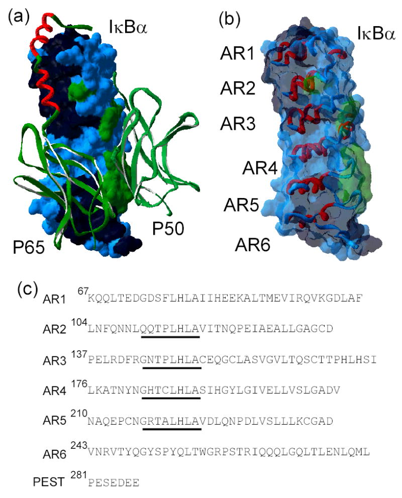
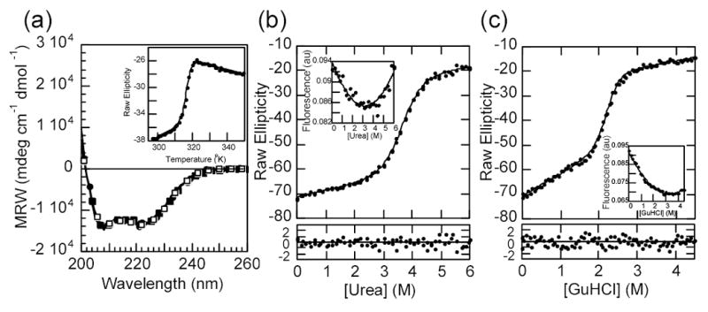
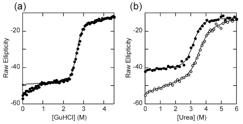
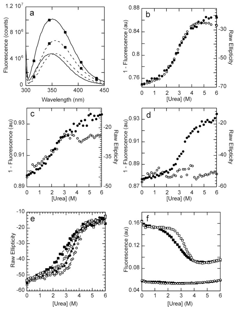
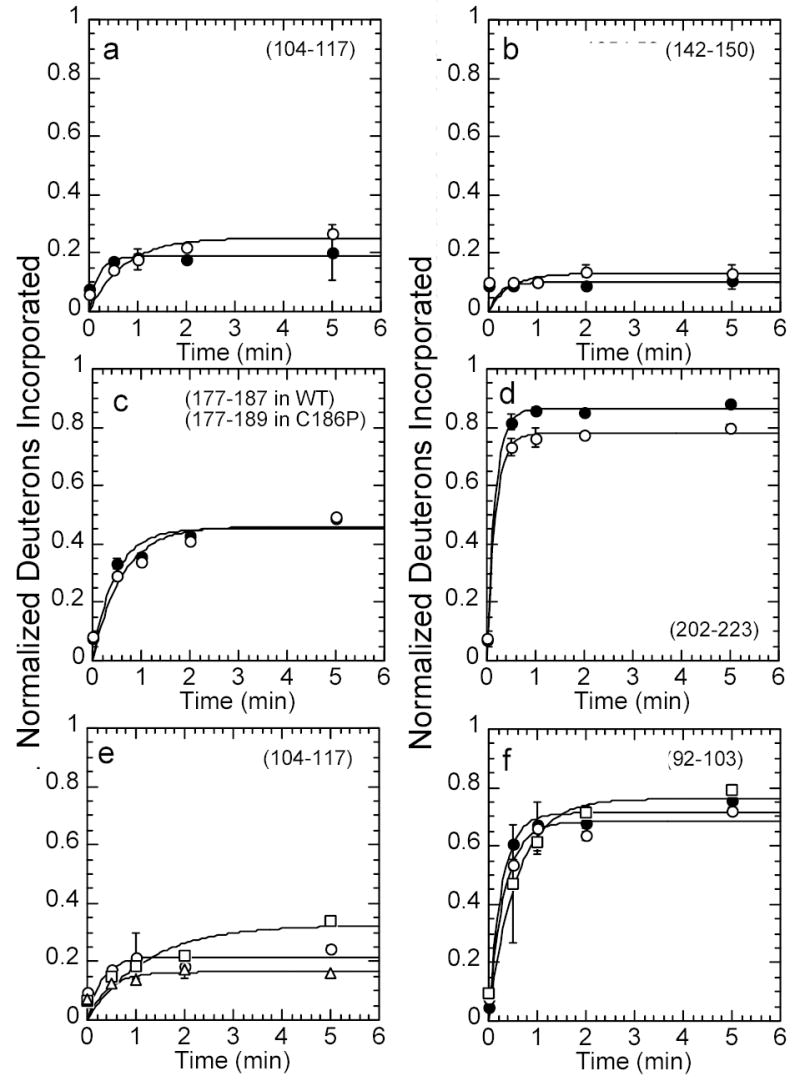
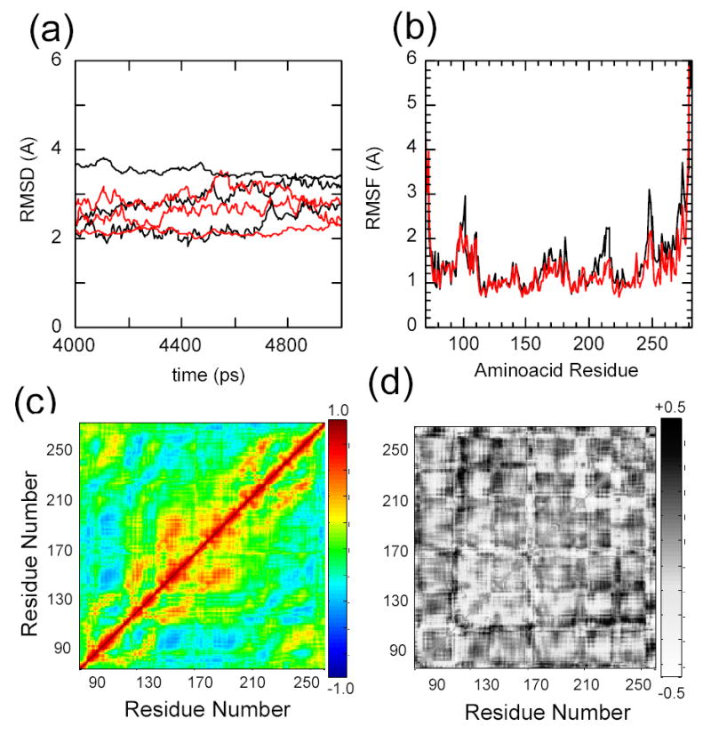
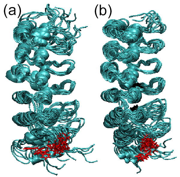
IkappaBalpha is the major regulator of transcription factor NF-kappaB function. The ankyrin repeat region of IkappaBalpha mediates specific interactions with NF-kappaB dimers, but ankyrin repeats 1, 5 and 6 display a highly dynamic character when not in complex with NF-kappaB. Using chemical denaturation, we show here that IkappaBalpha displays two folding transitions: a non-cooperative conversion under weak perturbation, and a major cooperative folding phase upon stronger insult. Taking advantage of a native Trp residue in ankyrin repeat (AR) 6 and engineered Trp residues in AR2, AR4 and AR5, we show that the cooperative transition involves AR2 and AR3, while the non-cooperative transition involves AR5 and AR6. The major structural transition can be affected by single amino acid substitutions converging to the "consensus" ankyrin repeat sequence, increasing the native state stability significantly. We further characterized the structural and dynamic properties of the native state ensemble of IkappaBalpha and the stabilized mutants by H/(2)H exchange mass spectrometry and NMR. The solution experiments were complemented with molecular dynamics simulations to elucidate the microscopic origins of the stabilizing effect of the consensus substitutions, which can be traced to the fast conformational dynamics of the folded ensemble.


Figures

Figure 1
Figure 1
a) The x-ray crystal structure of IκBα (surface representation) in complex with NF-κB (ribbon representation) (pdb accession number 1NFI) . The dimerization domains of NF-κB p50 and p65 are shown in green and the NF-κB p65-NLS polypeptide in red. The surface of IκBα is colored according to the NF-κB contact regions: p65 contacting residues in black, p50 contacting residues in green, non-contacting residues in blue. b) The high resolution structure of only IκBα from the structure of the complex in the same orientation as in a), with a ribbon representation of the ankyrin repeats (AR), and the same coloring scheme as in a) for the translucent surface. c) Amino acid sequence of the ankyrin repeat domain of IκBα(67 to 287), aligned according to each ankyrin repeat. The ‘consensus’ GxTPLHLA motif is underlined.
Figure 2
Figure 2
a) The far UV circular dichroism spectra of IκBα and selected mutants: wild type, continuous line; Q111G, open circles; C186P·A220P, closed circles, Q111G·C186P·A220P, closed squares. Inset: thermal unfolding of IκBα wild type followed by the CD signal at 225 nm. b) Urea denaturation curve of IκBα wild type at 3 μM total protein concentration, (conditions described in Materials and Methods) followed by the CD signal at 225nm. The line represents the best fit of the data to a two-state folding model with a linear drift in the native ensemble baseline. The residuals of the fit are shown in the bottom panel. Inset: intrinsic fluorescence of IκBα collected during the same experiment. The line is drawn through the points merely to guide the eye. c) GuHCl denaturation curve of IκBα wild type in the same conditions as in a). Insert: intrinsic fluorescence of IκBα collected during the same experiment.
Figure 3
Figure 3
a) GuHCl denaturation curve of IκBα Q111G·C186P·A220P at 2 μM under standard conditions. The line represents the best fit to a two-state folding model. The data points below 1M were left out of the fit. b) Urea denaturation curve of IκBα(67-206) (closed circles) with the urea denaturation curve for the wild type protein shown for comparison (open circles). The line represents the best fit to a two-state folding model.
Figure 4
Figure 4. Probing the local folding of AR2-AR3
a) Fluorescence emission spectra of wild type IκBα at 1 μM in standard conditions and the A133W mutant (marked by closed squares) under the same conditions. The continuous line shows the emission spectra under native conditions and the dashed line shows the emission spectra of the same proteins in 6M urea. The excitation wavelength was set to 295 nm. b) Urea denaturation curve of IκBα A133W showing the CD signal (closed circles) and the fluorescent signal (open circles). c) Urea denaturation curve of IκBα L205W showing the CD signal (closed circles) and the fluorescent signal (open circles). d) Urea denaturation curve of IκBα C239W showing the CD signal (closed circles) and the fluorescent signal (open circles). e) Urea denaturation curves followed by the CD signal at 225nm of IκBα wild type (closed circles), A133W (closed squares), C186P·A220P (open circles) and A133W·C186P·A220P (open squares). f) Urea denaturation curves followed by the fluorescent signal of the same proteins, with the same symbols as in e).
Figure 5
Figure 5
Kinetic plots of amide H/2H exchange (fit to a single exponential) in wild type IκBα, IκBα C186P·A220P, IκBα A133W·C186P·A220P, and IκBα Q111G·C186P·A220P. Deuterium incorporation was compared in wild type IκBα (filled circles) and IκBα C186P·A220P (open circles) in (a) the β-hairpin loop in AR2 (residues 104-117, 12 amides), (b) the β-hairpin loop in AR3 (residues 142-150, 7 amides), (c) the β-hairpin loop in AR4 (residues 177-187, 10 amides in wild type IκBα, residues 177-189, 11 amides in C186P), (d) the β-hairpin loop in AR5 and the end of the variable loop in AR4 (residues 202-223, 19 amides in wild type IκBα, 18 amides in A220P). Deuterium incorporation was also compared in IκBα C186P·A220P (open circles), IκBα A133W·C186P·A220P (open squares), and IκBα Q111G·C186P·A220P (open triangles) in (e) the β-hairpin loop in AR2 (residues 104-117, 12 amides), and (f) the variable loop in AR1 (residues 92-103, 11 amides). The amide H/2H exchange in Q111G·C186P·A220P was the same as that in C186P·A220P, within error, so it was omitted for clarity in panel (f).
Figure 6
Figure 6
The 1H 15N HSQC NMR spectrum of the wild type protein (black) overlaid with the spectrum of the C186P·A220P mutant (red). Spectra were recorded at 800 MHz in buffer containing 25 mM Tris , 50 mM NaCl, 50 mM arginine, 50 mM glutamic acid, 1 mM DTT, 5mM CHAPS and 0.5 mM EDTA in 90% H2O 10% D2O at pH 7.5 and 15 °C.
Figure 7
Figure 7
Molecular dynamics simulations of IκBα with the initial coordinates taken from the co-crystal structure of IκBα in complex with NF-κB (pdb accession 1NFI, chain E), in explicit solvent, relaxed for 5 nsec (black). The C186P·A220P mutations were computationally introduced prior to the relaxation (red). Three independent trajectories were run for each protein a) Root mean square deviation of the proteins relative to the co-crystal form. b) Average root mean square fluctuations of the individual amino acids. c) Average Cα covariance matrix of wild type IκBα. d) Cα covariance difference between wild type IκBα and the C186P·A220P mutant.
Figure 8
Figure 8
Three snapshots of the last nsec of each of the trajectories of the all-atom MD simulations superimposed on residues 112-198. a) Results for the wild type IκBα ensemble, b) Results for the IκBα C186P·A220P mutant ensemble. The Trp 258 is shown in red and Pro 186 and Pro 220 are shown in black.

References

    1. Baeuerle PA, Baltimore D. NF-kappa B: ten years after. Cell. 1996;87:13–20. - PubMed
    1. Baldwin AS., Jr Series introduction: the transcription factor NF-kappaB and human disease. J Clin Invest. 2001;107:3–6. - PMC - PubMed
    1. Baldwin AS., Jr The NF-kappa B and I kappa B proteins: new discoveries and insights. Ann Rev Immunol. 1996;14:649–83. - PubMed
    1. Ghosh S, May MJ, Kopp EB. NF-kappa B and Rel proteins: evolutionarily conserved mediators of immune responses. Ann Rev Immunol. 1998;16:225–60. - PubMed
    1. Hoffmann A, Levchenko A, Scott ML, Baltimore D. The IkappaB-NF-kappaB signaling module: temporal control and selective gene activation. Science. 2002;298:1241–5. - PubMed

Publication types