. 2007 Jul;21(5):686-98.
doi: 10.1016/j.bbi.2006.10.012. Epub 2006 Dec 15.

Intrathecal interleukin-10 gene therapy attenuates paclitaxel-induced mechanical allodynia and proinflammatory cytokine expression in dorsal root ganglia in rats

Affiliations

Intrathecal interleukin-10 gene therapy attenuates paclitaxel-induced mechanical allodynia and proinflammatory cytokine expression in dorsal root ganglia in rats

Annemarie Ledeboer et al. Brain Behav Immun. 2007 Jul.

Abstract

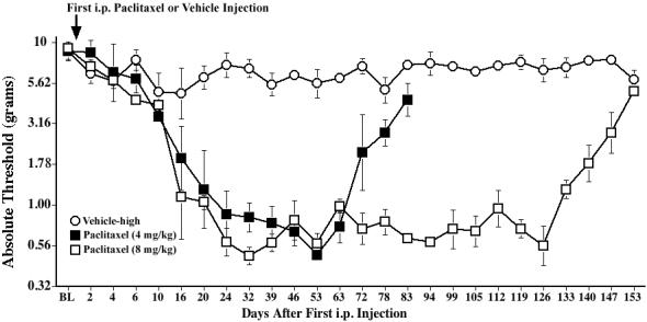
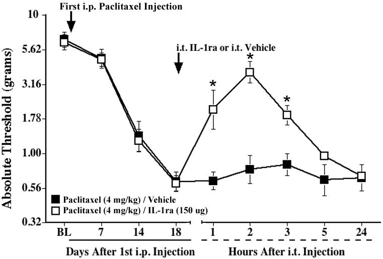
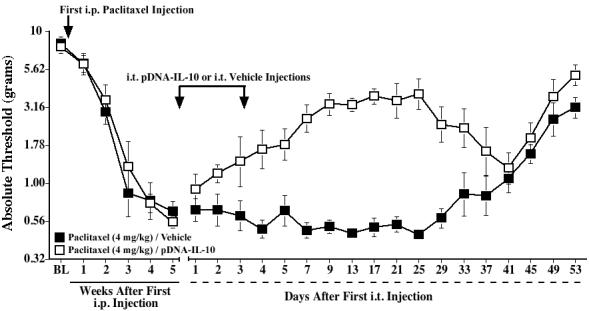
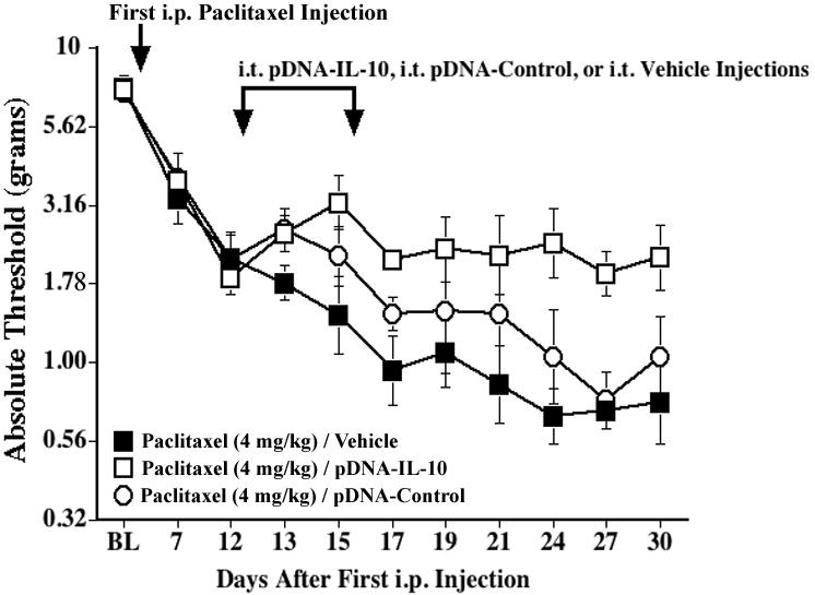
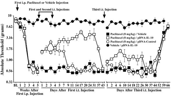
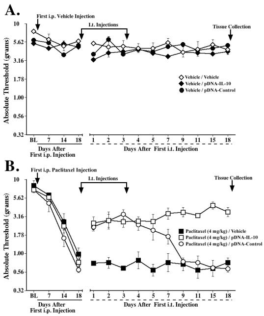
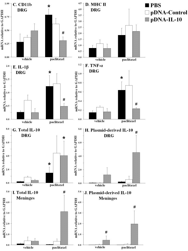
Paclitaxel is a commonly used cancer chemotherapy drug that frequently causes painful peripheral neuropathies. The mechanisms underlying this dose-limiting side effect are poorly understood. Growing evidence supports that proinflammatory cytokines, such as interleukin-1 (IL-1) and tumor necrosis factor (TNF), released by activated spinal glial cells and within the dorsal root ganglia (DRG) are critical in enhancing pain in various animal models of neuropathic pain. Whether these cytokines are involved in paclitaxel-induced neuropathy is unknown. Here, using a rat neuropathic pain model induced by repeated systemic paclitaxel injections, we examined whether paclitaxel upregulates proinflammatory cytokine gene expression, and whether these changes and paclitaxel-induced mechanical allodynia can be attenuated by intrathecal IL-1 receptor antagonist (IL-1ra) or intrathecal delivery of plasmid DNA encoding the anti-inflammatory cytokine, interleukin-10 (IL-10). The data show that paclitaxel treatment induces mRNA expression of IL-1, TNF, and immune cell markers in lumbar DRG. Intrathecal IL-1ra reversed paclitaxel-induced allodynia and intrathecal IL-10 gene therapy both prevented, and progressively reversed, this allodynic state. Moreover, IL-10 gene therapy resulted in increased IL-10 mRNA levels in lumbar DRG and meninges, measured 2 weeks after initiation of therapy, whereas paclitaxel-induced expression of IL-1, TNF, and CD11b mRNA in lumbar DRG was markedly decreased. Taken together, these data support that paclitaxel-induced neuropathic pain is mediated by proinflammatory cytokines, possibly released by activated immune cells in the DRG. We propose that targeting the production of proinflammatory cytokines by intrathecal IL-10 gene therapy may be a promising therapeutic strategy for the relief of paclitaxel-induced neuropathic pain.


Figures

Fig. 1
Fig. 1
Time course of mechanical allodynia induced by repeated administration of paclitaxel. Rats received 4 i.p. injections of paclitaxel (1 or 2 mg/kg) on alternate days for a cumulative dose of 4 or 8 mg/kg, respectively, and low-threshold mechanical sensitivity was assessed by the von Frey test, before (baseline, BL), and up to Day 153. Data represent means ± SE.
Fig. 2
Fig. 2
Repeated administration of paclitaxel increases expression of the microglial activation markers OX-42 and OX-6, but not of the astrocyte marker GFAP, in lumbar (L5/L6) spinal cord. Rats received i.p. injections of vehicle or paclitaxel (cumulative dose 8 mg/kg) on alternate days, and were sacrificed on Day 35 or 42 to collect tissues for immunohistochemistry analysis. Representative photomicrographs are shown of GFAP (A, B) and OX-42 (C, D) immunoreactivity in the dorsal horn, and of OX-42 (E, F) and OX-6 (G, H) immunoreactivity in the ventral part of the lumbar spinal cord of rats treated with vehicle (A, C, E, G) or paclitaxel (B, D, F, H). Scale bar is 100 μm in E and F, and 200 μm in A-D, G, H.
Fig. 3
Fig. 3
A single i.t. injection of IL-1ra transiently reverses paclitaxel-induced mechanical allodynia. Rats received i.p. injections of paclitaxel (cumulative dose 4 mg/kg) on alternate days, and IL-1ra (150 μg) or vehicle was administered intrathecally 18 days after the first paclitaxel injection (indicated by the arrow). Low-threshold mechanical sensitivity was assessed by the von Frey test, before (baseline, BL), on Day 18, and 1, 2, 3, 5, and 24 h after the i.t. injection. Data represent means ± SE. *p<0.05 vs. paclitaxel/vehicle.
Fig. 4
Fig. 4
Intrathecal IL-10 gene therapy partially reverses paclitaxel-induced mechanical allodynia. Rats received i.p. injections of paclitaxel (cumulative dose 4 mg/kg) on alternate days, and two i.t. injections of pDNA-IL-10 (100 and 25 μg, respectively) or vehicle were given 5 weeks after the first paclitaxel injection, 3 days apart (indicated by the arrows). Low-threshold mechanical sensitivity was assessed by the von Frey test, before (baseline, BL), weekly after the first paclitaxel injection, and up to 53 days after the first i.t. injection. Data represent means ± SE.
Fig. 5
Fig. 5
Intrathecal IL-10 gene therapy can arrest the development of mechanical allodynia induced by paclitaxel. Rats received i.p. injections of paclitaxel (cumulative dose 4 mg/kg) on alternate days, and two i.t. injections of pDNA-IL-10 (100 and 25 μg, respectively) or vehicle were given on Days 12 and 15 after the first paclitaxel injection (indicated by the arrows). Low-threshold mechanical sensitivity was assessed by the von Frey test, before (baseline, BL), and up to 30 days after the first paclitaxel injection. Data represent means ± SE.
Fig. 6
Fig. 6
Intrathecal IL-10 gene therapy partially reverses mechanical allodynia induced by a higher dose of paclitaxel. Rats received i.p. injections of paclitaxel (cumulative dose 8 mg/kg) or vehicle on alternate days, and two i.t. injections of pDNA-IL-10 (100 and 25 μg), pDNA-Control (100 and 25 μg), or vehicle were given 5 weeks after the first paclitaxel injection, 3 days apart (indicated by the arrows). Low-threshold mechanical sensitivity was assessed by the von Frey test, before (baseline, BL) and weekly after the first paclitaxel or vehicle injection, and up to 43 days after the first i.t. injection. A third i.t. injection of pDNA-IL-10 (25 μg), pDNA-Control (25 μg), or vehicle was given on Day 43 after the first i.t injection (indicated by the arrow), and von Frey responses were subsequently assessed up to 66 days later. Data represent means ± SE.
Fig. 7
Fig. 7
Effects of paclitaxel and IL-10 gene therapy on mRNA expression of immune/glial cell markers and proinflammatory cytokines in lumbar DRG and lumbar meninges. Rats received i.p. vehicle or paclitaxel (cumulative dose 4 mg/kg), and i.t. pDNA-IL-10, pDNA-Control, or vehicle (PBS) on Days 18 and 21 after the first i.p. injection. Assessments on the von Frey test confirmed that these treatments did not affect basal thresholds (A) and that paclitaxel induced mechanical allodynia (B), which was reversed by pDNA-IL-10 (long-term) and pDNA-Control (short-term). Tissues were collected on Day 36 for RT-PCR analysis; C-H show mRNA expression in lumbar DRG of CD11b, MHC class II, IL-1β, TNF-α, total IL-10 and plasmid-derived IL-10, respectively. I and J show mRNA levels in lumbar meninges of total IL-10 and plasmid-derived IL-10, respectively. * p<0.05 vs. respective vehicle group; # p<0.05 vs. paclitaxel/pDNA-Control and vs. paclitaxel/PBS groups. Data represent means ± SE.
Fig. 7
Fig. 7
Effects of paclitaxel and IL-10 gene therapy on mRNA expression of immune/glial cell markers and proinflammatory cytokines in lumbar DRG and lumbar meninges. Rats received i.p. vehicle or paclitaxel (cumulative dose 4 mg/kg), and i.t. pDNA-IL-10, pDNA-Control, or vehicle (PBS) on Days 18 and 21 after the first i.p. injection. Assessments on the von Frey test confirmed that these treatments did not affect basal thresholds (A) and that paclitaxel induced mechanical allodynia (B), which was reversed by pDNA-IL-10 (long-term) and pDNA-Control (short-term). Tissues were collected on Day 36 for RT-PCR analysis; C-H show mRNA expression in lumbar DRG of CD11b, MHC class II, IL-1β, TNF-α, total IL-10 and plasmid-derived IL-10, respectively. I and J show mRNA levels in lumbar meninges of total IL-10 and plasmid-derived IL-10, respectively. * p<0.05 vs. respective vehicle group; # p<0.05 vs. paclitaxel/pDNA-Control and vs. paclitaxel/PBS groups. Data represent means ± SE.

References

    1. Alessandri-Haber N, Dina OA, Yeh JJ, Parada CA, Reichling DB, Levine JD. Transient receptor potential vanilloid 4 is essential in chemotherapy-induced neuropathic pain in the rat. J Neurosci. 2004;24:4444–4452. - PMC - PubMed
    1. Authier N, Gillet JP, Fialip J, Eschalier A, Coudore F. Description of a short-term Taxol-induced nociceptive neuropathy in rats. Brain Res. 2000;887:239–249. - PubMed
    1. Bogdan C, Ding A. Taxol, a microtubule-stabilizing antineoplastic agent, induces expression of tumor necrosis factor alpha and interleukin-1 in macrophages. J Leukoc Biol. 1992;52:119–121. - PubMed
    1. Byrd-Leifer CA, Block EF, Takeda K, Akira S, Ding A. The role of MyD88 and TLR4 in the LPS-mimetic activity of Taxol. Eur J Immunol. 2001;31:2448–2457. - PubMed
    1. Cata JP, Weng HR, Lee BN, Reuben JM, Dougherty PM. Clinical and experimental findings in humans and animals with chemotherapy-induced peripheral neuropathy. Minerva Anestesiol. 2006;72:151–169. - PubMed

Publication types

MeSH terms