. 2007 Feb;34(2):219-30.
doi: 10.1016/j.mcn.2006.11.001. Epub 2006 Dec 15.

Localization of glutamate receptors to distal dendrites depends on subunit composition and the kinesin motor protein KIF17

Affiliations

Localization of glutamate receptors to distal dendrites depends on subunit composition and the kinesin motor protein KIF17

N Kayadjanian et al. Mol Cell Neurosci. 2007 Feb.

Abstract

Correct glutamate receptor localization in neurons is crucial for neurotransmission in the brain. Here we investigated the mechanisms underlying localization of kainate GluR5 receptors to dendrites in cultured hippocampal neurons. We find that the GluR5 distribution depends on association with GluR6 and KA2 subunits. The GluR5 subunit was expressed in distal dendrites only when GluR6 and KA2 subunits were present, whereas it was restricted to proximal dendrites in the absence of these subunits. The overlap between GluR5 distribution and the organization of microtubules in dendrites led us to examine whether KIF17, a microtubule motor protein expressed in distal dendrites, is involved in GluR5 localization to distal dendrites. We show here, for the first time that the microtubule motor protein KIF17 interacts with GluR6 and KA2 subunits and is required for GluR5 localization to distal dendrites, defining a novel mechanism that controls receptor localization in neurons.


Figures

Fig. 1
Fig. 1
GFP-R5 forms functional receptors in hippocampal neurons. (A) Schematic diagram of GFP-R5 structure. The GFP tag (green bar) was inserted in the N-terminal extracellular domain between the fifth and sixth amino acids after the predicted signal peptide (SP). (B) The punctate distribution of GFP-R5 subunits (left panel) in neurons contrasts with the diffuse distribution of GFP in all the neuronal compartments (right panel). These two confocal images represent a single focal plane taken at the level of the nucleus. Note the absence of GFP-R5 expression in the nucleus when compared to GFP-expressing neurons. Scale bar, 20 μm. (C) GFP-R5 subunits form functional channels in cultured primary hippocampal neurons. At DIV7, application of ATPA (30 μM), in the continuous presence of SYM2206 (100 μM) onto neurons originating from R5-R6-KA2−/− deficient mice, elicited no current in untransfected neurons (n=12) whereas an inward current was observed in the soma of a GFP-R5 transfected neuron (n=14). Similar results were obtained at DIV15 in untransfected (n= 2) and transfected cells (n= 4).
Fig. 2
Fig. 2
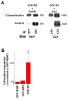
GFP –R5 shows normal assembly properties and surface expression. (A) GFP-R5 subunits co-assemble with GluR6 (left panel) and KA2 subunits (right panel). HEK 293T cells were cotransfected with the combinations of kainate receptor subunits indicated above each lane. Lysates were immunoprecipitated (IP) and immunoblotted with antibodies as indicated. Lower panel shows the corresponding control, where we checked that mixing lysates from cells expressing individual subunits (as indicated above) only resulted in background coimmunoprecipitation (n=5 independent experiments). (B) GFP-R5 subunits are expressed at the cell surface as measured by flow-cytometry (see Methods). Values represent the mean (± S.E.M) of 4 independent experiments. ** p < 0.01 when compared to GFP-R2NF (Mann-Whitney post-hoc test).
Fig. 3
Fig. 3
The distribution of GFP-R5 is polarized to the somato-dendritic compartment. Panel A and B illustrate neurons taken from control (A) and R5-R6-KA2−/− cultures (B), expressing GFP-R5 in the somato-dendritic compartment (yellow = colocalization of MAP2 and GFP). Scale bar, 20 μm. (Lower panel) Endogenous GluR6 and KA2 subunits are expressed in control but absent in R5-R6-KA2−/− hippocampal cultures. Lysates from hippocampal cultures were immunoblotted directly (crude) or IP with antibodies as indicated and then immunoblotted with the same antibody used for IP. 0.3 % of lysates from hippocampal cultures (2500 × 103 hippocampal cells) was loaded directly on the western blot gel (crude) while the remaining lysates were immunoprecipitated (IP) with the same antibody used for immunoblotting. Note in R5-R6-KA2−/− primary cultures the absence of KA2 subunits. Note also that the remaining immunoreactive band detected in R5-R6-KA2−/− cultures with the anti-GluR6/7 antibody corresponds to GluR7 subunits. No GluR6/7 immunoreactivity was detected in GluR6/7−/− knock-out mice (data not shown). Molecular size markers (in kilodaltons) are shown on the left. Scale bar, 20 μm. (C) The quantitative analysis of GFP-R5 dendrite/axon polarity in control and R5-R6-KA2−/− hippocampal cultures. The graph indicates the percent of transfected neurons exhibiting dendritic labeling only (red) or dendritic with axon labeling (green) (n = 50 neurons, 9 independent experiments).
Fig. 4
Fig. 4
The dendritic distribution of GFP-R5 depends on GluR6 and KA2 subunits. (A) The total dendritic length, as measured by the extent of GFP-labeling in dendrites, does not differ between hippocampal neurons cultured from control and R5-R6-KA2−/− mice. Data are derived from the same cultures as in Figure 3C and represent the mean (± S.E.M) somato-dendritic distance measured for each neuron. (B) Quantitative analysis of the dendritic location of GFP-R5. Data are derived from the same cultures reported in panel A. The distance from soma was reported as a percentage of the mean maximal dendritic length calculated from panel A. *** p < 0.001 when compared to control hippocampal cultures (Dunnett post-hoc test). A representation of proximal and distal dendrites defined from GFP-R5 distribution is also depicted on the right axis. (C) Both GluR6 and KA2 subunits are required for GFP-R5 localization to distal dendrites. The dendritic location of GFP-R5 was similar in neurons originating from R5-R6-KA2−/−, KA2−/− and R5-R6−/− knock-out mice (p > 0.05), and the mean ± S.E.M dendritic location was reported for each condition as a percentage of the maximal dendritic length (n = 70–120 neurons per condition from 3 independent experiments). (D) GFP-R5 receptors expressed in the proximal dendrites of R5-R6-KA2−/− hippocampal neurons are functional. To ensure that only dendritic GFP-R5 receptors were activated by ATPA, the tip of the application pipette was positioned less than 5 μm away from the dendrite with the soma located upstream of the application pipette. Whole-cell currents were evoked by local application of ATPA (30 μM) in the presence of SYM2206 (100 μM), to the proximal dendrite (100 μm from the soma) of hippocampal neurons expressing GFP-R5 (n=6).
Fig. 5
Fig. 5
Colocalization of GFP-R5 and KIF17 in hippocampal neurons. Immunocytochemistry in control neurons shows colocalization (arrows) of GFP-R5 (green) and endogenous KIF17 (red) proteins. Scale bar, 20 μm.
Fig. 6
Fig. 6
In vivo interaction of GluR6 and KA2 subunits with KIF17 in mouse hippocampi. (A) KIF17 coimmunoprecipitates GluR6 and KA2 subunits. Membrane proteins from 6 hippocampi were IP with an anti-IgG or anti-KIF17 antibody (20 μl) and immunoblotted with anti-GluR6/7 antibodies (upper panel) or anti-KA2 antibodies (lower panel). No signal was detected when membranes were IP with an anti-IgG antibody. (B) GluR6 and KA2 subunits coimmunoprecipitate KIF17 proteins. Hippocampi membrane proteins were IP with anti-R6/7 (5 μg, left panel) or anti-KA2 antibodies (10 μg, right panel) and immunoblotted with anti-KIF17 antibodies (1:1000, upper panel). The same immunoblot was then reprobed with anti-KA2 antibodies (lower panel) revealing that GluR6/7 subunits interact with both KIF17 and KA2 proteins. (C) KIF17 interaction with GluR6 and KA2 subunits is specific. Immunoprecipitation from KA2−/− hippocampi with anti-KIF17 antibodies yielded GluR6/R7 subunits but no band immunoreactive for KA2 was detected. In GluR7−/− mice, KIF17 coimmunoprecipitated KA2 subunits and GluR6 subunits. In GluR6/7−/− preparations, KIF17 coimmunoprecipitated KA2 subunits but no band immunoreactive for GluR6 or GluR7 was detected. We also verified that KIF17 did not bind to the AMPA GluR2 subunits by showing that no immunoreactive band for GluR2 antibodies (1:200, Chemicon) could be detected after immunoprecipitation of KIF17 from wild-type (WT) and knock-out mice.
Fig. 7
Fig. 7
KIF17 inhibition blocks GFP-R5 localization in distal dendrites. (A) Illustration of a neuron coexpressing GFP-R5 and KIF17 (left panel) or GFP-R5 and KIF17 Δ306 (right panel). GFP-R5 distribution in dendrites immunoreactive for MAP2 (red) extends in proximal and distal dendrites in neurons coexpressing KIF17, whereas it is restricted to proximal dendrites in neurons coexpressing the dominant-negative KIF17 Δ306. Scale bar, 20 μm. (B) Quantitative analysis of the dendritic location of GFP-R5 when coexpressed with KIF17 or KIF17 Δ306. *** p < 0.001 when compared to GFP-R5 coexpressed with KIF17 (n= 30–50 neurons from 2 independent experiments).

References

    1. Ahmari SE, Buchanan J, Smith SJ. Assembly of presynaptic active zones from cytoplasmic transport packets. Nat Neurosci. 2000;3:445–451. - PubMed
    1. Andrasfalvy BK, Magee JC. Distance-dependent increase in AMPA receptor number in the dendrites of adult hippocampal CA1 pyramidal neurons. J Neurosci. 2001;21:9151–9159. - PMC - PubMed
    1. Baas PW, Deitch JS, Black MM, Banker GA. Polarity orientation of microtubules in hippocampal neurons: uniformity in the axon and nonuniformity in the dendrite. Proc Natl Acad Sci USA. 1988;85:8335–8339. - PMC - PubMed
    1. Bah J, Quach H, Ebstein RP, Segman RH, Melke J, Jamain S, Rietschel M, Modai I, Kanas K, Karni O, Lerer B, Gourion D, Krebs MO, Etain B, Schurhoff F, Szoke A, Leboyer M, Bourgeron T. Maternal transmission disequilibrium of the glutamate receptor GRIK2 in schizophrenia. Neuroreport. 2004;15:1987–1991. - PubMed
    1. Bliss TV, Collingridge GL. A synaptic model of memory: long-term potentiation in the hippocampus. Nature. 1993;361:31–39. - PubMed

Publication types