. 2006 Dec 15;127(6):1167-78.
doi: 10.1016/j.cell.2006.09.050.

Single Holliday junctions are intermediates of meiotic recombination

Affiliations

Single Holliday junctions are intermediates of meiotic recombination

Gareth A Cromie et al. Cell. .

Abstract

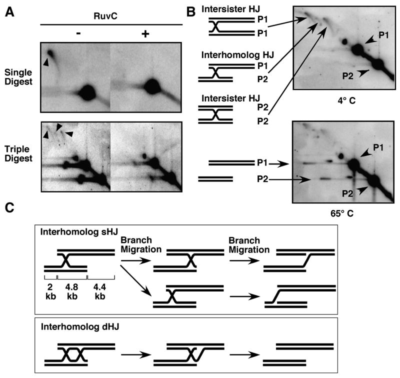
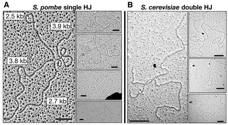
Crossing-over between homologous chromosomes facilitates their accurate segregation at the first division of meiosis. Current models for crossing-over invoke an intermediate in which homologs are connected by two crossed-strand structures called Holliday junctions. Such double Holliday junctions are a prominent intermediate in Saccharomyces cerevisiae meiosis, where they form preferentially between homologs rather than between sister chromatids. In sharp contrast, we find that single Holliday junctions are the predominant intermediate in Schizosaccharomyces pombe meiosis. Furthermore, these single Holliday junctions arise preferentially between sister chromatids rather than between homologs. We show that Mus81 is required for Holliday junction resolution, providing further in vivo evidence that the structure-specific endonuclease Mus81-Eme1 is a Holliday junction resolvase. To reconcile these observations, we present a unifying recombination model applicable for both meiosis and mitosis in which single Holliday junctions arise from single- or double-strand breaks, lesions postulated by previous models to initiate recombination.


Figures

Figure 1
Figure 1. Recombination Models
(A) A double-strand break (DSB) repair model of recombination (after Szostak et al., 1983; Sun et al., 1991). (i) A DSB is formed and processed to give 3′ single-stranded ends. (ii) Invasion of one DNA end into an intact homologous duplex forms a D-loop. (iii) The second end anneals to the left side of the D-loop, and branch migration at both ends of the D-loop forms a double Holliday junction (dHJ). Gaps and strand discontinuities are repaired by DNA synthesis and ligation. Mismatch correction can lead to gene conversion. Each HJ can be resolved by strand cleavage in orientation 1 or 2; if orientation is random, non-crossover and crossover products are formed at equal frequency. In this figure the left HJ is cleaved in orientation 1. (iv) Cleavage of the right HJ in orientation 1 produces a non-crossover, whereas (v) cleavage in orientation 2 produces a crossover. (B) A unifying recombination model initiated by a DSB or a single-strand nick and proceeding through a single Holliday junction (after Radding, 1982; Szostak et al., 1983). (i) Recombination is initiated from a DSB (left) or a nick or single-strand gap (right). (ii) DNA unwound from the DSB or the nick or gap produces a D-loop as in A. In contrast to A, the D-loop is cleaved at the left end, and (iii) the newly generated end anneals with the duplex initially cleaved. Mismatch correction and HJ resolution produce gene conversion without (iv) or with (v) crossing-over as in A.
Figure 2
Figure 2. In a Physical Assay, Meiotic Crossovers Are Greatly Reduced In a mus81 Mutant
(A) The mbs1 region of chromosome I. PvuII, PmlI (“L”) and XbaI (“R”) digestion and probing as shown reveals two parental (9.2 and 6.8 kb) and two recombinant (11.2 and 4.8 kb) fragments. (B) Recombinant DNA fragments arise during meiosis. DNA from meiotic timecourses of strains GP5086 (rec12+ mus81+), GP5082 (mus81Δ), GP5659 (rec12Δ), and GP5662 (rec12Δ mus81Δ),, was isolated, and analyzed by gel electrophoresis and Southern blotting with digestion and probing at mbs1 as in 2A. Asterisks indicate cross-hybridizing DNA not specific to meiosis. (C) Crossover frequencies, calculated from Phosphorimager analysis of Southern blots as in (B), equal 2xR2/total. Values are mean ± SEM (n ≥ 3). In rec12Δ or rec12Δ mus81Δ R2 was undetectable (<0.4%). (D) Timing of meiotic replication, DSB formation, crossing-over and the meiosis I division. Percentages of GP5086 (rec12+ mus81+) cells with replicated DNA were quantitated using flow cytometry. Transient [GP5086 (rad50+)] and accumulated [GP5411 (rad50S)] meiotic DSBs were quantitated from Southern blots using a maximum value of 10% for GP5086. Crossover formation in strain GP5086 (mus81+ rec12+) is from Figure 2C. All data above are the average of at least two independent inductions. The percentage of GP5086 cells with 1 (pre-MI) and >1 (post-MI) Hoechst 33342-staining bodies (nuclei) was determined from >100 scorable cells for each time-point with 100% as the maximum value.
Figure 3
Figure 3. Recombination Intermediates Accumulate In a mus81 Mutant and Are Most Frequent at DSB Hotspots
(A) Predicted migration of PvuII-digested mbs1 DNA during 2D gel electrophoresis (Brewer and Fangman, 1987). Parental (linear) DNA lies on an arc of linear molecules. Y-shaped intermediates arise during replication, and X-shaped intermediates during replication or recombination. (B) DNA from meiotic timecourses of strains GP5086 (rec12+ mus81+), GP5082 (mus81Δ), GP5659 (rec12Δ), and GP5662 (rec12Δ mus81Δ) was digested with PvuII, separated by 2D gel electrophoresis, Southern blotted, and probed for mbs1. (C) Frequencies of X-form species at mbs1, calculated from Phosphorimager analysis of Southern blots as in B. Values are the means of at least two independent experiments. Error bars, shown for clarity only for selected points, are SEM (n ≥ 3) for all points except for strain GP5082 at 4 and 7 hr, where 1/2 the difference between the two values is used (n = 2). (D) DNA isolated 5 hr after meiotic induction of strain GP5082 (mus81Δ) was analyzed as in (B) but probed for the mbs1 and mbs2 DSB hotspots and for two DSB-free intervals of comparable sizes located on the same cosmids. Restriction digestion was with PvuII (SPAC4G8 intervals) and BamHI (SPAC1D4 intervals). Frequencies of JMs are mean ± SEM (n = 3) for mbs1 and mean ± 1/2 the difference between the two values for others.
Figure 4
Figure 4. Intersister Joint Molecules Are More Frequent Than Interhomolog Joint Molecules, and Both Accumulate In a mus81 Mutant
(A) Predicted migration during 2D gel electrophoresis of mbs1-probed DNA digested with PvuII, PmlI, and XbaI (Figure 2A). Y- and X-shaped molecules are expected during replication, but only X-shaped molecules during recombination. Intersister JMs arising from either parent (P1 X or P2 X) migrate in a spike, whereas interhomolog JMs (IH X) migrate as two species joined by an arc (see Figure 5C). (B) DNA from 3 and 5 hr after meiotic induction of strains GP5086 (mus81+) and GP5082 (mus81Δ) was analyzed as in A. Black arrowheads indicate X-form JM species, and white arrowheads indicate interhomolog JMs with heteroduplex DNA preventing restriction at the R site. Both species are inferred to contain single HJs by their resistance to heating (Figure 5B). Partial digestion products are identified by thin arrows. (C) Quantitation of intersister and interhomolog recombination JMs at 5 hr at mbs1. The frequencies of JMs were calculated by Phosphorimager analysis of Southern blots as in B. Values are mean ± SEM (n ≥ 3). In strains GP5659 (rec12Δ) and GP5662 (rec12Δ mus81Δ) JMs were undetectable.
Figure 5
Figure 5. RuvC-treatment Resolves Both Intersister and Interhomolog Joint Molecules, but Only Intersister Joint Molecules Are Resolved to Linear Forms by Mild Heat-treatment
(A) DNA isolated 5 hr after meiotic induction of strain GP5082 (mus81Δ) was digested either with PvuII alone (upper panel) or with PvuII, PmlI, and XbaI (lower panel), treated with RuvC (+) or left untreated (−), separated by gel electrophoresis in two dimensions, Southern blotted, and probed for mbs1. Black arrows indicate intersister and interhomolog JM species. (B) DNA isolated 5 hr after meiotic induction of strain GP5082 (mus81Δ) was digested with PvuII, PmlI, and XbaI, separated by gel electrophoresis in the first dimension, heated to 65° or left at 4°, separated in the second dimension, and analyzed as in A. (C) Two distinct, stable DNA structures are formed by branch migration of a single HJ in an interhomolog joint molecule. Branch migration of a single HJ to the right (top) or left (bottom) stops when a Y junction is formed. These two structures should be unable to reverse due to high activation energy and, despite having the same mass, are expected to separate upon 2D gel electrophoresis because of their different shapes Branch migration of a double HJ in either direction generates separate linear molecules; only rightward branch migration is shown.
Figure 6
Figure 6. S. pombe Recombination Joint Molecules Contain Single Holliday Junctions
(A) Electron micrographs of HJ-containing molecules isolated from S. pombe strains GP5082 (mus81Δ) (main picture and three upper right) and GP5086 (rec12+ mus81+) (bottom right) 5 hr after meiotic induction. Note the partially denatured (open centered) HJs in the left and third (from top) right panels. Scale bars are 0.2 μm. (B) Electron micrographs of HJ-containing molecules isolated from S. cerevisiae strains NHY1226 (SPO11 MUS81 ndt80) and NHY1296 (SPO11 MUS81 NDT80) 4.5 hr after meiotic induction. Note the separate (double) HJs in the left and middle right panels and the fused (double) HJs in the upper and lower right panels. Scale bars are 0.2 μm.

Comment in

References

    1. Allers T, Lichten M. Differential timing and control of noncrossover and crossover recombination during meiosis. Cell. 2001;106:47–57. - PubMed
    1. Argueso JL, Wanat J, Gemici Z, Alani E. Competing crossover pathways act during meiosis in Saccharomyces cerevisiae. Genetics. 2004;168:1805–1816. - PMC - PubMed
    1. Bell LR, Byers B. Homologous association of chromosomal DNA during yeast meiosis. Cold Spring Harb Symp Quant Biol. 1983;47:829–840. - PubMed
    1. Benson FE, West SC. Substrate specificity of the Escherichia coli RuvC protein. Resolution of three- and four-stranded recombination intermediates. J Biol Chem. 1994;269:5195–5201. - PubMed
    1. Boddy MN, Gaillard PH, McDonald WH, Shanahan P, Yates JR, 3rd, Russell P. Mus81-Eme1 are essential components of a Holliday junction resolvase. Cell. 2001;107:537–548. - PubMed

Publication types

MeSH terms