. 2006 Dec 15;127(6):1179-91.
doi: 10.1016/j.cell.2006.09.049.

A Bir1-Sli15 complex connects centromeres to microtubules and is required to sense kinetochore tension

Affiliations

A Bir1-Sli15 complex connects centromeres to microtubules and is required to sense kinetochore tension

Sharsti Sandall et al. Cell. .

Abstract

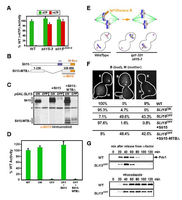
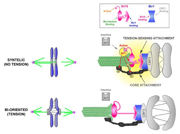
Proper connections between centromeres and spindle microtubules are of critical importance in ensuring accurate segregation of the genome during cell division. Using an in vitro approach based on the sequence-specific budding yeast centromere, we identified a complex of the chromosomal passenger proteins Bir1 and Sli15 (Survivin and INCENP) that links centromeres to microtubules. This linkage does not require Ipl1/Aurora B kinase, whose targeting and activation are controlled by Bir1 and Sli15. Ipl1 is the tension-dependent regulator of centromere-microtubule interactions that ensures chromosome biorientation on the spindle. Elimination of the linkage between centromeres and microtubules mediated by Bir1-Sli15 phenocopies mutations that selectively cripple Ipl1 kinase activation. These findings lead us to propose that the Bir1-Sli15-mediated linkage, which bridges centromeres and microtubules and includes the Aurora kinase-activating domain of INCENP family proteins, is the tension sensor that relays the mechanical state of centromere-microtubule attachments into local control of Ipl1 kinase activity.


Figures

Fig. 1
Fig. 1
A quantitative in vitro assay for the binding of budding yeast CEN DNA to microtubules. (A) Schematic of the in vitro assay. Adaptations to the original scheme important for quantitative biochemical analysis are emphasized here and include: 1) stable adsorption of microtubules using tubulin covalently modified with digoxigenin; 2) multiplexing of flow cells on a single slide; and 3) automated image analysis to measure number of bound beads. For quantitation, 10 fields at 20X magnification are photographed per sample and averaged. (B) Linkage of beads to microtubules is observed with wild-type but not mutant CEN DNA. The mutant harbors a deletion of the central CCG in CDEIII that prevents binding of the CBF3 complex and abolishes centromere activity in vivo. Error bars=SD. (C) Partial purification of CBF3 using a CEN DNA gel-shift assay. The flowchart describes the chromatography steps and the gel panel shows enrichment of the CEN DNA bandshift relative to starting extract in the partially purified (PP) fraction. The arrowhead marks the CEN DNA-CBF3 complex. (D) Partially purified CBF3 does not link CEN DNA beads to microtubules. Note that the volume of starting extract used to prepare the CBF3 added to the (PP)CBF3 reaction is ∼25-fold greater than that assayed in the extract reaction. If equivalent extract volumes are assayed, no binding is observed with (PP)CBF3. Error bars=SD.
Figure 2
Figure 2
Conventional purification of an activity that complements CBF3 in the in vitro assay. (A) Schematic of the in vitro complementation approach. (B) Example of a standard curve used to quantitatively monitor fractionation of the complementing activity. The starting material, in this case the gel filtration load, is serially diluted into a constant amount of (PP)CBF3 and the points fitted to a polynomial curve. Complementing activity in each fraction measured after adding the same amount of (PP)CBF3 is converted to a percentage of total loaded activity. Error bars=SD. (C) Column profile of Sephacryl S400HR gel filtration. The percentage of loaded activity calculated from the standard curve and the percentage of total loaded protein is plotted for each fraction. (D) Summary of the complementing activity purification. The activity column lists the percentage yield, relative to the starting extract, after each step. (E) Fractions from the MonoS gradient elution stained with Coomassie Blue. The complementing activity is indicated with the gray bars above each fraction. No activity is detected in the column flowthrough. Asterisks denote the two fractions that constitute the MonoS Pool. (F) The MonoS Pool complements (PP)CBF3. Error bars=SD. (G) Annotation-based classification of proteins identified by mass spectrometry of the MonoS pool. The 247 proteins that showed >10% sequence coverage are represented in the pie chart (see also Suppl. Table 2).
Figure 3
Figure 3
Bir1 is required for linking CEN DNA to microtubules in vitro. (A) bir1∆ cells lack Bir1 protein. Western blot of extracts prepared from WT (ODY49), bir1∆(ODY65), and bir1∆+pCEN-BIR1 (ODY114) strains probed with an anti-Bir1 antibody. Asterisks indicate background bands that serve as loading controls. (B) Bir1 is required for linking CEN DNA to microtubules. Extracts indicated in (A) were analyzed using the bead assay. Activity was normalized relative to the wild-type extract. Error bars=SD. (C) Bir1 does not affect the ability of CBF3 to bind to CEN-DNA. Arrowhead indicates position of the CBF3-CEN DNA complex. (D) Bir1 and Sli15, but not Ipl1, co-fractionate with the complementing activity. Gel filtration fractions of extracts prepared from BIR1:6HA; SLI15:13Myc (ODY97) were analyzed by western blotting using anti-HA, anti-Myc and anti-Ipl1 antibodies. The blot signal intensity for all 3 proteins, as well as activity in the bead assay, is plotted as a percentage of the respective peak fractions (12/13 for Sli15, Bir1, and activity; 18 for Ipl1). (E) Bir1 and Sli15 continue to co-fractionate with the complementing activity during the cation exchange step. The activity peak from gel filtration (fractions 12/13 in (D)) was further fractionated using a MonoS cation exchange column and analyzed as in (D).
Figure 4
Figure 4
Sli15, but not Ipl1, is required for the complementing activity. (A) Extracts prepared from SLI15:6HA (ODY54) and IPL1:6HA (ODY55) cells were immunodepleted using an anti-HA affinity resin. The depleted supernatants were serially diluted and complementing activity measured with constant (PP)CBF3. Comparing a standard curve of the input extract to the depleted extract using anti-HA immunoblotting assessed depletion efficiency. Both Sli15 and Ipl1 were successfully depleted by >90% but only the Sli15 depletion resulted in a severe activity reduction. (B) Ipl1 kinase activity regulates the binding observed in the in vitro assay. ipl1-321 mutant extracts are insensitive to ATP addition, which is in contrast to wild-type extracts. Addition of purified GST-Ipl1 protein restores ATP sensitivity to the mutant extracts. The MonoS pool combined with (PP)CBF3 is ATP-insensitive in the bead assay but becomes sensitive following addition of GST-Ipl1. (C) Flowchart describing the purification and use of the Bir1-TAP complex. (D) Immunoblot of Bir1-TAP after elution by TEV cleavage. Both Sli15 and Bir1 are present in the elution. (E) Bir1-TAP complements (PP)CBF3. (F) Bir1-TAP complements loss of activity in bir1∆ extracts. In both (E) and (F), the amount of Bir1-TAP added is similar to that present in 2 μl of extract, which is the standard amount analyzed in the bead assay (D).
Figure 5
Figure 5
Sli15 mutants that eliminate CEN DNA-microtubule interactions in vitro phenocopy Ipl1 kinase activation mutants in vivo. (A) sli15-3 extracts behave similarly to Ipl1 kinase mutant extracts. sli15L656>S is an engineered mutant with only the IN box amino acid change in sli15-3 (see Suppl. Fig. 2). (B) Schematic of Sli15 and the Sli15-MTB-binding domain. The C-terminal region used to generate the anti-Sli15 antibody is underlined. (C) Immunoblotting of ODY155 (pGAL-SLI15), ODY192 (pGAL-SLI15+Sli15) and ODY193 (pGAL-SLI15+Sli15-MTB∆) in galactose (ON) or glucose (OFF) medium. All SLI15OFF samples were prepared from cells arrested with nocodazole to maintain viability. Asterisk denotes the background band that serves as a loading control. See also Suppl. Fig. 4 and Suppl. Fig. 5. (D) Depletion of Sli15 or deletion of its microtubule-binding domain eliminate the CEN DNA-microtubule linkage in vitro. (E) Ipl1/Aurora B is required for the correction of attachment errors where both sister chromatids are connected to the same spindle pole (syntelic attachment; upper panels). Mutants that perturb Ipl1 kinase activity (ipl1-321, ipl1-2, sli15-3), fail to correct syntelic attachments, resulting in sister chromatid missegregation (lower panels). (F) SLI15OFF and SLI15OFF+Sli15-MTB∆ both phenocopy Ipl1 kinase activity mutants in vivo. Segregation of a marked Chr IV was monitored 120 minutes after release from alpha factor arrest. Between 100-200 cells for each strain and growth condition were analyzed. The mother cell (M) was identified by residual alpha factor-induced shmoos (projections in cell outline). (G) SLI15OFF cells do not arrest in the cell cycle despite having a functional checkpoint. WT (SBY818) and pGAL:SLI15 cells expressing Pds1-Myc18 (ODY181) were grown as described above. Nocodazole was added to one set of cultures at 10 μg/ml upon release from alpha factor. Lysates were immunoblotted with an anti-Myc antibody.
Figure 6
Figure 6
A model for tension-regulated Ipl1 activation by the Bir1-Sli15 mediated linkage between centromeres and microtubules. The central tenet of the model is based on the finding that the Bir1-Sli15 mediated linkage between CBF3-CEN DNA and microtubules is required for correction of syntely but not for the core attachment between centromeres and microtubules in vivo. We propose that this linkage constitutes a tension-sensing attachment that is modulated by the primary force-generating (or force-transducing) core attachment such that syntely (no tension) promotes Ipl1 activation. Active Ipl1 phosphorylates multiple targets (red arrows) to dissociate the centromere from the microtubule. Bi-orientation (tension) silences Ipl1 activation, stabilizing the correct configuration.

Similar articles

Cited by

References

    1. Adams RR, Maiato H, Earnshaw WC, Carmena M. Essential roles of Drosophila inner centromere protein (INCENP) and aurora B in histone H3 phosphorylation, metaphase chromosome alignment, kinetochore disjunction, and chromosome segregation. J Cell Biol. 2001;153:865–880. - PMC - PubMed
    1. Adams RR, Wheatley SP, Gouldsworthy AM, Kandels-Lewis SE, Carmena M, Smythe C, Gerloff DL, Earnshaw WC. INCENP binds the Aurora-related kinase AIRK2 and is required to target it to chromosomes, the central spindle and cleavage furrow. Curr Biol. 2000;10:1075–1078. - PubMed
    1. Andrews PD, Knatko E, Moore WJ, Swedlow JR. Mitotic mechanics: the auroras come into view. Curr Opin Cell Biol. 2003;15:672–683. - PubMed
    1. Biggins S, Murray AW. The budding yeast protein kinase Ipl1/Aurora allows the absence of tension to activate the spindle checkpoint. Genes Dev. 2001;15:3118–3129. - PMC - PubMed
    1. Biggins S, Severin FF, Bhalla N, Sassoon I, Hyman AA, Murray AW. The conserved protein kinase Ipl1 regulates microtubule binding to kinetochores in budding yeast. Genes Dev. 1999;13:532–544. - PMC - PubMed

Publication types

MeSH terms