Comparative Study
. 2007 Feb;224(1-2):34-50.
doi: 10.1016/j.heares.2006.11.005. Epub 2006 Dec 18.

Genetic dependence of cochlear cells and structures injured by noise

Affiliations
Comparative Study

Genetic dependence of cochlear cells and structures injured by noise

Kevin K Ohlemiller et al. Hear Res. 2007 Feb.

Abstract

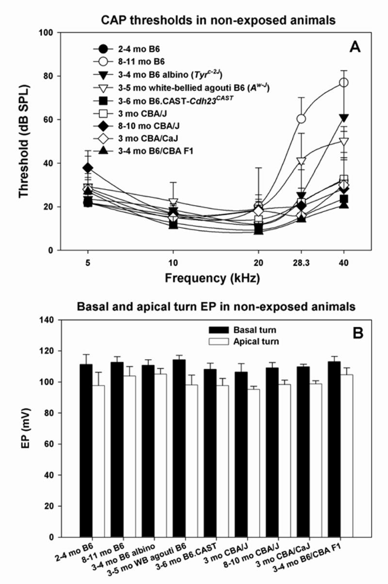
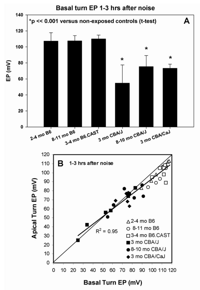
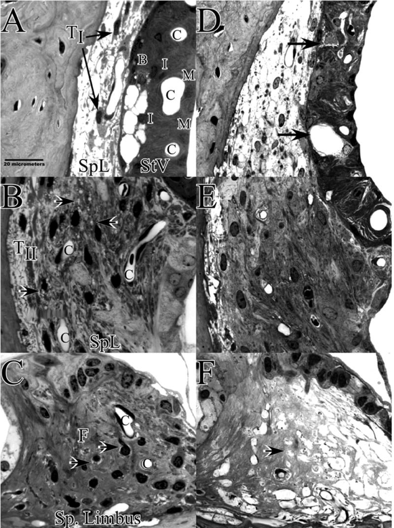
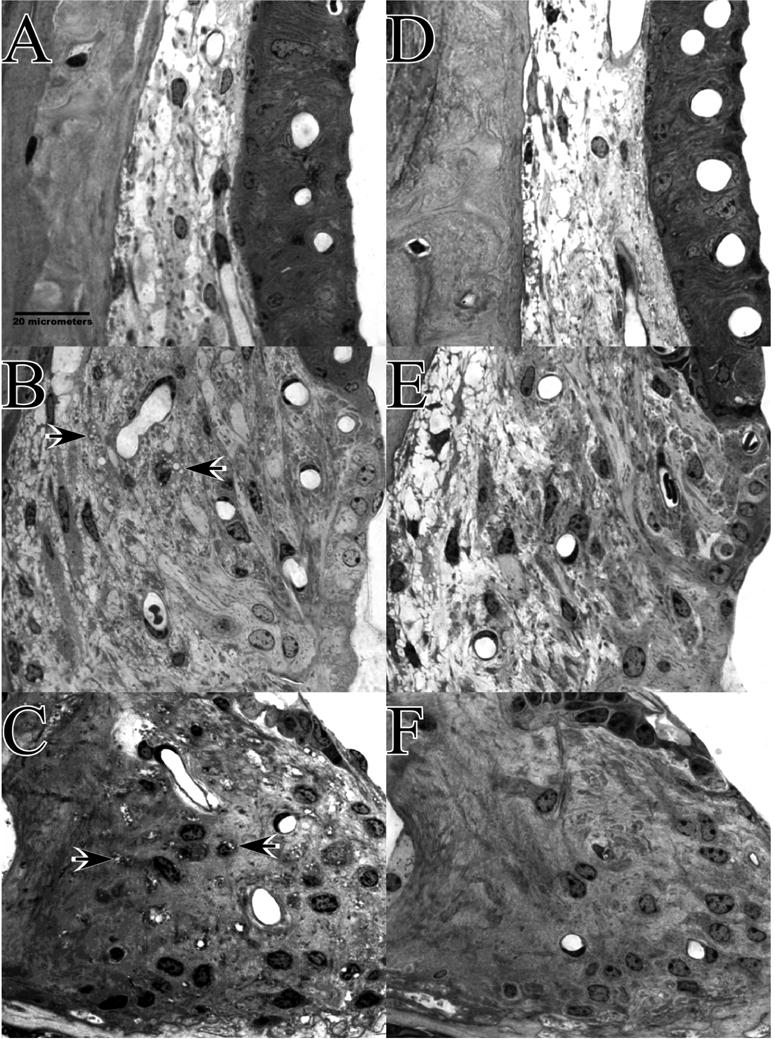
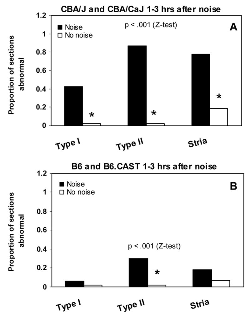
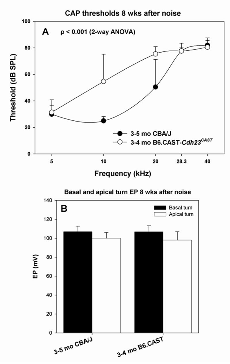
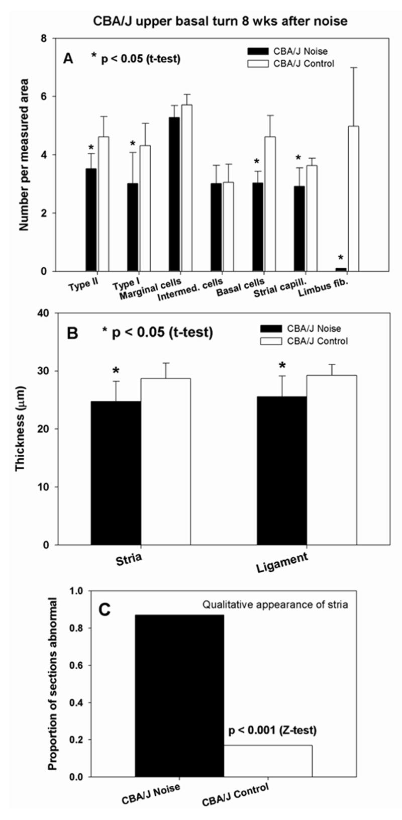
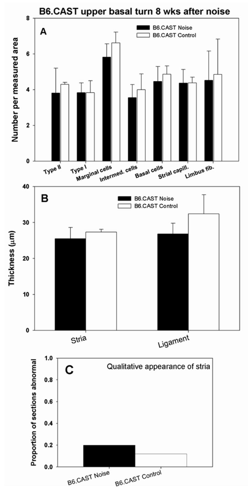
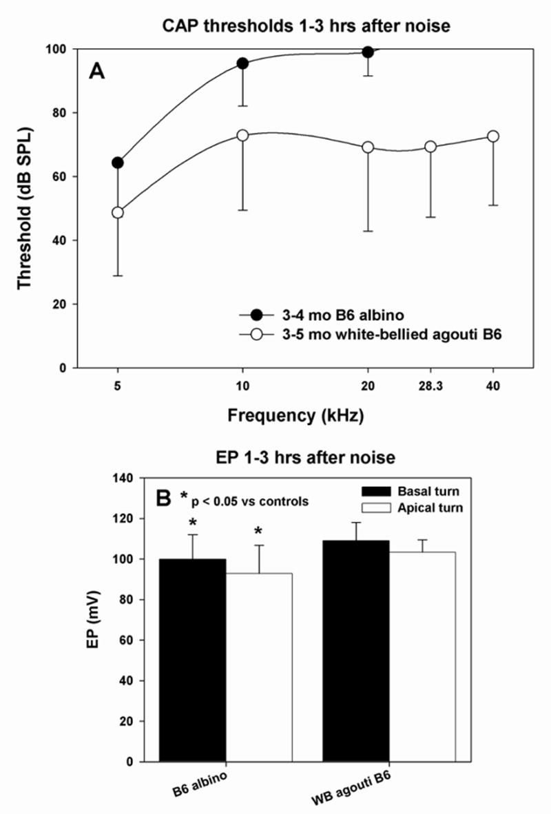
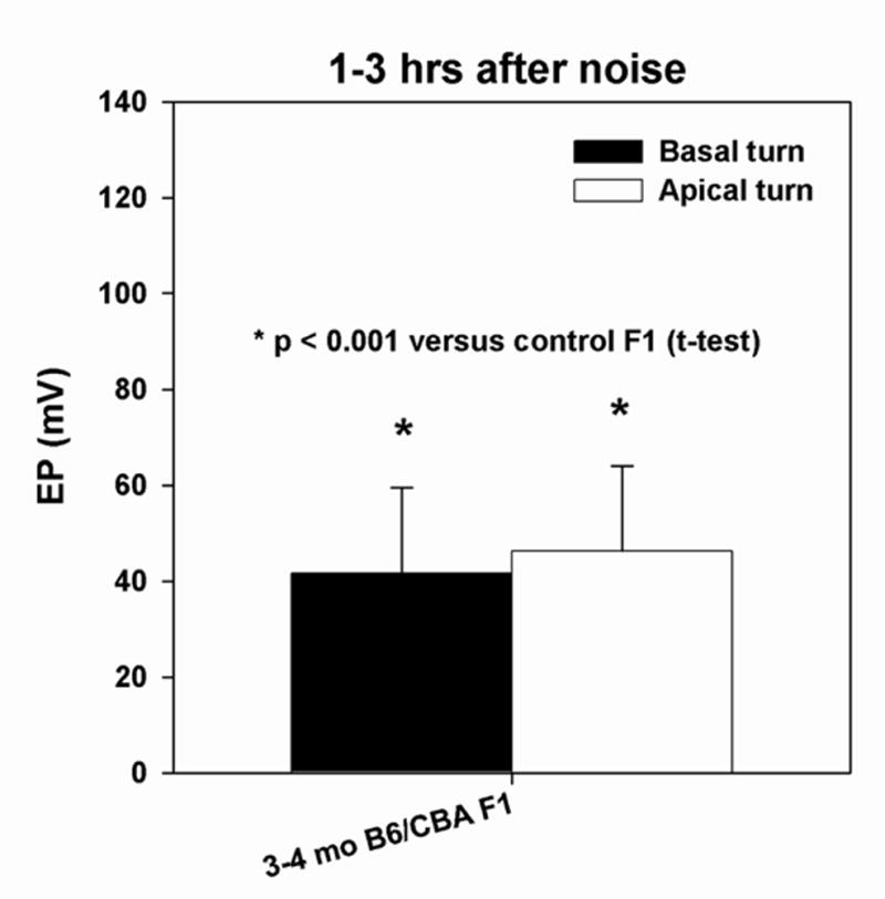
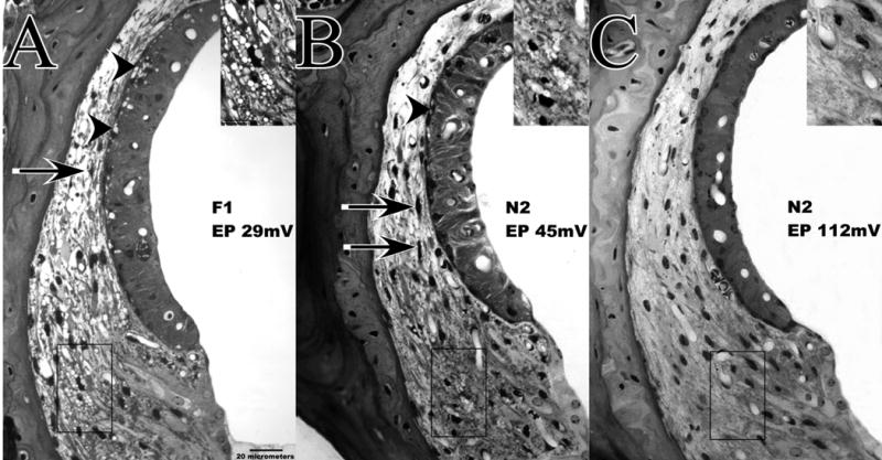
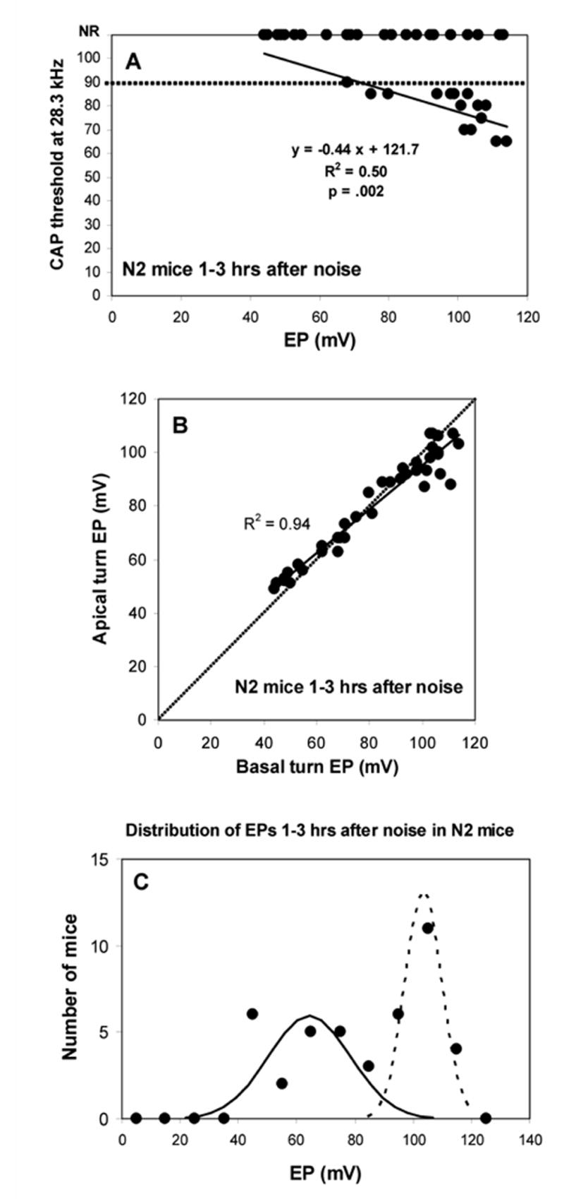
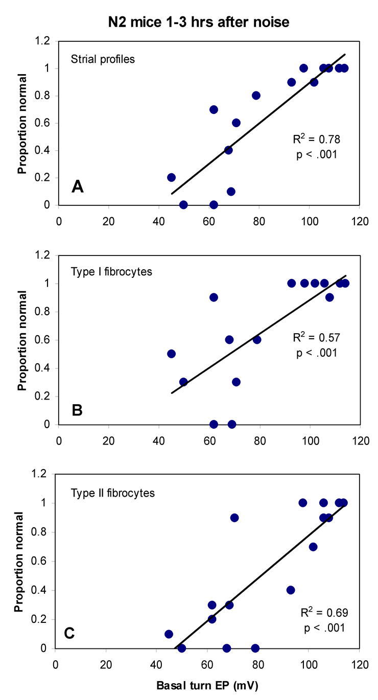
The acute and permanent effects of a single damaging noise exposure were compared in CBA/J, C57BL/6 (B6), and closely related strains of mice. Two hours of broadband noise (4-45 kHz) at 110 dB SPL led to temporary reduction in the endocochlear potential (EP) of CBA/J and CBA/CaJ (CBA) mice and acute cellular changes in cochlear stria vascularis and spiral ligament. For the same exposure, B6 mice showed no EP reduction and little of the pathology seen in CBA. Eight weeks after exposure, all mice showed a normal EP, but only CBA mice showed injury and cell loss in cochlear lateral wall, despite the fact that B6 sustained larger permanent threshold shifts. Examination of noise injury in B6 congenics carrying alternate alleles of genes encoding otocadherin (Cdh23), agouti protein, and tyrosinase (albinism) indicated that none of these loci can account for the strain differences observed. Examination of CBA x B6 F1 mice and N2 backcross mice to B6 further indicated that susceptibility to noise-related EP reduction and associated cell pathology are inherited in an autosomal dominant manner, and are established by one or a few large effect quantitative trait loci. Findings support a common genetic basis for an entire constellation of noise-related cochlear pathologies in cochlear lateral wall and spiral limbus. Even within species, cellular targets of acute and permanent cochlear noise injury may vary with genetic makeup.


Figures

Figure 1
Figure 1
A. Mean(+SD) CAP thresholds for unexposed controls of all mouse lines examined in the study. B. Mean (+SD) basal and apical turn EP values for the mice depicted in A.
Figure 2
Figure 2
A. Mean (+SD) basal turn endocochlear potentials measured 1–3 hrs after noise exposure in young and old B6, B6.CAST-Cdh23CAST congenics, young and old CBA/J, and young CBA/CaJ mice. Only CBA mice showed significant EP reduction versus non-exposed controls of the same strain. B. Basal turn EP versus apical turn EP for the mice depicted in A. The normal spatial gradient for EP is preserved in B6 mice, but eliminated or reversed in CBAs, which show almost complete separation from B6.
Figure 3
Figure 3
Cochlear upper basal turn lateral wall and spiral limbus in example CBA/J mice 1–3 hrs after noise (A–C), or 8 wks after noise (D–F). A. Acutely, CBAs show shrinkage of Type I fibrocytes (arrows) and vacuolized basal cells within stria vascularis. B. Type II fibrocytes in spiral ligament contain vacuoles (arrows). C. Spiral limbus shows shrinkage of fibrocytes in the central zone (arrows). This was seen in both CBA and B6 and was present to some extent in control mice. D. At 8 wks the most obvious feature of the lateral wall is appearance of voids and extracellular debris within the stria (arrows). Stria is also notably thinner. E. Type II region of spiral ligament appears generally normal. F. Spiral limbus is completely devoid of fibrocytes. TI and TII: Type I and II fibrocytes; SpL: Spiral ligament; StV: Stria vascularis; B: Basal cell layer; I: Intermediate cell layer; M: Marginal cell layer; C: Capillary.
Figure 4
Figure 4
Appearance of cochlear upper basal turn lateral wall and spiral limbus in example B6.CAST mice 1–3 hrs after noise (A–C), or 8 wks after noise (D–F). A. Acutely, stria vascularis and adjacent spiral ligament appear normal. B. Some Type II fibrocytes in spiral ligament contain vacuoles (arrows) but to a much more limited extent than in CBAs. C. Spiral limbus shows vacuolization of fibrocytes in the central zone (arrows). Like the shrinkage shown in Figure 3C, this was seen in both CBA and B6 and was present to some extent in control mice. D. At 8 wks stria and adjacent ligament appear normal. E. Type II region of spiral ligament appears generally normal. F. Spiral limbus shows no significant loss of fibrocytes.
Figure 5
Figure 5
Incidence of abnormalities of spiral ligament and stria vascularis in CBA (A) and B6 mice (B) 1–3 hrs after noise exposure. Sections were blindly scored based on appearance of Type I fibrocytes (shrinkage) and Type II fibrocytes (vacuoles) in ligament and presence of vacuoles in the stria. A. Noise-exposed CBA mice showed significant increase in the presence of all three types of anomalies versus non-exposed controls. B. Compared to non-exposed controls, exposed B6 mice showed significant increase only in the appearance of vacuoles in Type II fibrocytes, although at about 1/3 the incidence seen in CBAs.
Figure 6
Figure 6
Permanent physiological changes in CBA/J and B6.CAST mice with respect to CAP threshold (A) and basal and apical turn EP (B) measured 8 wks after noise exposure. A. B6.CAST mice showed significantly greater permanent threshold shifts than CBA/J (2-way ANOVA). B. Both strains showed EPs indistinguishable from non-exposed controls (t-test, p>0.05), indicating complete recovery.
Figure 7
Figure 7
Quantitative analysis of stria vascularis, spiral ligament, and spiral limbus in cochlear upper basal turn of CBA/J mice 8 wks after noise exposure. A. Versus non-exposed controls, noise exposed mice showed significant loss of Type I and II fibrocytes, strial basal cells, strial capillaries, and (nearly complete) loss of limbus fibrocytes. B. Stria and ligament were significantly thinner than in non-exposed controls. C. The incidence of strial abnormalities was significantly greater in noise exposed mice, increasing more than 4-fold.
Figure 8
Figure 8
Quantitative analysis of stria vascularis, spiral ligament, and spiral limbus in cochlear upper base of B6.CAST mice 8 wks after noise exposure. No anomalies were significant versus non-exposed controls (Compare with Figure 7.) Significant thinning of spiral ligament cannot be ruled out due to scatter of data (B).
Figure 9
Figure 9
Acute physiological changes in co-isogenic mice to B6 carrying alleles for albinism (C57BL/6-Tyrc-2J) and white-bellied agouti (C57BL/6-Aw-J) with respect to CAP threshold (A) and basal and apical turn EP (B). A. Albinos showed acute threshold shifts similar to B6 parent strain, while agouti mice were resistant to threshold shifts. B. Versus non-exposed controls, noise-exposed albinos showed modest but significant reduction in EP in both cochlear turns. Agouti mice showed a statistically normal EP.
Figure 10
Figure 10
Basal and apical turn endocochlear potential in B6xCBA F1 mice measured 1–3 hrs after noise exposure. The EP in both turns was significantly reduced compared to unexposed F1s.
Figure 11
Figure 11
Lateral wall of cochlear upper basal turn and EPs in F1 (A) and N2 (B,C) mice examined 1–3 hrs after noise exposure. Insets show expanded view of indicated Type II fibrocyte region of spiral ligament. N2 mice were chosen to illustrate the range of EP values and acute injury seen in these. Both the F1 featured in A and the N2 having a depressed EP (B) show CBA-characteristic acute pathology of spiral ligament and stria vascularis, including vacuolized Type II fibrocytes, shrunken Type I fibrocytes (arrows), and vacuolized strial basal cells (arrowheads). The N2 showing a normal EP (C) shows none of these features.
Figure 12
Figure 12
Physiological measures in N2 backcross mice measured 1–3 hrs after noise exposure. A. CAP threshold at 28.3 kHz versus basal turn EP. Roughly half the animals showed no CAP response, largely independent of EP. Among mice showing a response, there was a significant negative correlation between threshold and EP, indicating an impact of the EP on acute threshold shifts. The dotted line indicates that maximum output of the sound system at 28.3 kHz. B. Basal turn EP versus apical turn EP for the animals depicted in A. As also shown in Figure 2, the normal spatial gradient for the EP is altered when the EP is reduced by noise exposure. C. Distribution of basal turn EP values for the same animals. EPs were sorted into 10 mV bins. 21 of 42 mice showed EPs below 90 mV; 21 showed higher EPs. Distribution was not normal, but was well fit by two Gaussians with mean±SD of 64.3±14.1 mV (‘depressed EP’ cluster) and 103.5±6.3 mV (‘normal EP’ cluster).
Figure 13
Figure 13
Incidence of acute pathology of Type I and II fibrocytes and strial anomalies in N2 mice versus basal turn EP. Incidence of pathology (given as the proportion of sections that scored as normal) of stria vascularis (A), Type I (B), and Type II (C) was significantly correlated with EP by Pearson correlation.
Figure 14
Figure 14
Cochlear pathology observed 24 hrs post-noise exposure in example F1 hybrid mouse. The basal turn EP at the time of sacrifice was 50 mV. Lateral wall of the upper base (A) and spiral limbus of the lower apex (B) are shown. Note swollen intra-strial space, and pyknotic nuclei and dense cytoplasm of Type I and II fibrocytes (upper and lower insets, respectively, in A). B shows loss of cells and condensed nuclei of remaining cells in central zone of the limbus.

Similar articles

Cited by

References

    1. Axelsson A, Borg E, Hornstrand C. Noise effects on the cochlear vasculature in normotensive and spontaneously hypertensive rats. Acta Otolaryngol. 1983;96:215–225. - PubMed
    1. Barrenas ML, Holgers KM. Ototoxic interaction between noise and pheomelanin: Distortion product otoacoustic emissions after acoustical trauma in chloroquine-treated red, black, and albino guinea pigs. Audiology. 2000;39:238–246. - PubMed
    1. Barrenas ML, Lindgren F. The influence of eye color on susceptibility to TTS in humans. Br J Audiol. 1991;25:303–307. - PubMed
    1. Barrenas M. Hair cell loss from acoustic trauma in chloroquine-treated red, black and albino guinea pigs. Audiology. 1997;36:187–201. - PubMed
    1. Beagley HA. Acoustic trauma in the guinea pig. I Electrophysiology and Histology. Acta Otolaryngol. 1965;60:437–451. - PubMed

Publication types