. 2006 Dec 26;45(51):15670-83.
doi: 10.1021/bi061718f.

Cytoskeletal tension regulates both expression and degradation of h2-calponin in lung alveolar cells

Affiliations

Cytoskeletal tension regulates both expression and degradation of h2-calponin in lung alveolar cells

M Moazzem Hossain et al. Biochemistry. .

Abstract

Calponin is an actin filament-associated regulatory protein, and its h2 isoform is expressed in lung alveolar epithelial cells under postnatal upregulation during lung development corresponding to the commencement of respiratory expansion. Consistent with this correlation to mechanical tension, the expression of h2-calponin in alveolar cells is dependent on substrate stiffness and cytoskeleton tension. The function of h2-calponin in the stability of actin cytoskeleton implicates a role in balancing the strength and compliance of alveoli. An interesting finding is a rapid degradation of h2-calponin in lung after prolonged deflation, which is prevented by inflation of the lung to the in situ expanded volume. Decreasing mechanical tension in cultured alveolar cells by reducing the dimension of culture matrix reproduced the degradation of h2-calponin. Inhibition of myosin II ATPase also resulted in the degradation of h2-calponin in alveolar cells, showing a determining role of the tension in the actin cytoskeleton. Alveolar cells statically cultured on silicon rubber membrane build high tension in the cytoskeleton corresponding to a high expression of h2-calponin. Chronic cyclic stretching of cells on the membrane did not increase but decreased the expression of h2-calponin. This finding suggests that when cellular structure adapts to the stretched dimension, cyclic relaxations periodically release cytoskeleton tension and lower the total amount of tension that the cell senses over time. Therefore, the isometric tension, other than tension dynamics, determines the expression of h2-calponin. The tension regulation of h2-calponin synthesis and degradation demonstrates a novel mechanical regulation of cellular biochemistry.


Figures

Figure 1
Figure 1
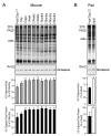
Expression of h2-calponin in representative tissues. Total protein extracts from major organs of the mouse were analyzed by Western blots using the anti-h2-calponin polyclonal antibody RAH2 and mAb CP21 together with the anti-h1-calponin mAb CP3. Purified mouse h1- and h2-calponin proteins were used as control. The sample loading was normalized by the level of actin (separately in the non-muscle and muscle tissue groups). The blots detected h2-calponin in smooth muscle and several non-muscle organs with high levels of expression in spleen and lung.
Figure 2
Figure 2
Expression of h2-calponin in lung alveolar epithelial cells. (A) Frozen sections of adult mouse lung were incubated with of anti-h2-calponin mAb CP22 hybridoma cultural supernatant or SP2/0 myeloma control supernatant followed by horseradish peroxidase-labeled anti-mouse IgG second antibody and H2O2-daminobenzidin substrate reaction. Significant amounts of h2-calponin were detected in alveolar epithelial cells. (B) Total protein extracts from SW1573 human alveolar cells were analyzed by SDS-PAGE and Western blots using anti-h2 calponin polyclonal antibody RAH2, mAb CP22 and mAb 1D2 (raised against cloned human h2-calponin, 15) together with the anti-h1-calponin mAb CP3. Purified mouse h1- and h2-calponin proteins were used as controls. The results show that the alveolar cells express h2-calponin and no h1-calponin was detected.
Figure 3
Figure 3
Postnatal up-regulation of h2-calponin in the lung. (A) Mouse lung tissues were obtained at a series of developmental time points and examined by Western blots using anti-h2-calponin antibody RAH2. Normalized by the level of actin or total protein, densitometry analysis demonstrated a rapid postnatal up-regulation of h2-calponin in the developing lung with a correlation to the commencement of respiratory activity. (B) Fetal versus adult rat lungs were examined by Western blot using RAH2 antibody. Densitometry quantification against actin or total cellular protein showed significantly higher levels of h2-calponin expressed in the adult versus fetal lung. *P<0.001 as compared with the plateau levels.
Figure 4
Figure 4
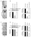
Substrate anchorage- and stiffness-dependent expression of h2-calponin in alveolar cells. (A) SW1573 human alveolar cells were cultured on plastic dishes steadily or with vibration. Phase contrast microscopic images show that in contrast to the monolayer formed in the steady culture, continuous vibration prevented the cells from anchoring to the cultural dish and produced cell aggregates that could attach to the dish when vibration was stopped. (B) Total protein extracts from the cells were examined by SDS-PAGE and Western blotting using anti-h2-calponin polyclonal antibody RAH2. Normalized by the level of actin, total cellular protein, or histones (representing the number of cells), the results show that H2-calponin expression in alveolar cells decreased significantly when growing as floating aggregates in comparison with that in monolayer cells attached to plastic dish (*P<0.001). The expression of h2-calponin was up-regulated to resume the control level after the floating cells attached to the plastic substrate to form a monolayer. The results were summarized from 4 individual experiments. (C) SW1573 human alveolar cells were cultured on plastic dish or polyacrylamide gels of high or low stiffness for 3 days. (D) The levels of h2-calponin were determined by Western blot analysis using anti-h2-calponin antibody RAH2. Normalized by the amount of actin, total cellular protein, or histones, densitometry quantification shows a significant decrease in the expression of h2-calponin in alveolar cells grown on soft polyacrylamide gel in comparison with that in the hard gel and plastic dish cultures (*P<0.005, **P<0.001). The results were summarized from 3 individual experiments.
Figure 5
Figure 5
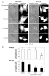
Role of h2-calponin in the stability of actin cytoskeleton in alveolar cells. SW1573 human alveolar cells cultured on hard or soft gel substrate were treated with cytochalasin B. (A) The effects on cellular structure were examined by phase contrast microscopy and the actin cytoskeleton was stained with rhodamine-labeled phalloidin. The results demonstrate that cytochalasin B treatment disrupted the actin cytoskeleton to result in slack appearance of the cell in a concentration dependent manner. Cells cultured on the gel substrates contained very fine actin stress fibers compared with that in cells cultured on plastic substrate. The cells cultured on soft gel showed collapses of actin cytoskeleton at lower concentrations of cytochalasin B in comparison to the hard gel controls, indicating a lower stability of the actin filaments in correspondence to the decreased h2-calponin content (Figure 4, C and D). (B) Cell spreading area was measured to quantitatively compare the destructing effects of cytochalasin B on cytoskeleton structure. The results are consistent with a decreased stability of the actin filaments in cells cultured on soft gel substrate as compared with the hard gel control. *P<0.001. The data are summarized from measurements of 30 cells in each group.
Figure 6
Figure 6
Rapid degradation of h2-calponin in lung tissue after prolonged relaxation. (A) Total protein extracts from infant and adult human lung autopsy tissues were analyzed by Western blot using the anti-h2-calponin antibody RAH2. The results revealed various degrees of h2-calponin degradation in the postmortem lung samples. (B) Adult rat lung tissue samples, freshly prepared or stored on ice for 24h, were examined by Western blotting using RAH2 antibody. The results showed a decrease in h2-calponin after the storage. (C) Deflated adult mouse lung was incubated at 37 °C for 6 hours and analyzed by 15% high cross linker SDS-gel and Western blotting using the RAH2 antibody. H2-calponin degradation with a fragment similar to that found in the human autopsy lung samples was detected. (D) Deflated adult mouse lung were incubated at 37°C and tissue samples were harvested at a series of time points for SDS-PAGE and RAH2 Western blot analysis. Normalized by the levels of actin or total protein, densitometry of the Coomassie Brilliant Blue R250-stained SDS-gels and Western blots demonstrated a rapid selective degradation of h2-calponin. Actin and other major proteins (MHC, myosin heavy chain) did not show significant changes in the SDS-gel and densitometry scans. *P<0.05; **P<0.005; ***P<0.001. The results are summarized from 4 individual experiments.
Figure 7
Figure 7
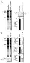
Tension release in cultured alveolar cells resulted in h2-calponin degradation. RLE-6TN rat alveolar cells were cultured on rubber membrane that was bi-axially stretched and held at 108% of its original surface size for three days before the stretching was released to relax the attached cells. SDS-PAGE and Western blot analysis using RAH2 antibody (A) and densitometry quantification against the levels of actin or total cellular protein (B) detected decreases in h2-calponin after the reduction of cellular tension. In contrast, the levels of tropomyosin did change as shown by the Western blot quantification using mAb LC24 (). The data are summarized as SEM from six sets of samples, *P<0.05. (C) The phase contrast micrographs and rhodamine-phalloidin fluorescence stains of the actin cytoskeleton demonstrate a decreased resistance of actin stress fibers to cytochalasin B treatment in alveolar cells after the reduction of tension. This is shown by the collapses of actin stress fibers at lower concentrations of cytochalasin B than that needed for the high tension control, indicating a lower stability of the actin filaments corresponding to the decreased level of h2-calponin.
Figure 8
Figure 8
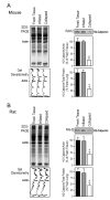
Inflation-distension of the lung prevents the degradation h2-calponin. Mouse (A) and rat (B) lungs were inflated with air and incubated at 37°C for 6 hours together with collapsed control lungs. Total protein extracts were examined by Western blot with the RAH2 antibody. The densitometry quantification of Western blots and Coomassie blue stained SDS-gel showed a effective prevention of h2-calponin proteolysis in the inflated lung (normalized by the levels of actin or total protein, *P<0.001). The results are summarized from 4 individual experiments.
Figure 9
Figure 9
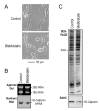
Regulation of h2-calponin in alveolar cells by myosin motor-based cytoskeleton tension. (A) Monolayer cultures of RLE-6TN rat alveolar cells were treated with 100 μM blebbistatin or DMSO solvent control for 6 hours or 3 days. Morphology changes of the cells can be seen in the lower magnification phase contrast images taken from the culture dish. The cells on cover slips were fixed with cold acetone and the actin stress fibers were visualized by staining with rhodamine-conjugated phalloidin under higher magnification. The phase contrast and fluorescence images showed that the blebbistatin-treated cells became slack, indicating a reduced cytoskeleton tension. (B) Parallel cultures were analyzed by SDS-PAGE and RAH2 Western blot. The densitometry analysis of the h2-calponin blots against the level of actin, total cellular protein or histones showed significant decreases of h2-calponin after short or long time blebbistatin treatments, suggesting that myosin II-based cytoskeleton tension affects both proteolytic and transcriptional regulations of h2-calponin in alveolar cells. The data are summarized from two sets of the experiment. *P<0.05; **P<0.001.
Figure 10
Figure 10
Inhibition of myosin motor decreases h2-calponin gene expression in NIH 3T3 cells. NIH 3T3 mouse fibroblasts cultured on plastic substrate were treated with 100 μM blebbistatin for 3 days. The control cells were treated with 0.2% DMSO solvent control. (A) Phase contrast images show the slack morphology of the blebbistatin-treated cells. (B) Northern blotting was carried out with 32P-labeled h2-calponin specific cDNA probe on total RNA extracted from the cells. Normalized by the ribosomal RNA levels, the autoradiograph shows decreased expression of h2-calponin mRNA in the blebbistatin-treated cells. (C) The corresponding decrease in h2-calponin protein is demonstrated by Western blot using the anti-h2-calponin antibody RAH2.
Figure 11
Figure 11
Cyclic stretching decreases h2-calponin expression in alveolar cells. (A) SW1573 human alveolar cells were cultured on plastic dish or rubber membranes without stretching for 3 days. Total protein extracts were examined by SDS-PAGE and RAH2 antibody Western blot. The SDS-gel and Western blots densitometry quantifications against the level of actin show that alveolar cells statically cultured on plastic dish and rubber membrane had similar protein contents and high h2-calponin expression, indicating that they both provide cultural substrates non-deformable by the force generated in alveolar cells. (B) RLE-6TN rat alveolar cells cultured on rubber membranes were treated with cyclic stretching for 3 days. Total protein extracts were examined as above and SDS-gel and Western blots densitometry quantifications against the level of actin, total cellular protein, or histones showed that cyclic stretch significantly decreased the level of h2-calponin expression in the cells (*P<0.005, **P<0.001). The results are summarized from 6 individual experiments.
Figure 11
Figure 11
Cyclic stretching decreases h2-calponin expression in alveolar cells. (A) SW1573 human alveolar cells were cultured on plastic dish or rubber membranes without stretching for 3 days. Total protein extracts were examined by SDS-PAGE and RAH2 antibody Western blot. The SDS-gel and Western blots densitometry quantifications against the level of actin show that alveolar cells statically cultured on plastic dish and rubber membrane had similar protein contents and high h2-calponin expression, indicating that they both provide cultural substrates non-deformable by the force generated in alveolar cells. (B) RLE-6TN rat alveolar cells cultured on rubber membranes were treated with cyclic stretching for 3 days. Total protein extracts were examined as above and SDS-gel and Western blots densitometry quantifications against the level of actin, total cellular protein, or histones showed that cyclic stretch significantly decreased the level of h2-calponin expression in the cells (*P<0.005, **P<0.001). The results are summarized from 6 individual experiments.

Similar articles

Cited by

References

    1. Chicurel ME, Chen CS, Ingber DE. Cellular control lies in the balance of forces. Curr Opin Cell Biol. 1998;10:232–239. - PubMed
    1. Eckes B, Krieg T. Regulation of connective tissue homeostasis in the skin by mechanical forces. Clin Exp Rheumatol. 2004;22:S73–S76. - PubMed
    1. Hamill OP, Martinac B. Molecular basis of mechanotransduction in living cells. Physiol Rev. 2001;81:685–740. - PubMed
    1. Lehoux S, Tedgui A. Cellular mechanics and gene expression in blood vessels. J Biomech. 2003;36:631–643. - PubMed
    1. Walker JL, Fournier AK, Assoian RK. Regulation of growth factor signaling and cell cycle progression by cell adhesion and adhesion-dependent changes in cellular tension. Cytokine Growth Factor Rev. 2005;16:395–405. - PubMed

Publication types

MeSH terms

Substances