Comparative Study
. 2006 Dec 26;45(51):15921-30.
doi: 10.1021/bi061626q. Epub 2006 Dec 19.

Specific and efficient binding of xeroderma pigmentosum complementation group A to double-strand/single-strand DNA junctions with 3'- and/or 5'-ssDNA branches

Affiliations
Comparative Study

Specific and efficient binding of xeroderma pigmentosum complementation group A to double-strand/single-strand DNA junctions with 3'- and/or 5'-ssDNA branches

Zhengguan Yang et al. Biochemistry. .

Abstract

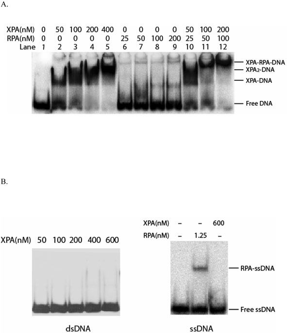
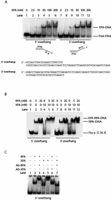
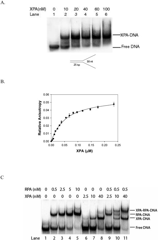
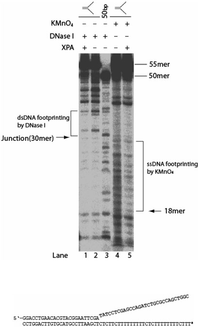
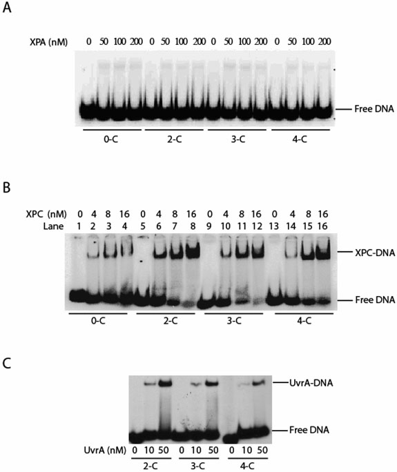
Human XPA is an important DNA damage recognition protein in nucleotide excision repair (NER). We previously observed that XPA binds to the DNA lesion as a homodimer [Liu, Y., Liu, Y., Yang, Z., Utzat, C., Wang, G., Basu, A. K., and Zou, Y. (2005) Biochemistry 44, 7361-7368]. Herein we report that XPA recognized undamaged DNA double-strand/single-strand (ds-ssDNA) junctions containing ssDNA branches with binding affinity (Kd = 49.1 +/- 5.1 nM) much higher than its ability to bind to DNA damage. The recognized DNA junction structures include the Y-shape junction (with both 3'- and 5'-ssDNA branches), 3'-overhang junction (with a 3'-ssDNA branch), and 5'-overhang junction (with a 5'-ssDNA branch). Using gel filtration chromatography and gel mobility shift assays, we showed that the highly efficient binding appeared to be carried out by the XPA monomer and that the binding was largely independent of RPA. Furthermore, XPA efficiently bound to six-nucleotide mismatched DNA bubble substrates with or without DNA adducts including C8 guanine adducts of AF, AAF, and AP and the T[6,4]T photoproducts. Using a set of defined DNA substrates with varying degrees of DNA bending, we also found that the XPC-HR23B complex recognized DNA bending, whereas neither XPA nor the XPA-RPA complex could bind to bent DNA. We propose that, besides DNA damage recognition, XPA may also play a novel role in stabilizing, via its high affinity to ds-ssDNA junctions, the DNA strand opening surrounding the lesion for stable formation of preincision NER intermediates. Our results provide a plausible mechanistic interpretation for the indispensable requirement of XPA for both global genome and transcription-coupled repairs. Since ds-ssDNA junctions are common intermediates in many DNA metabolic pathways, the additional potential role of XPA in cellular processes is discussed.


Figures

Figure 1
Figure 1
XPA binding to DNA duplex containing a G[8,5-Me]T crosslink lesion. Panel A: Homogeneous XPA protein purified from baculovirus-infected insect cells showed high specific affinity to 50-bp DNA substrate containing a γ-radiation-induced G[8,5-Me]T crosslink lesion in the presence and absence of RPA in gel mobility shift assays. RPA protein itself displayed no affinity for the DNA crosslink adduct (lanes 6-9), although the presence of RPA resulted in a super-shift of XPA-DNA bands corresponding to the XPA-RPA-DNA interaction (lanes 10-12). Panel B: XPA protein showed little or no affinity for undamaged DNA duplex or ssDNA. Gel mobility shift assays were performed on 3.5% native polyacrylamide gel electrophoresed at 4°C.
Figure 2
Figure 2
Binding of XPA protein to partially mismatched DNA bubble substrates with or without a lesion. Panel A: XPA bound to six base-mismatched DNA bubble substrates with or without AF, AAF, AP or T[6,4]T adduct at varying concentrations. ND-B6 stands for undamaged DNA substrate containing a six-base mismatched bubble. Panel B: XPA bound to DNA substrates with various sizes of bubble. The underline in the sequences indicates the mismatched bases for bubble formation.
Figure 3
Figure 3
Interactions of XPA and/or RPA with 3′- and 5′-single strand overhang DNA substrates. Panel A: XPA bound to 3′-ssDNA overhang and 5′-ssDNA overhang DNA substrates at various concentrations. Panel B: XPA and RPA interacted with the 3′-overhang and 5′-overhang DNA substrates. Panel C: XPA and RPA interacted with 5′-overhang DNA substrate, and the complexes were supershifted with corresponding antibodies, respectively.
Figure 4
Figure 4
The binding of XPA to Y-shape DNA substrate in the presence and absence of RPA. Panel A: XPA bound to the Y-shape DNA substrate in an XPA-concentration dependent manner. Panel B: Fluorescence anisotropy measurements of XPA binding to ds-ssDNA junction substrate. The binding isotherms were generated by titrating the substrate with increasing concentrations of XPA. Panel C: XPA and/or RPA bound to Yshape DNA. RPA bound to the single strand regions of the Y-shape substrate (lanes 2-5), while XPA bound to the ds-ssDNA junction of the substrate (lanes 6-8). When XPA and RPA both were present, formation of three complexes was observed corresponding to XPA binding to ds-ssDNA junction (XPA-DNA), RPA binding to the ssDNA regions of the substrate (RPA-DNA), and XPA and RPA concurrently binding to the junction and ssDNA regions of the same substrate molecules (XPA-RPA-DNA), respectively (lanes 10 and 11).
Figure 5
Figure 5
Footprinting of XPA binding to the ds-ssDNA junctions in the Y-shape substrate with 3′- and 5′-ssDNA overhangs. Lanes 1-3, dsDNA footprinting by DNase I. Lanes 4 and 5, ssDNA footprinting by KMnO4. Y-shape substrates with 32P-labeled 5′-end of the bottom strand (indicated by the asterisk) were incubated at 30 °C for 15 min in binding buffer with or without XPA. After the incubation, substrates were digested with 0.7 ng DNase I (lanes 1-3) or probed with 0.2 mM KMnO4 (lanes 5 and 6), followed by hot piperidine treatment. Both footprinting reactions were performed at room temperature for 1 min. The samples were analyzed on a 12% denaturing polyacrylamide gel.
Figure 6
Figure 6
Gel filtration-scintillation analysis for determination of the stoichiometry of XPA binding to Y-shape DNA. XPA of 40 and 1000 nM was incubated with radiolabeled Y-shape and AAF-50bp DNA substrates, respectively, and analyzed by gel filtration chromatography on Superdex® 200 column. The eluted fractions were then subjected to scintillation counting. Radioactivity profiles of the XPA–DNA complexes versus retention volume show that XPA-Y-shape DNA complex was eluted at 13.2 mL (▼), as compared to XPA-AAF DNA complex at 12.2 mL (●) (1). Free DNA was eluted at ~22 mL (◦). Determination of apparent molecular weights of the XPA-DNA complexes based on a linear relationship between the molecular weights of protein markers and their retention times suggests that XPA may bind to the Y-shape DNA substrate as a monomer.
Figure 7
Figure 7
Recognition of DNA bending by XPA or XPC-HR23B complex. Pane A: XPA displayed no significant affinity for any of the substrates with DNA bending induced by a two carbon (2-C), three carbon (3-C), or four carbon (4-C) tether crosslinking two adjacent guanines. The 0-C stands for unmodified DNA with no bending (no carbon tether). Pane B: XPC-HR23B complex (labeled as XPC) had high affinities to DNA bending with the following order: 2-C > 3-C > 4-C > 0-C, which is consistent with the order of their bending-angles: 2-C > 3-C > 4-C > 0-C. Panel C: Binding of E. coli UvrA to the same DNA bending substrates (27-29).

References

    1. Liu Y, Liu Y, Yang Z, Utzat C, Wang G, Basu AK, Zou Y. Cooperative interaction of human XPA stabilizes and enhances specific binding of XPA to DNA damage. Biochemistry. 2005;44:7361–7368. - PMC - PubMed
    1. Wood RD. DNA damage recognition during nucleotide excision repair in mammalian cells. Biochimie. 1999;81:39–44. - PubMed
    1. Thoma BS, Vasquez KM. Critical DNA damage recognition functions of XPC-hHR23B and XPA-RPA in nucleotide excision repair. Mol Carcinog. 2003;38:1–13. - PubMed
    1. Sancar A, Lindsey-Boltz LA, Unsal-Kacmaz K, Linn S. Molecular mechanisms of mammalian DNA repair and the DNA damage checkpoints. Annu Rev Biochem. 2004;73:39–85. - PubMed
    1. Reardon JT, Sancar A. Nucleotide excision repair. Prog Nucleic Acid Res Mol Biol. 2005;79:183–235. - PubMed

Publication types

MeSH terms