. 2006 Dec 18:5:41.
doi: 10.1186/1475-2859-5-41.

Secretion and properties of a hybrid Kluyveromyces lactis-Aspergillus niger beta-galactosidase

Affiliations

Secretion and properties of a hybrid Kluyveromyces lactis-Aspergillus niger beta-galactosidase

Angel Pereira Rodríguez et al. Microb Cell Fact. .

Abstract

Background: The beta-galactosidase from Kluyveromyces lactis is a protein of outstanding biotechnological interest in the food industry and milk whey reutilization. However, due to its intracellular nature, its industrial production is limited by the high cost associated to extraction and downstream processing. The yeast-system is an attractive method for producing many heterologous proteins. The addition of a secretory signal in the recombinant protein is the method of choice to sort it out of the cell, although biotechnological success is not guaranteed. The cell wall acting as a molecular sieve to large molecules, culture conditions and structural determinants present in the protein, all have a decisive role in the overall process. Protein engineering, combining domains of related proteins, is an alternative to take into account when the task is difficult. In this work, we have constructed and analyzed two hybrid proteins from the beta-galactosidase of K. lactis, intracellular, and its Aspergillus niger homologue that is extracellular. In both, a heterologous signal peptide for secretion was also included at the N-terminus of the recombinant proteins. One of the hybrid proteins obtained has interesting properties for its biotechnological utilization.

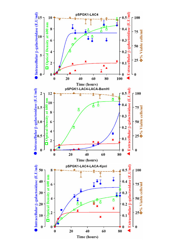
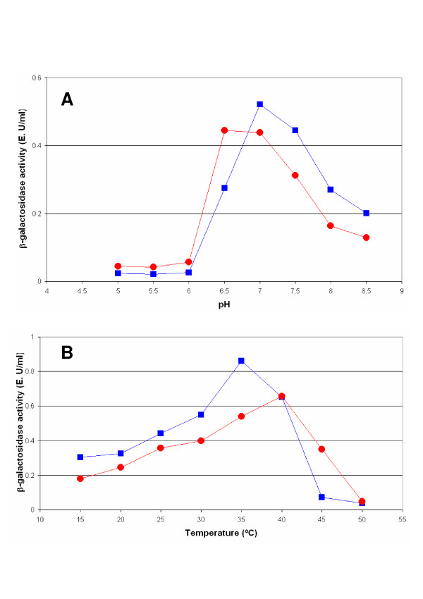
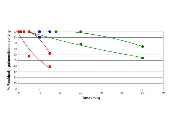
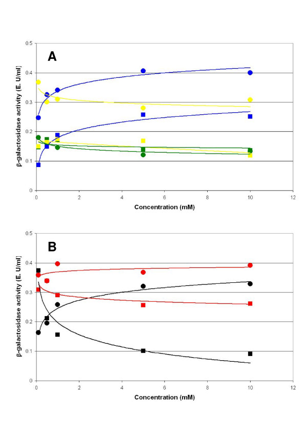
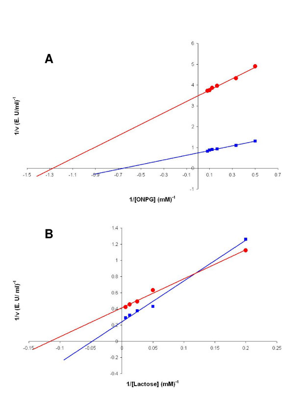
Results: The highest levels of intracellular and extracellular beta-galactosidase were obtained when the segment corresponding to the five domain of K. lactis beta-galactosidase was replaced by the corresponding five domain of the A. niger beta-galactosidase. Taking into account that this replacement may affect other parameters related to the activity or the stability of the hybrid protein, a thoroughly study was performed. Both pH (6.5) and temperature (40 degrees C) for optimum activity differ from values obtained with the native proteins. The stability was higher than the corresponding to the beta-galactosidase of K. lactis and, unlike this, the activity of the hybrid protein was increased by the presence of Ni2+. The affinity for synthetic (ONPG) or natural (lactose) substrates was higher in the hybrid than in the native K. lactis beta-galactosidase. Finally, a structural-model of the hybrid protein was obtained by homology modelling and the experimentally determined properties of the protein were discussed in relation to it.

Conclusion: A hybrid protein between K. lactis and A. niger beta-galactosidases was constructed that increases the yield of the protein released to the growth medium. Modifications introduced in the construction, besides to improve secretion, conferred to the protein biochemical characteristics of biotechnological interest.


Figures

Figure 1
Figure 1
Amino acid sequence alignment of E. coli β-galactosidase with the K. lactis and A. niger β-galactosidase. Multiple sequence alignment of Escherichia coli β-galactosidase (ECLACZ), Kluyveromyces lactis β-galactosidase (KLLAC4) and Aspergillus niger β-galacatosidase (ANLACA). "*" means that the residues in that column are identical in all sequences in the alignment. ":" means that conserved substitutions have been observed. "." means that semi-conserved substitutions are observed. Acid (blue colour) and basic (red colour) amino acids of K. lactis and A. niger β-galactosidase are marked. The coloured bar below the E. coli β-galactosidase represents the five different domains structurally determined in the protein (Domain 1: green; Domain 2: yellow; Domain 3: red; Domain 4: light blue; Domain 5: dark blue). The secondary structure of E. coli β-galactosidase was obtained from the Protein Data Bank [42]. The localization of the restriction sites BamHI (residues underlined and pink) and KpnI (residues underlined and blue) are indicated. The conserved residues in E. coli β-galactosidase and K. lactis β-galactosidase important for catalytic function in E. coli β-galactosidase are shown in green. The residues of A. niger signal sequence are in yellow and underlined.
Figure 2
Figure 2
Kinetics of growth and secretion. Growth (Optical Density at 600 nm), percentage of viable cells per ml, extracellular and intracellular β-galactosidase production (E. U. mL-1) by the MW190-9B strain transformed with the corresponding plasmids. Values represent the mean of 5 different cultures.
Figure 3
Figure 3
Determination of the pH and temperature optimum. Optimum pH (A) and optimum temperature (B) for the hybrid enzyme between the β-galactosidase of K. lactis and A. niger (red) and the β-galactosidase of K. lactis (blue). Experimental variations are less than10% of the value of the point. Data are the mean of three independent experiments.
Figure 4
Figure 4
Determination of the thermal stability. Determination of the thermal stability at 30°C (green), 42°C (blue) and 50°C (red) for the hybrid enzyme between the β-galactosidase of K. lactis and A. niger (circles) and the native β-galactosidase of K. lactis (square). Experimental variations are less than 10% of the value of the point. Results are the average of two independent experiments.
Figure 5
Figure 5
Determination of the effects of the divalentcations. Determination of the effects of the divalent cations Mg2+ (blue), Ca2+ (green) and Zn2+ (yellow) (A) and Mn2+ (red) and the Ni2+ (black) (B) on the enzymatic activity of the β-galactosidase hybrid between K. lactis and A. niger (circles) and the native β-galactosidase of K. lactis (square). Experimental variations are less than 10% of the value of the point. Results are the average of two independent experiments.
Figure 6
Figure 6
Lineweaver-Burk plots. Lineweaver-Burk plot of the reaction catalyzed by the β-galactosidase hybrid between K. lactis and A. niger (red circles) and the native β-galactosidase of K. lactis (square blue) in the presence of the synthetic substrate ONPG (A) or the natural substrate lactose (B). Experimental variations are less than 10% of the value of the point. Results are the average of two independent experiments.
Figure 7
Figure 7
Ribbon representations. Ribbon diagram corresponding to the prediction of the tertiary structure of K. lactis β-galactosidase (A), A. niger β-galactosidase (B) and hybrid β-galactosidase (C) using the Swiss-Model program. The residues mentioned in Figure 1 have been drawn as spheres of colours (E482 blue, M522 green, Y523 yellow, E551 red). D1–D5 identify the five domains of K. lactis β-galactosidase (A) predicted by alignment in comparison with the sequence of the E. coli β-galactosidase (Figure 1). The fifth domain of the A. niger β-galactosidase is coloured in green (B and C). The white arrow (C) shows the slight opening of the third domain in the hybridβ-galactosidase.

References

    1. Becerra M, Rodríguez-Belmonte ME, Cerdán ME, González Siso MI. Engineered autolytic yeast strains secreting Kluyveromyces lactis β-galactosidase for production of heterologous proteins in lactose media. J Biotechnol. 2004;109:131–137. doi: 10.1016/j.jbiotec.2003.10.030. - DOI - PubMed
    1. González Siso MI. The biotechnological utilization of cheese whey: a review. Biores Technol. 1996;57:1–11. doi: 10.1016/0960-8524(96)00036-3. - DOI
    1. Becerra M, Díaz-Prado S, González Siso MI, Cerdán ME. New secretory strategies for Kluyveromyces lactis β-galactosidase. Prot Eng. 2001;14:379–386. doi: 10.1093/protein/14.5.379. - DOI - PubMed
    1. Becerra M, Cerdán ME, González Siso MI. Microescale purification of the enzyme β-galactosidase from Kluyveromyces lactis reveals that dimeric and tetrameric forms are active. Biotechnol Techniques. 1998;12:253–256. doi: 10.1023/A:1008885827560. - DOI
    1. Rossini D, Porro D, Brambilla L, Venturini M, Ranzi BM, Vanoni M, Porro D. In Saccharomyces cerevisiae, protein secretion into the growth medium depends on environmental factors. Yeast. 1993;9:77–84. doi: 10.1002/yea.320090110. - DOI - PubMed

LinkOut - more resources