

doi: 10.1021/ja0665592.
Optical imaging of bacterial infection in living mice using a fluorescent near-infrared molecular probe
Affiliations
- PMID: 17177377
- PMCID: PMC2531239
- DOI: 10.1021/ja0665592
Item in Clipboard
Optical imaging of bacterial infection in living mice using a fluorescent near-infrared molecular probe
J Am Chem Soc.
.
Abstract
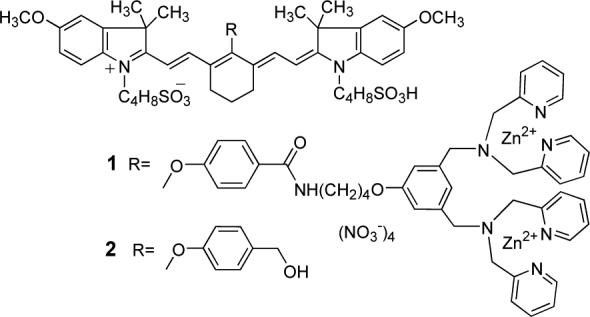
An optical imaging probe was synthesized by attaching a near-infrared carbocyanine fluorophore to an affinity group containing two zinc(II) dipicolylamine (Zn-DPA) units. The probe has a strong and selective affinity for the surfaces of bacteria, and it was used to image infections of Gram-positive S. aureus and Gram-negative E. coli bacteria in living nude mice. After intravenous injection, the probe selectively accumulates at the sites of localized bacterial infections in the thigh muscles of the mice.
Figures
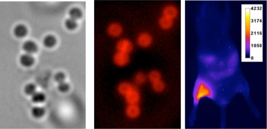
Left: Phase contrast image of S. aureus cells treated with probe 1 (10 μM) and viewed at 1500X. Middle: Fluoresence image of the same cells acquired with a Cy7 filter set. Right: Fluorescence image of a live mouse after thigh muscle injections of S. aureus cells that were pre-incubated for 5 min with 10 μM of either 1 or 2 (left and right leg, respectively). Scale represents relative fluorescence intensity in arbitrary units.
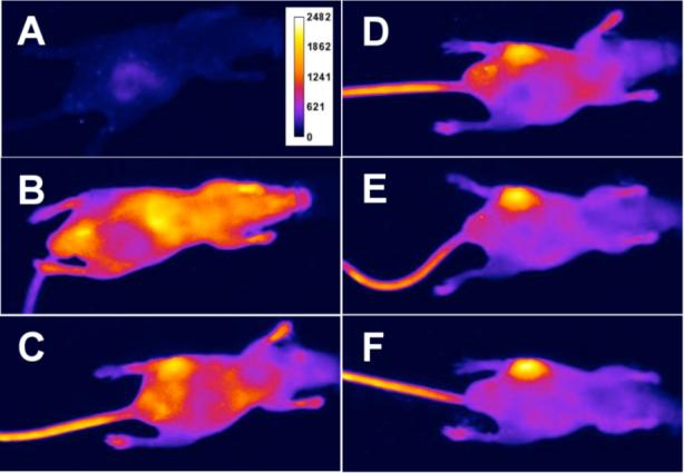
Optical images of a mouse with a S. aureus infection in the left rear thigh muscle. Images were acquired before (A), and immediately following (B), intravenous injection of probe 1, and at 6 h (C), 12 h (D), 18 h (E), and 21 h (F). Scale represents the same relative fluorescence intensity for all six images in arbitrary units.
Similar articles
-
Real-time imaging of bacteria in living mice using a fluorescent dye.Biotech Histochem. 2011 Apr;86(2):104-7. doi: 10.3109/10520295.2010.498295. Epub 2010 Jul 7. Biotech Histochem. 2011. PMID: 20608773
-
Studies on acedan-based mononuclear zinc complexes toward selective fluorescent probes for pyrophosphate.Org Biomol Chem. 2012 Nov 14;10(42):8410-7. doi: 10.1039/c2ob26000j. Epub 2012 Sep 24. Org Biomol Chem. 2012. PMID: 23001147
-
Optical imaging of bacterial infection in living mice using deep-red fluorescent squaraine rotaxane probes.Bioconjug Chem. 2010 Jul 21;21(7):1297-304. doi: 10.1021/bc1000998. Bioconjug Chem. 2010. PMID: 20536173 Free PMC article.
-
Imaging and therapeutic applications of zinc(ii)-dipicolylamine molecular probes for anionic biomembranes.Chem Commun (Camb). 2016 Jul 7;52(57):8787-801. doi: 10.1039/c6cc03669d. Chem Commun (Camb). 2016. PMID: 27302091 Free PMC article. Review.
-
Cy7-Bis-dipicolylamine-zinc.2008 Oct 8 [updated 2008 Nov 12]. In: Molecular Imaging and Contrast Agent Database (MICAD) [Internet]. Bethesda (MD): National Center for Biotechnology Information (US); 2004–2013. 2008 Oct 8 [updated 2008 Nov 12]. In: Molecular Imaging and Contrast Agent Database (MICAD) [Internet]. Bethesda (MD): National Center for Biotechnology Information (US); 2004–2013. PMID: 20641625 Free Books & Documents. Review.
Cited by
-
Non-invasive in vivo fluorescence imaging of apoptotic retinal photoreceptors.Sci Rep. 2019 Feb 7;9(1):1590. doi: 10.1038/s41598-018-38363-z. Sci Rep. 2019. PMID: 30733587 Free PMC article.
-
Targeting apoptosis for optical imaging of infection.Mol Imaging Biol. 2012 Apr;14(2):163-71. doi: 10.1007/s11307-011-0490-6. Mol Imaging Biol. 2012. PMID: 21538153 Free PMC article.
-
Squaraine rotaxanes: superior substitutes for Cy-5 in molecular probes for near-infrared fluorescence cell imaging.Angew Chem Int Ed Engl. 2007;46(29):5528-31. doi: 10.1002/anie.200701491. Angew Chem Int Ed Engl. 2007. PMID: 17585399 Free PMC article. No abstract available.
-
Structure-Activity Studies of Nitroreductase-Responsive Near-Infrared Heptamethine Cyanine Fluorescent Probes.European J Org Chem. 2022 Jun 20;2022(23):e202200270. doi: 10.1002/ejoc.202200270. Epub 2022 Mar 23. European J Org Chem. 2022. PMID: 38322783 Free PMC article.
-
Maltodextrin-based imaging probes detect bacteria in vivo with high sensitivity and specificity.Nat Mater. 2011 Jul 17;10(8):602-7. doi: 10.1038/nmat3074. Nat Mater. 2011. PMID: 21765397 Free PMC article.
References
Publication types
MeSH terms
Substances
Grants and funding
LinkOut - more resources
Full Text Sources
Other Literature Sources
Medical
