. 2006 Dec;3(12):e485.
doi: 10.1371/journal.pmed.0030485.

Epidermal growth factor receptor activation in glioblastoma through novel missense mutations in the extracellular domain

Affiliations

Epidermal growth factor receptor activation in glioblastoma through novel missense mutations in the extracellular domain

Jeffrey C Lee et al. PLoS Med. 2006 Dec.

Abstract

Background: Protein tyrosine kinases are important regulators of cellular homeostasis with tightly controlled catalytic activity. Mutations in kinase-encoding genes can relieve the autoinhibitory constraints on kinase activity, can promote malignant transformation, and appear to be a major determinant of response to kinase inhibitor therapy. Missense mutations in the EGFR kinase domain, for example, have recently been identified in patients who showed clinical responses to EGFR kinase inhibitor therapy.

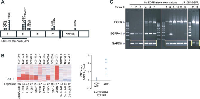
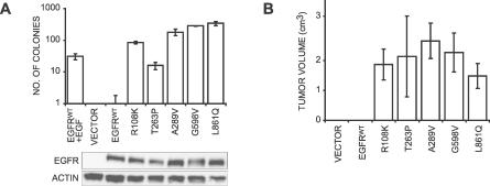
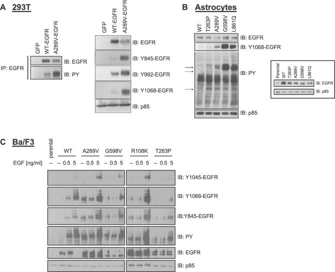
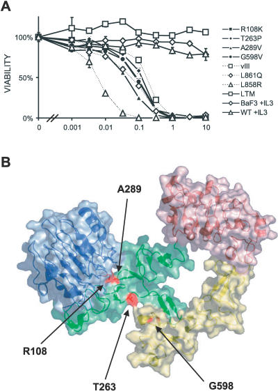
Methods and findings: Encouraged by the promising clinical activity of epidermal growth factor receptor (EGFR) kinase inhibitors in treating glioblastoma in humans, we have sequenced the complete EGFR coding sequence in glioma tumor samples and cell lines. We identified novel missense mutations in the extracellular domain of EGFR in 13.6% (18/132) of glioblastomas and 12.5% (1/8) of glioblastoma cell lines. These EGFR mutations were associated with increased EGFR gene dosage and conferred anchorage-independent growth and tumorigenicity to NIH-3T3 cells. Cells transformed by expression of these EGFR mutants were sensitive to small-molecule EGFR kinase inhibitors.

Conclusions: Our results suggest extracellular missense mutations as a novel mechanism for oncogenic EGFR activation and may help identify patients who can benefit from EGFR kinase inhibitors for treatment of glioblastoma.


Conflict of interest statement

Competing Interests: RB owns shares in AstraZeneca, maker of gefitinib. MM does consulting for Novartis Pharmaceuticals and receives research support from Novartis Pharmaceuticals and Genentech. WRS is an employee of Novartis Pharmaceuticals.

Figures

Figure 1
Figure 1. EGFR Missense Mutations in Glioblastoma Cluster in the Extracellular Domain and Are Associated with Increased EGFR Gene Dose
(A) Location of missense mutations within the EGFR protein in a panel of 151 gliomas (132 glioblastomas, 11 WHO grade III gliomas, and eight glioblastoma cell lines). Each diamond represents one sample harboring the indicated mutation. Amino acid (AA) numbers are based on the new convention for EGFR numbering, which starts at the initiator methionine of pro-EGFR. Ligand-binding domains (I and III), cysteine-rich domains (II and IV), kinase domain (kinase), and the extracellular deletion mutant EGFRvIII [45] are indicated as reference. (B) Increased EGFR gene dose in tumors harboring EGFR missense mutations. The array (left) shows a high-resolution view of Affymetrix 100K SNP array at the EGFR gene locus for ten glioblastoma tumors and three normal controls (sample numbers are indicated above each column). EGFR mutation and log2 ratio (see Methods) are indicated below each column. The plot (left) shows a comparison of EGFR gene copy number determination by SNP array (y-axis, EGFR log2 ratios) and FISH (x-axis). AMP, amplified; NON-AMP, non amplified. (C) RT-PCR for EGFRvIII and full-length EGFR in 14 fresh-frozen glioblastoma tumors (see Methods). The upper band represents full-length EGFR (1,044 bp), the lower band EGFRvIII (243 bp), and the inset shows glyceraldehyde-3-phosphate dehydrogenase (GAPDH) RT-PCR results.
Figure 2
Figure 2. EGFR Missense Mutations Are Transforming and Tumorigenic
(A) Anchorage-independent growth of NIH-3T3 cells expressing various EGFR alleles as mean number of colonies ± standard deviation (bar graph, above). The lanes (below) show EGFR and actin immunoblots of whole cell lysates from NIH-3T3 subclones plated in soft agar. EGF (10 ng/ml) was added to the top agar where indicated. (B) Tumorigenicity of NIH-3T3 cells stably expressing the indicated EGFR alleles in nude mice. Mean tumor size ± standard deviation was determined 3–4 wk after subcutaneous inoculation into nude mice (n = 6 per cell line).
Figure 3
Figure 3. Basal Activation and Ligand Response of EGFR Ectodomain Mutants
(A) Increased EGFR tyrosine phosphorylation of A289V-EGFR. 293T cells were transiently transfected with green fluorescent protein (GFP; control), wild-type EGFR, or A289V-EGFR. At 24 h after transfection, 24 cells were serum starved for 12 h and then lysed. Shown are immunoblots of immunoprecipitated EGFR (left blots) and whole cell lysates (right blots). (B) Increased basal activity of EGFR missense mutants in human astrocytes. Immortalized human astrocytes were stably infected with wild-type EGFR or the indicated EGFR missense mutants. Shown are total phosphotyrosine (PY), Y1068-EGFR, total EGFR, and PI3K p85 (loading control) immunoblots of whole cell lysates from cells following 12 h of serum starvation. The solid arrow at the PY position represents tyrosine-phosphorylated EGFR, and the interrupted arrows indicated other differentially tyrosine-phosphorylated proteins. The inset shows an anti-EGFR immunoblot of parental astrocytes (far-left lane) and stable astrocyte subclones (designated in remaining five lanes) growing in full serum. (C) Basal receptor phosphorylation and EGF-responsiveness of wild-type EGFR and four different EGFR ectodomain mutants stably expressed in Ba/F3 murine hematopoietic cells. Shown are immunoblots of stable Ba/F3 subclones after 12 h of serum starvation (− EGF) and 15 min following EGF-induction (0.5 or 5 ng/ml EGF).
Figure 4
Figure 4. EGFR Missense Mutations Sensitize Cells to EGFR Kinase Inhibitors
(A) Effect of increasing concentrations of the EGFR inhibitor erlotinib (0–10 μM) on the viability of IL-3 independent Ba/F3 subclones expressing EGFR ectodomain mutants (R108K, T263P, A289V, G598V, and EGFRvIII), the EGFR kinase domain mutants (L858R and L861Q), or the erlotinib-resistant EGFR double mutant L858R-T790M (LTM). Parental Ba/F3 cells and Ba/F3 cells expressing wild-type EGFR are not IL-3 independent and were included as controls. Viability (a mean percent of control ± standard deviation) was determined after exposure to erlotinib for 48 h. (B) Oncogenic EGFR ectodomain mutations map to interdomain interfaces. Shown are ribbon and surface diagrams of the EGFR [46] with sites of amino acid substitutions highlighted. Blue, domain I; green, domain II; red, domain III; and yellow, domain IV. Sites of the most prevalent amino acid substitutions are shown in red. Images were created with PyMOL (http://pymol.sourceforge.net/). P596 is not visible in this view.

References

    1. Hynes NE, Lane HA. ERBB receptors and cancer: The complexity of targeted inhibitors. Nat Rev Cancer. 2005;5:341–354. - PubMed
    1. Kleihues P, Louis DN, Scheithauer BW, Rorke LB, Reifenberger G, et al. The WHO classification of tumors of the nervous system. J Neuropathol Exp Neurol. 2002;61:215–225. 226–219. discussion. - PubMed
    1. Reardon DA, Rich JN, Friedman HS, Bigner DD. Recent advances in the treatment of malignant astrocytoma. J Clin Oncol. 2006;24:1253–1265. - PubMed
    1. Libermann TA, Nusbaum HR, Razon N, Kris R, Lax I, et al. Amplification, enhanced expression and possible rearrangement of EGF receptor gene in primary human brain tumours of glial origin. Nature. 1985;313:144–147. - PubMed
    1. Yamazaki H, Fukui Y, Ueyama Y, Tamaoki N, Kawamoto T, et al. Amplification of the structurally and functionally altered epidermal growth factor receptor gene (c-erbB) in human brain tumors. Mol Cell Biol. 1988;8:1816–1820. - PMC - PubMed

Publication types

MeSH terms