. 2006 Dec;3(12):e491.
doi: 10.1371/journal.pmed.0030491.

Elevated serum levels of interferon-regulated chemokines are biomarkers for active human systemic lupus erythematosus

Affiliations

Elevated serum levels of interferon-regulated chemokines are biomarkers for active human systemic lupus erythematosus

Jason W Bauer et al. PLoS Med. 2006 Dec.

Abstract

Background: Systemic lupus erythematosus (SLE) is a serious systemic autoimmune disorder that affects multiple organ systems and is characterized by unpredictable flares of disease. Recent evidence indicates a role for type I interferon (IFN) in SLE pathogenesis; however, the downstream effects of IFN pathway activation are not well understood. Here we test the hypothesis that type I IFN-regulated proteins are present in the serum of SLE patients and correlate with disease activity.

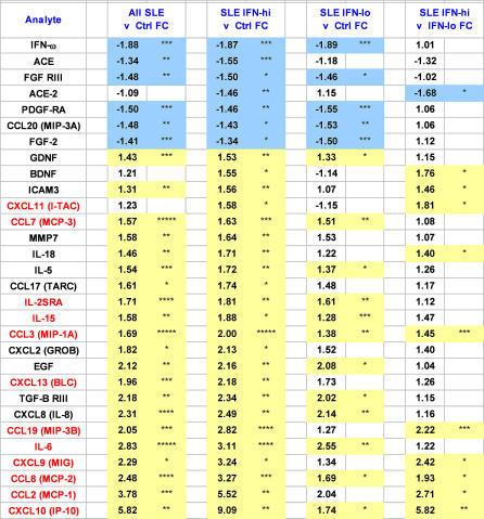
Methods and findings: We performed a comprehensive survey of the serologic proteome in human SLE and identified dysregulated levels of 30 cytokines, chemokines, growth factors, and soluble receptors. Particularly striking was the highly coordinated up-regulation of 12 inflammatory and/or homeostatic chemokines, molecules that direct the movement of leukocytes in the body. Most of the identified chemokines were inducible by type I IFN, and their levels correlated strongly with clinical and laboratory measures of disease activity.

Conclusions: These data suggest that severely disrupted chemokine gradients may contribute to the systemic autoimmunity observed in human SLE. Furthermore, the levels of serum chemokines may serve as convenient biomarkers for disease activity in lupus.


Conflict of interest statement

Competing Interests: DDP was a member of the Scientific Advisory Board for Molecular Staging, a company that performed the protein microarray experiments. DDP has received grant funding from the National Institutes of Health (NIH) and Beckman Coulter to perform work that is broadly related to the area of interest in this paper. DDP has received travel grants and honoraria from numerous institutions and the NIH for speaking and participation in meetings on the subject matter of this paper. DDP is a member of professional societies, including the American College of Rheumatology, the American Academy of Allergy, Asthma and Immunology, and the Clinical Immunology Society, that have lobbying activities.

Figures

Figure 1
Figure 1. IFN Gene Expression Scores for IFN-hi SLE, IFN-lo SLE, and Control Participants
Whole blood gene expression microarrays were used to identify 82 type I IFN-regulated genes that distinguished 81 SLE cases from 42 controls. From these genes, a normalized IFN gene score was derived. Plotted are IFN gene scores for 15 IFN-hi SLE (mean ± SD, 41.0 ± 4.8), 15 IFN-lo SLE (14.4 ± 2.8), and 15 controls (12.1 ± 1.9).
Figure 2
Figure 2. Thirty Protein Analytes Dysregulated in SLE Serum
Sera from 15 IFN-hi SLE, 15 IFN-lo SLE, and 15 controls were assayed for the presence of 160 protein analytes by antibody microarrays. CXCL10 (IP-10) was measured by Luminex bead immunoassays. Shown are 30 analytes that demonstrated significant differences in at least one inter-group comparison, using the following criteria: mean fold change (FC) ≥ 1.5, and p < 0.05 by unpaired t-test. Data are presented as mean fold change of the group comparisons. Analytes regulated by type I IFN, as determined by in vitro gene expression microarray experiments, are highlighted in red font. * p < 0.05; ** p < 0.01; *** p < 0.001; **** p < 0.0001; ***** p < 0.00001. Comparisons highlighted in yellow represent significant positive associations, and blue indicates significant negative associations. The levels of statistical significance shown represent nominal p-values and are not corrected for the multiple hypotheses tested. The results obtained when these p-values were corrected using the Benjamini-Hochberg adjustment for multiple testing are provided in Table S1.
Figure 3
Figure 3. Coordinate Dysregulation of Serum Protein Levels in SLE
Hierarchical clustering was applied to protein levels of the identified analytes (“Serum Protein Levels”). Individual data points represent the log2 ratio of the analyte concentration to the mean of control concentrations (scale shows linear fold-differences). “In Vitro Control PBMC Gene Expression” columns present gene expression microarray data obtained by incubating normal control PBMCs in vitro with medium alone or with type I IFN for 6 and 24 h. Data are normalized to control medium-alone conditions (scale depicts linear fold-differences). Grey boxes indicate missing data. Analytes induced by type I IFN in vitro (>2-fold and >500 expression unit mean difference) are highlighted in red font.
Figure 4
Figure 4. IFN-Regulated Chemokines Are Associated with SLE Disease Activity
Shown are 20 serum analytes that exhibited significant positive (highlighted in yellow) or negative (highlighted in blue) correlation coefficients with clinical measures of SLE. The IFN gene score was calculated from 82 IFN-inducible transcripts measured by concurrent whole blood gene expression microarrays. The chemokine protein “score” was calculated using the seven IFN-regulated CC and CXC chemokines highlighted in red. * p < 0.05; ** p < 0.01; *** p < 0.001; **** p < 0.0001. p-Values were obtained by permutation testing.

References

    1. Wallace DJ. The clinical presentation of systemic lupus erythematosus. In: Wallace DJ, Hahn BH, editors. Dubois' lupus erythematosus. 5th Ed. Baltimore (Maryland): Williams & Wilkins; 1997. pp. 627–633.
    1. Baechler EC, Batliwalla FM, Karypis G, Gaffney PM, Ortmann WA, et al. Interferon-inducible gene expression signature in peripheral blood cells of patients with severe lupus. Proc Natl Acad Sci U S A. 2003;100:2610–2615. - PMC - PubMed
    1. Bennett L, Palucka AK, Arce E, Cantrell V, Borvak J, et al. Interferon and granulopoiesis signatures in systemic lupus erythematosus blood. J Exp Med. 2003;197:711–723. - PMC - PubMed
    1. Kirou KA, Lee C, George S, Louca K, Papagiannis IG, et al. Coordinate overexpression of interferon-alpha-induced genes in systemic lupus erythematosus. Arthritis Rheum. 2004;50:3958–3967. - PubMed
    1. Isaacs A, Lindenmann J. Virus interference. 1 The interferon. Proc R Soc B. 1957;147:258–273. - PubMed