. 2006;8(6):R187.
doi: 10.1186/ar2099.

The role of synovial macrophages and macrophage-produced cytokines in driving aggrecanases, matrix metalloproteinases, and other destructive and inflammatory responses in osteoarthritis

Affiliations

The role of synovial macrophages and macrophage-produced cytokines in driving aggrecanases, matrix metalloproteinases, and other destructive and inflammatory responses in osteoarthritis

Jan Bondeson et al. Arthritis Res Ther. 2006.

Abstract

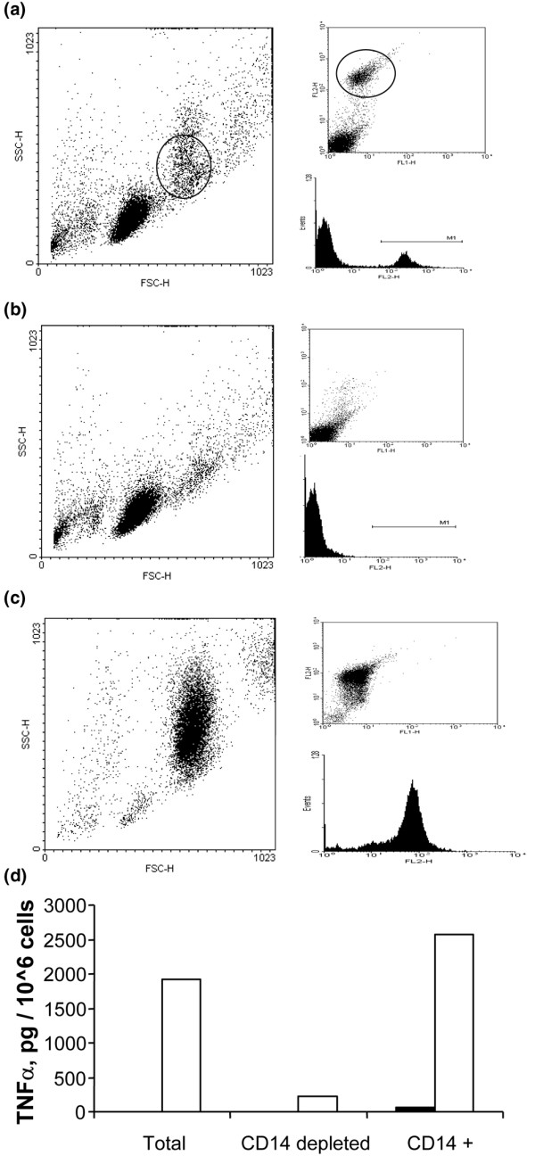
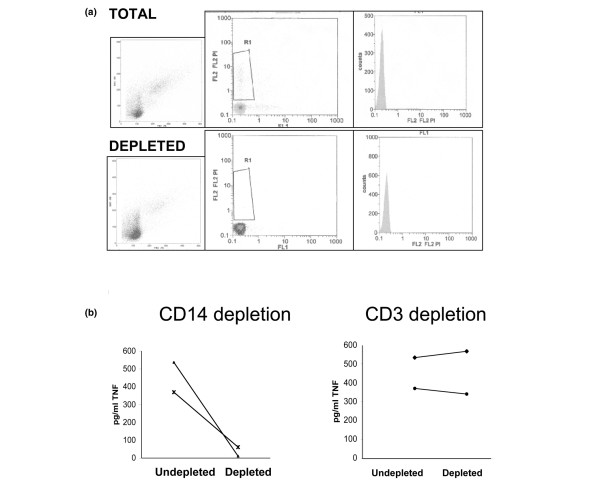
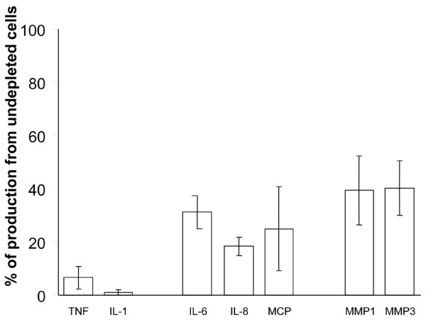
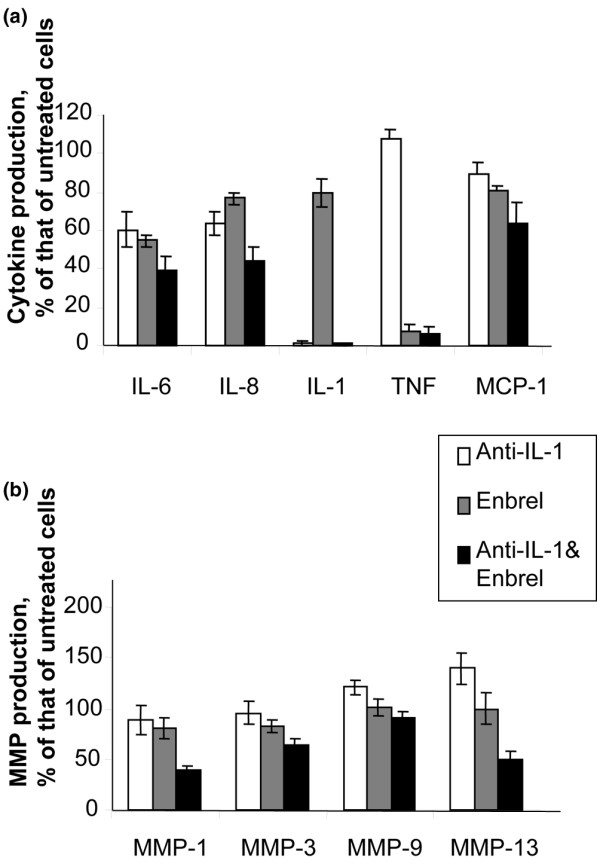
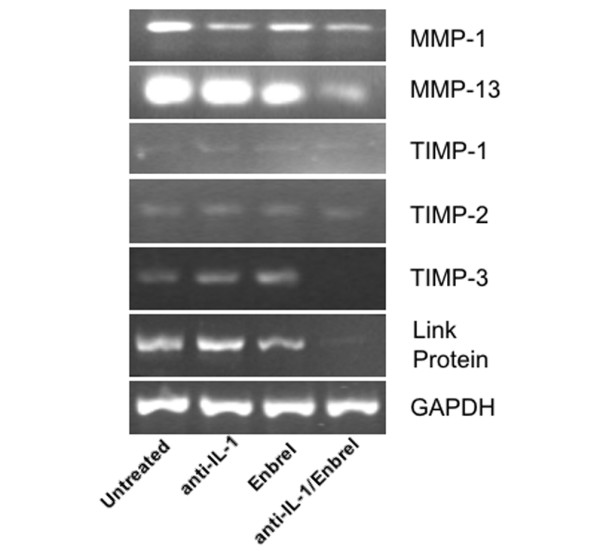
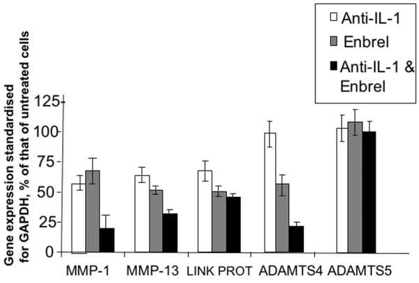
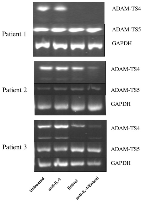
There is an increasing body of evidence that synovitis plays a role in the progression of osteoarthritis and that overproduction of cytokines and growth factors from the inflamed synovium can influence the production of degradative enzymes and the destruction of cartilage. In this study, we investigate the role of synovial macrophages and their main proinflammatory cytokines, interleukin (IL)-1 and tumour necrosis factor-alpha (TNF-alpha), in driving osteoarthritis synovitis and influencing the production of other pro- and anti-inflammatory cytokines, production of matrix metalloproteinases, and expression of aggrecanases in the osteoarthritis synovium. We established a model of cultures of synovial cells from digested osteoarthritis synovium derived from patients undergoing knee or hip arthroplasties. By means of anti-CD14-conjugated magnetic beads, specific depletion of osteoarthritis synovial macrophages from these cultures could be achieved. The CD14+-depleted cultures no longer produced significant amounts of macrophage-derived cytokines like IL-1 and TNF-alpha. Interestingly, there was also significant downregulation of several cytokines, such as IL-6 and IL-8 (p < 0.001) and matrix metalloproteinases 1 and 3 (p < 0.01), produced chiefly by synovial fibroblasts. To investigate the mechanisms involved, we went on to use specific downregulation of IL-1 and/or TNF-alpha in these osteoarthritis cultures of synovial cells. The results indicated that neutralisation of both IL-1 and TNF-alpha was needed to achieve a degree of cytokine (IL-6, IL-8, and monocyte chemoattractant protein-1) and matrix metalloproteinase (1, 3, 9, and 13) inhibition, as assessed by enzyme-linked immunosorbent assay and by reverse transcription-polymerase chain reaction (RT-PCR), similar to that observed in CD14+-depleted cultures. Another interesting observation was that in these osteoarthritis cultures of synovial cells, IL-1beta production was independent of TNF-alpha, in contrast to the situation in rheumatoid arthritis. Using RT-PCR, we also demonstrated that whereas the ADAMTS4 (a disintegrin and metalloprotease with thrombospondin motifs 4) aggrecanase was driven mainly by TNF-alpha, ADAMTS5 was not affected by neutralisation of IL-1 and/or TNF-alpha. These results suggest that, in the osteoarthritis synovium, both inflammatory and destructive responses are dependent largely on macrophages and that these effects are cytokine-driven through a combination of IL-1 and TNF-alpha.


Figures

Figure 1
Figure 1
Fluorescence-activated cell sorting (FACS) analysis of monocyte/macrophage depletion in peripheral blood mononuclear cells (PBMC). (a) The left panel shows FACS analysis of forward scatter (FSC) and side scatter (SSC) of unlabeled PBMC, with the approximate monocyte population indicated with a circle. In the right panels, cells have been incubated with an anti-CD14 phycoerythrin antibody, clearly showing the distinct population of CD14+ cells on FACS (upper panel) and in a histogram (lower panel). (b) The corresponding panels show monocyte-depleted PBMC. In the left panel, the monocyte population is reduced, although the debris and cell clusters blur this effect. In the right upper panel, the CD14+ population virtually disappears, as confirmed by the histogram in the right lower panel. (c) The corresponding panels show the enriched monocytes/macrophages. In the left panel, although there is still a fair amount of debris and cell clusters, the monocyte population is considerably enriched. In the right upper panel, the CD14+ population is also very much enriched, as confirmed by the histogram in the panel below. (d) A functional assay using enzyme-linked immunosorbent assay analysis of spontaneous and lipopolysaccharide-stimulated (open bars) tumour necrosis factor-alpha (TNF-α) production in monocyte/macrophage-depleted and -undepleted PBMCs verifies that the monocyte/macrophage depletion is effective.
Figure 2
Figure 2
FACS analysis of OA synovial cells. (a) Fluorescence-activated cell sorting (FACS) analysis of total (upper three panels) and CD14+-depleted (lower three panels) osteoarthritis (OA) cultures of synovial cells. The upper three panels show FACS analysis of forward scatter/side scatter (FSC/SSC) (left panel), analysis of FL2+ cells after incubation with the anti-CD14 phycoerythrin antibody with the CD14+ population indicated in region R2 (middle panel), and the CD14+ cells in this region showing as FL2+ in a histogram (right panel). The corresponding lower three panels show diminution of the macrophage population on FSC/SSC (left panel), disappearance of CD14+ cells (middle panel), and a histogram (right panel) after depletion of CD14+ cells. (b) A functional assay using the spontaneous tumour necrosis factor-alpha (TNF-α) production from the OA synovial cells shows effective inhibition of TNF-α production in CD14+-depleted cells from two patients (left panel), whereas CD3+ cell depletion has no effect.
Figure 3
Figure 3
Effect of macrophage depletion on cytokine and matrix metalloproteinase (MMP) production in osteoarthritis (OA) synovial cells. OA cultures of synovial cells were either left intact or macrophage-depleted, as described in Materials and methods. Cells were left to adhere for 24 hours before the supernatants were removed for enzyme-linked immunosorbent assay analysis of cytokines and MMPs. Data are expressed as the percentage of cytokine/MMP production in the depleted culture as compared with the undepleted one. The standard error of the mean is given (n = 7–9). IL, interleukin; MCP, monocyte chemoattractant protein; TNF, tumour necrosis factor.
Figure 4
Figure 4
Effect of neutralisation of tumour necrosis factor (TNF)-α and/or interleukin (IL)-1 on cytokine and matrix metalloproteinase (MMP) production in osteoarthritis synovial cells. In these experiments, 2 × 10⁶ cells per well were plated into 4 wells on a 24-well plate in 1 ml of RPMI 1640 supplemented with 10% foetal calf serum. The cells in these 4 wells were left untreated, incubated with the p75 TNF-soluble receptor-immunoglobulin fusion protein etanercept (Enbrel), incubated with a neutralising anti-IL-1β antibody, or incubated with a combination of etanercept and anti-IL-1β, as described in Materials and methods. After incubation for 48 hours, the supernatants were removed for enzyme-linked immunosorbent assay analysis of various cytokines (a) and MMPs (b). The data are expressed as percentage of the production of untreated cells, and the standard error of the mean is given (n = 6–7).
Figure 5
Figure 5
Effect of neutralisation of tumour necrosis factor (TNF)-α and/or interleukin (IL)-1 on the expression of matrix metalloproteinases (MMPs), tissue inhibitors of MMP (TIMPs), and link protein in osteoarthritis synovial cell cultures. Two million cells per well were plated into 4 wells on a 24-well plate and left untreated, incubated with the p75 TNF-soluble receptor-immunoglobulin fusion protein etanercept (Enbrel), incubated with a neutralising anti-IL-1β antibody, or incubated with a combination of etanercept and anti-IL-1β, as described in Materials and methods. After incubation for 48 hours, the cells were washed with phosphate-buffered saline and the RNA extracted using Tri-reagent for reverse transcription-polymerase chain reaction analysis with oligonucleotide primers specific for MMP-1, MMP-3, MMP-13, TIMP-1, TIMP-2, TIMP-3, and link protein. In all panels, analysis of GAPDH (glyceraldehyde-3-phosphate dehydrogenase) was used for comparison of gene expression.
Figure 6
Figure 6
Polymerase chain reaction data from matrix metalloproteinase (MMP), link protein, and ADAMTS analysis, derived as described in Materials and methods and in Figures 5 and 7. Data are expressed as percentage of the gene expression in untreated cells, as standardised for GAPDH (glyceraldehyde-3-phosphate dehydrogenase). The standard error of the mean is given (n = 4–5). ADAMTS, a disintegrin and metalloprotease with thrombospondin motifs; IL, interleukin.
Figure 7
Figure 7
Effect of neutralisation of tumour necrosis factor (TNF)-α and/or interleukin (IL)-1 on the expression of the ADAMTS4 and ADAMTS5 aggrecanases in osteoarthritis synovial cell cultures. Two million cells per well were plated into 4 wells on a 24-well plate and left untreated, incubated with the p75 TNF-soluble receptor-immunoglobulin fusion protein etanercept (Enbrel), incubated with a neutralising anti-IL-1β antibody, or incubated with a combination of etanercept and anti-IL-1β, as described in Materials and methods. After incubation for 48 hours, the cells were washed with phosphate-buffered saline and the RNA extracted using Tri-reagent for reverse transcription-polymerase chain reaction analysis with oligonucleotide primers specific for ADAMTS4 and ADAMTS5 in three patients. In all panels, analysis of GAPDH (glyceraldehyde-3-phosphate dehydrogenase) was used for comparison of gene expression. ADAMTS, a disintegrin and metalloprotease with thrombospondin motifs.

Similar articles

Cited by

References

    1. Pelletier JP, Martel-Pelletier J, Abramson SB. Osteoarthritis, an inflammatory disease: potential implication for the selection of new therapeutic targets. Arthritis Rheum. 2001;44:1237–1247. doi: 10.1002/1529-0131(200106)44:6<1237::AID-ART214>3.0.CO;2-F. - DOI - PubMed
    1. Benito MJ, Veale DJ, FitzGerald O, van den Berg WB, Bresnihan B. Synovial tissue inflammation in early and late osteoarthritis. Ann Rheum Dis. 2005;64:1263–1267. doi: 10.1136/ard.2004.025270. - DOI - PMC - PubMed
    1. Farahat MN, Yanni G, Poston R, Panayi GS. Cytokine expression in synovial membranes of patients with rheumatoid arthritis and osteoarthritis. Ann Rheum Dis. 1993;52:870–875. - PMC - PubMed
    1. Clark AG, Jordan JM, Vilim V, Renner JB, Dragomir AD, Luta G, Kraus VB. Serum cartilage oligomeric protein reflects osteoarthritis presence and severity: the Johnston County Osteoarthritis Project. Arthritis Rheum. 1999;42:2356–2364. doi: 10.1002/1529-0131(199911)42:11<2356::AID-ANR14>3.0.CO;2-R. - DOI - PubMed
    1. Sharif M, Shepstone L, Elson CJ, Dieppe PA, Kirwan JR. Increased serum C reactive protein may reflect events that precede radiographic progression in osteoarthritis of the knee. Ann Rheum Dis. 2000;59:71–74. doi: 10.1136/ard.59.1.71. - DOI - PMC - PubMed

Publication types

MeSH terms