

Lung cancer induced in mice by the envelope protein of jaagsiekte sheep retrovirus (JSRV) closely resembles lung cancer in sheep infected with JSRV
- PMID: 17177996
- PMCID: PMC1764900
- DOI: 10.1186/1742-4690-3-94
Lung cancer induced in mice by the envelope protein of jaagsiekte sheep retrovirus (JSRV) closely resembles lung cancer in sheep infected with JSRV
Abstract
Background: Jaagsiekte sheep retrovirus (JSRV) causes a lethal lung cancer in sheep and goats. Expression of the JSRV envelope (Env) protein in mouse lung, by using a replication-defective adeno-associated virus type 6 (AAV6) vector, induces tumors resembling those seen in sheep. However, the mouse and sheep tumors have not been carefully compared to determine if Env expression alone in mice can account for the disease features observed in sheep, or whether additional aspects of virus replication in sheep are important, such as oncogene activation following retrovirus integration into the host cell genome.
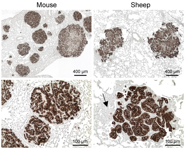
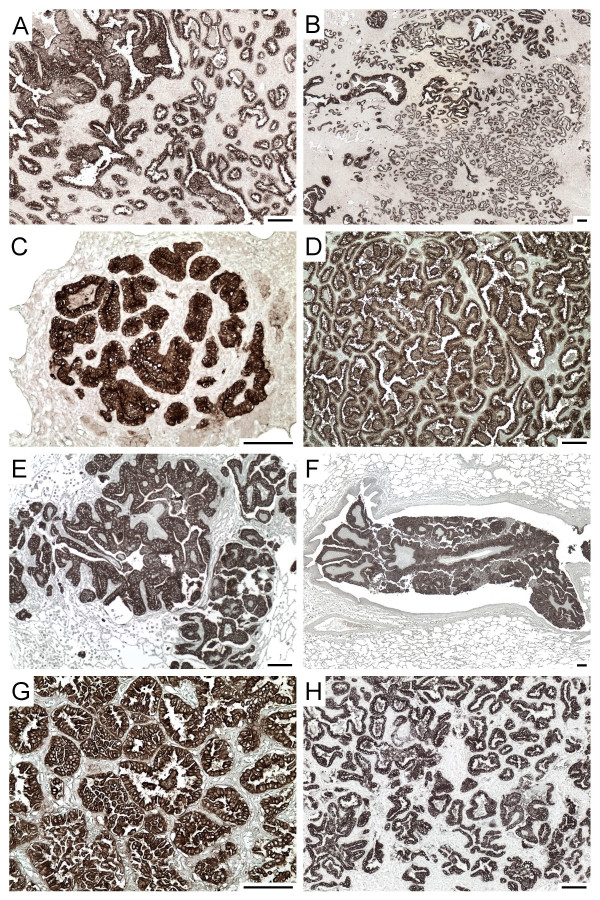
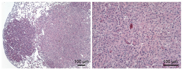
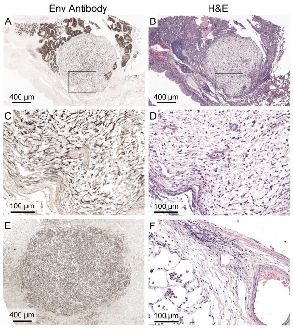
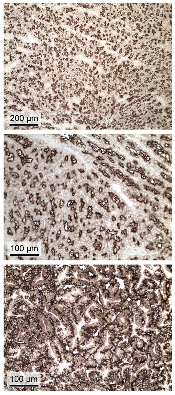
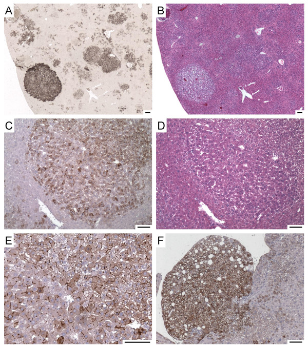
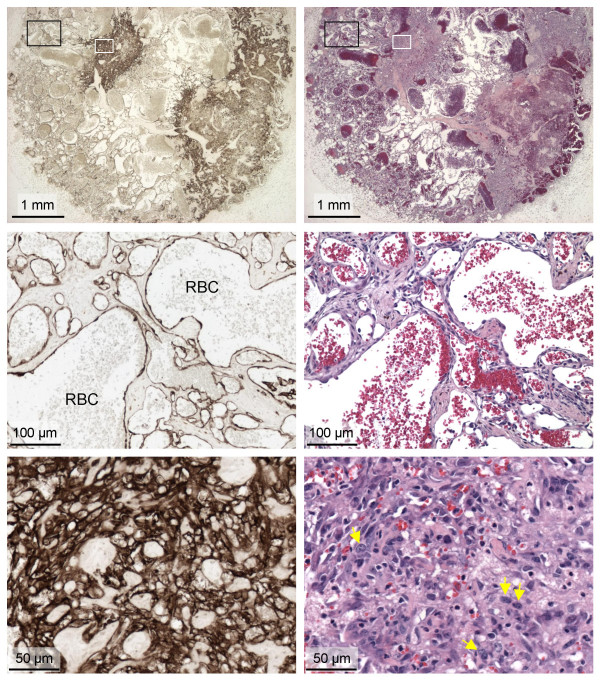
Results: We have generated mouse monoclonal antibodies (Mab) against JSRV Env and have used these to study mouse and sheep lung tumor histology. These Mab detect Env expression in tumors in sheep infected with JSRV from around the world with high sensitivity and specificity. Mouse and sheep tumors consisted mainly of well-differentiated adenomatous foci with little histological evidence of anaplasia, but at long times after vector exposure some mouse tumors did have a more malignant appearance typical of adenocarcinoma. In addition to epithelial cell tumors, lungs of three of 29 sheep examined contained fibroblastic cell masses that expressed Env and appeared to be separate neoplasms. The Mab also stained nasal adenocarcinoma tissue from one United States sheep, which we show was due to expression of Env from ovine enzootic nasal tumor virus (ENTV), a virus closely related to JSRV. Systemic administration of the AAV6 vector encoding JSRV Env to mice produced numerous hepatocellular tumors, and some hemangiomas and hemangiosarcomas, showing that the Env protein can induce tumors in multiple cell types.
Conclusion: Lung cancers induced by JSRV infection in sheep and by JSRV Env expression in mice have similar histologic features and are primarily characterized by adenomatous proliferation of peripheral lung epithelial cells. Thus it is unnecessary to invoke a role for insertional mutagenesis, gene activation, viral replication, or expression of other viral gene products in sheep lung tumorigenesis, although these processes may play a role in other clinically less important sequelae of JSRV infection such as metastasis observed with variable frequency in sheep.
Figures
Similar articles
-
Expression of the jaagsiekte sheep retrovirus envelope glycoprotein is sufficient to induce lung tumors in sheep.J Virol. 2006 Aug;80(16):8030-7. doi: 10.1128/JVI.00474-06. J Virol. 2006. PMID: 16873259 Free PMC article.
-
Detection of Jaagsiekte sheep retrovirus in the peripheral blood during the pre-clinical period of ovine pulmonary adenomatosis.Genet Mol Res. 2016 Aug 29;15(3). doi: 10.4238/gmr.15038521. Genet Mol Res. 2016. PMID: 27706644
-
Envelope proteins of Jaagsiekte sheep retrovirus and enzootic nasal tumor virus induce similar bronchioalveolar tumors in lungs of mice.J Virol. 2006 Sep;80(18):9322-5. doi: 10.1128/JVI.00865-06. J Virol. 2006. PMID: 16940543 Free PMC article.
-
Pathology and pathogenesis of ovine pulmonary adenocarcinoma.J Comp Pathol. 2010 May;142(4):260-83. doi: 10.1016/j.jcpa.2009.12.013. Epub 2010 Feb 16. J Comp Pathol. 2010. PMID: 20163805 Review.
-
Jaagsiekte Sheep Retrovirus (JSRV): from virus to lung cancer in sheep.Vet Res. 2007 Mar-Apr;38(2):211-28. doi: 10.1051/vetres:2006060. Epub 2007 Jan 25. Vet Res. 2007. PMID: 17257570 Review.
Cited by
-
Adeno-associated virus vector mediated expression of an oncogenic retroviral envelope protein induces lung adenocarcinomas in immunocompetent mice.PLoS One. 2012;7(12):e51400. doi: 10.1371/journal.pone.0051400. Epub 2012 Dec 10. PLoS One. 2012. PMID: 23251519 Free PMC article.
-
Neoplasia-Associated Wasting Diseases with Economic Relevance in the Sheep Industry.Animals (Basel). 2021 Feb 3;11(2):381. doi: 10.3390/ani11020381. Animals (Basel). 2021. PMID: 33546178 Free PMC article. Review.
-
Hyaluronidase 2 and its intriguing role as a cell-entry receptor for oncogenic sheep retroviruses.Semin Cancer Biol. 2008 Aug;18(4):296-301. doi: 10.1016/j.semcancer.2008.03.010. Epub 2008 Mar 26. Semin Cancer Biol. 2008. PMID: 18485731 Free PMC article. Review.
-
Receptor binding and low pH coactivate oncogenic retrovirus envelope-mediated fusion.J Virol. 2009 Nov;83(22):11447-55. doi: 10.1128/JVI.00748-09. Epub 2009 Sep 2. J Virol. 2009. PMID: 19726505 Free PMC article.
-
The U3 and Env Proteins of Jaagsiekte Sheep Retrovirus and Enzootic Nasal Tumor Virus Both Contribute to Tissue Tropism.Viruses. 2019 Nov 14;11(11):1061. doi: 10.3390/v11111061. Viruses. 2019. PMID: 31739606 Free PMC article.
References
-
- Sharp JM, DeMartini JC. Natural history of JSRV in sheep. Curr Top Microbiol Immunol. 2003;275:55–79. - PubMed
-
- De las Heras M, Gonzalez L, Sharp JM. Pathology of ovine pulmonary adenocarcinoma. Curr Top Microbiol Immunol. 2003;275:25–54. - PubMed
-
- Rai SK, Duh FM, Vigdorovich V, Danilkovitch-Miagkova A, Lerman MI, Miller AD. Candidate tumor suppressor HYAL2 is a glycosylphosphatidylinositol (GPI)-anchored cell-surface receptor for jaagsiekte sheep retrovirus, the envelope protein of which mediates oncogenic transformation. Proc Natl Acad Sci USA. 2001;98:4443–4448. doi: 10.1073/pnas.071572898. - DOI - PMC - PubMed
Publication types
MeSH terms
Substances
Grants and funding
LinkOut - more resources
Full Text Sources
Medical
Molecular Biology Databases
