

Structure and dynamics of UDP-glucose pyrophosphorylase from Arabidopsis thaliana with bound UDP-glucose and UTP
- PMID: 17178129
- PMCID: PMC1847403
- DOI: 10.1016/j.jmb.2006.11.059
Structure and dynamics of UDP-glucose pyrophosphorylase from Arabidopsis thaliana with bound UDP-glucose and UTP
Abstract
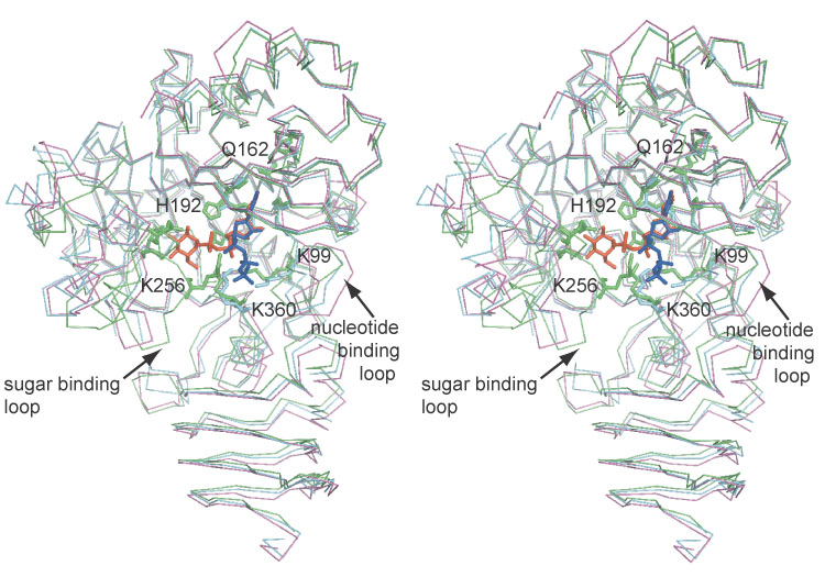
The structure of the UDP-glucose pyrophosphorylase encoded by Arabidopsis thaliana gene At3g03250 has been solved to a nominal resolution of 1.86 Angstroms. In addition, the structure has been solved in the presence of the substrates/products UTP and UDP-glucose to nominal resolutions of 1.64 Angstroms and 1.85 Angstroms. The three structures revealed a catalytic domain similar to that of other nucleotidyl-glucose pyrophosphorylases with a carboxy-terminal beta-helix domain in a unique orientation. Conformational changes are observed between the native and substrate-bound complexes. The nucleotide-binding loop and the carboxy-terminal domain, including the suspected catalytically important Lys360, move in and out of the active site in a concerted fashion. TLS refinement was employed initially to model conformational heterogeneity in the UDP-glucose complex followed by the use of multiconformer refinement for the entire molecule. Normal mode analysis generated atomic displacement predictions in good agreement in magnitude and direction with the observed conformational changes and anisotropic displacement parameters generated by TLS refinement. The structures and the observed dynamic changes provide insight into the ordered mechanism of this enzyme and previously described oligomerization effects on catalytic activity.
Figures
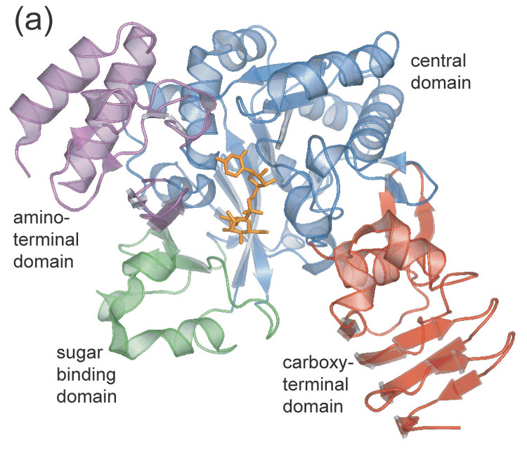
Ribbon diagram of UDPGP monomer. Domains are color coded as follows: central domain (blue), carboxy-terminal domain (red), amino-terminal domain (magenta), sugar binding domain (green).
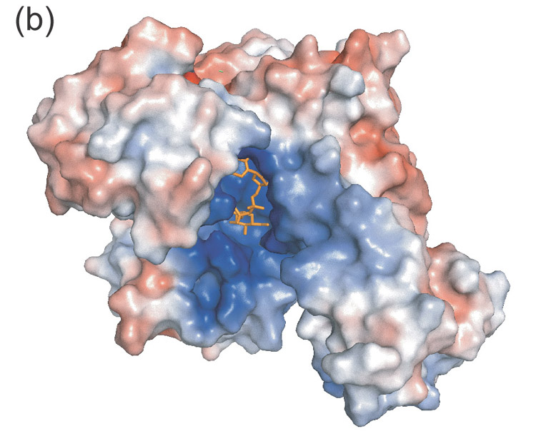
Electrostatic surface diagram illustrating the positively charged substrate cavity.
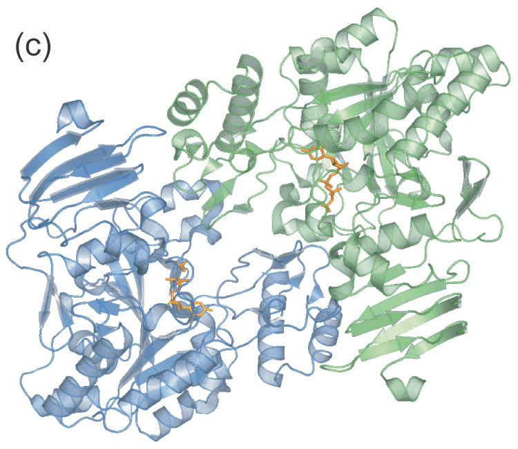
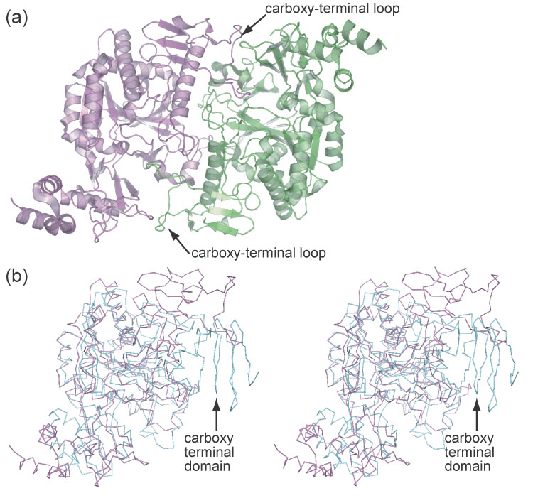
Apparent UDPGP dimer. The amino-terminal domain of each monomer resides near the active site of the other respective monomer.
Ribbon diagram of UDPGP monomer. Domains are color coded as follows: central domain (blue), carboxy-terminal domain (red), amino-terminal domain (magenta), sugar binding domain (green).
Electrostatic surface diagram illustrating the positively charged substrate cavity.
Apparent UDPGP dimer. The amino-terminal domain of each monomer resides near the active site of the other respective monomer.

UDP-glucose complex (2icy).
UTP complex (2icx).
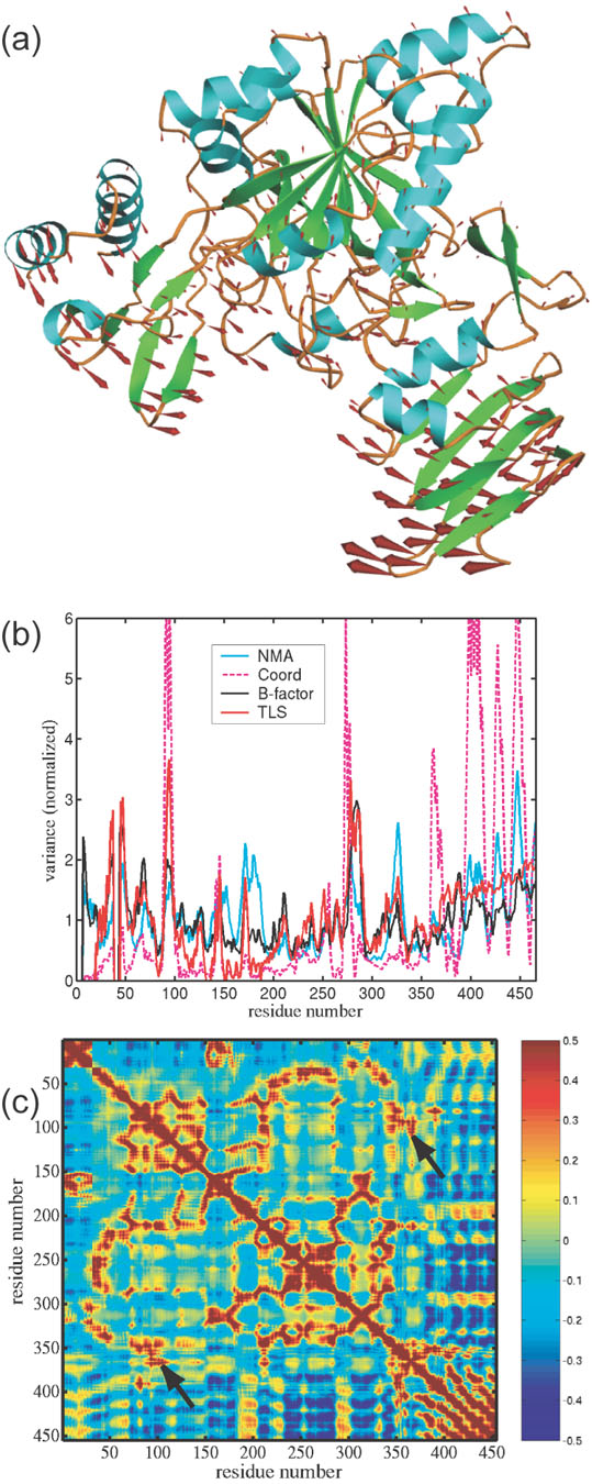
Motion in the Arabidopsis UDPGP as predicted by normal mode analysis. Displacement vectors are represented by red arrows. Trajectories were obtained from model 1 chain A of the UDP-glucose complex (2icy).
Plot of normalized positional variance for UDPGP from the B-factors in the final refinement (blue), TLS refinement (red), Normal Mode Analysis model (black) and observed atomic displacements (dashed magenta). The correlation coefficient is 0.76 between TLS and B-factors, 0.81 between TLS and NMA, 0.56 between NMA and B-factors, 0.58 between TLS and observed atomic displacements, and 0.34 between B-factors and observed atomic displacements.
Matrix of correlations in positional deviation for UDPGP computed from 100 modes of NMA. Red indicates a positive correlation between residues, blue indicated anti-correlation, and green indicates lack of correlation. Arrows point to strong correlation between the nucleotide binding loop (residues 87–100) and residues around Lys360 of the carboxy-terminal domain.
The AGX1 dimer.
Overlay of the AGX1 (magenta) and UDPGP (cyan) monomers. The β-helix of UDPGP prevents dimerization similar to that of AGX1.
Similar articles
-
Substrate kinetics and substrate effects on the quaternary structure of barley UDP-glucose pyrophosphorylase.Phytochemistry. 2012 Jul;79:39-45. doi: 10.1016/j.phytochem.2012.04.002. Epub 2012 May 1. Phytochemistry. 2012. PMID: 22552276
-
Glucose-1-phosphate uridylyltransferase from Erwinia amylovora: Activity, structure and substrate specificity.Biochim Biophys Acta Proteins Proteom. 2017 Nov;1865(11 Pt A):1348-1357. doi: 10.1016/j.bbapap.2017.08.015. Epub 2017 Aug 24. Biochim Biophys Acta Proteins Proteom. 2017. PMID: 28844747
-
A quaternary mechanism enables the complex biological functions of octameric human UDP-glucose pyrophosphorylase, a key enzyme in cell metabolism.Sci Rep. 2015 Apr 10;5:9618. doi: 10.1038/srep09618. Sci Rep. 2015. PMID: 25860585 Free PMC article.
-
Exploring the active site in UDP-glucose pyrophosphorylase by affinity labelling and site-directed mutagenesis.Biotechnol Appl Biochem. 1993 Oct;18(2):209-16. Biotechnol Appl Biochem. 1993. PMID: 8251118 Review.
-
A common structural blueprint for plant UDP-sugar-producing pyrophosphorylases.Biochem J. 2011 Nov 1;439(3):375-9. doi: 10.1042/BJ20110730. Biochem J. 2011. PMID: 21992098 Review.
Cited by
-
Effects of Magnesium, Pyrophosphate and Phosphonates on Pyrophosphorolytic Reaction of UDP-Glucose Pyrophosphorylase.Plants (Basel). 2022 Jun 20;11(12):1611. doi: 10.3390/plants11121611. Plants (Basel). 2022. PMID: 35736762 Free PMC article.
-
Identification of a UDP-glucose pyrophosphorylase from cotton (Gossypium hirsutum L.) involved in cellulose biosynthesis in Arabidopsis thaliana.Plant Cell Rep. 2011 Jul;30(7):1303-12. doi: 10.1007/s00299-011-1042-x. Epub 2011 Mar 5. Plant Cell Rep. 2011. PMID: 21373794
-
Oligomerization, membrane association, and in vivo phosphorylation of sugarcane UDP-glucose pyrophosphorylase.J Biol Chem. 2014 Nov 28;289(48):33364-77. doi: 10.1074/jbc.M114.590125. Epub 2014 Oct 15. J Biol Chem. 2014. PMID: 25320091 Free PMC article.
-
Identification, subcellular localization, biochemical properties, and high-resolution crystal structure of Trypanosoma brucei UDP-glucose pyrophosphorylase.Glycobiology. 2010 Dec;20(12):1619-30. doi: 10.1093/glycob/cwq115. Epub 2010 Aug 19. Glycobiology. 2010. PMID: 20724435 Free PMC article.
-
Characterization, Expression Profile Analysis, and Functional Prediction of UGP Gene Family in Dendrocalamus brandisii.Plants (Basel). 2025 May 14;14(10):1458. doi: 10.3390/plants14101458. Plants (Basel). 2025. PMID: 40431023 Free PMC article.
References
-
- Sonnhammer EL, Eddy SR, Durbin R. Pfam: a comprehensive database of protein domain families based on seed alignments. Proteins. 1997;28:405–420. - PubMed
-
- Sheu KF, Frey PA. UDP-glucose pyrophosphorylase. Stereochemical course of the reaction of glucose 1-phosphate with uridine-5′[1-thiotriphosphate] J Biol Chem. 1978;253:3378–3380. - PubMed
-
- Tsuboi KK, Fukunaga K, Petricciani JC. Purification and specific kinetic properties of erythrocyte uridine diphosphate glucose pyrophosphorylase. J Biol Chem. 1969;244:1008–1015. - PubMed
-
- Lamerz AC, Haselhorst T, Bergfeld AK, von Itzstein M, Gerardy-Schahn R. Molecular cloning of the Leishmania major UDP-glucose pyrophosphorylase, functional characterization, and ligand binding analyses using NMR spectroscopy. J Biol Chem. 2006;281:16314–16322. - PubMed
Publication types
MeSH terms
Substances
Associated data
- Actions
Grants and funding
LinkOut - more resources
Full Text Sources
Molecular Biology Databases
