. 2006 Dec 21;52(6):997-1009.
doi: 10.1016/j.neuron.2006.10.028.

Staufen- and FMRP-containing neuronal RNPs are structurally and functionally related to somatic P bodies

Affiliations

Staufen- and FMRP-containing neuronal RNPs are structurally and functionally related to somatic P bodies

Scott A Barbee et al. Neuron. .

Abstract

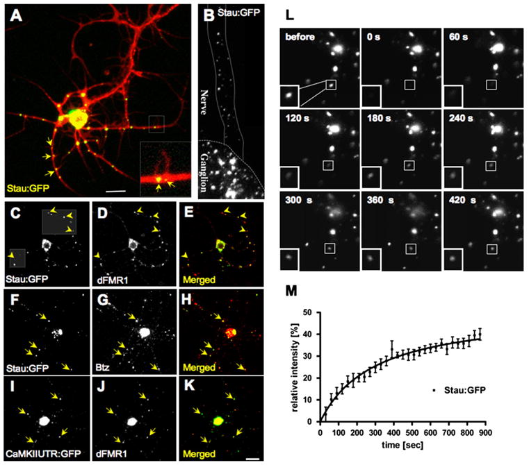
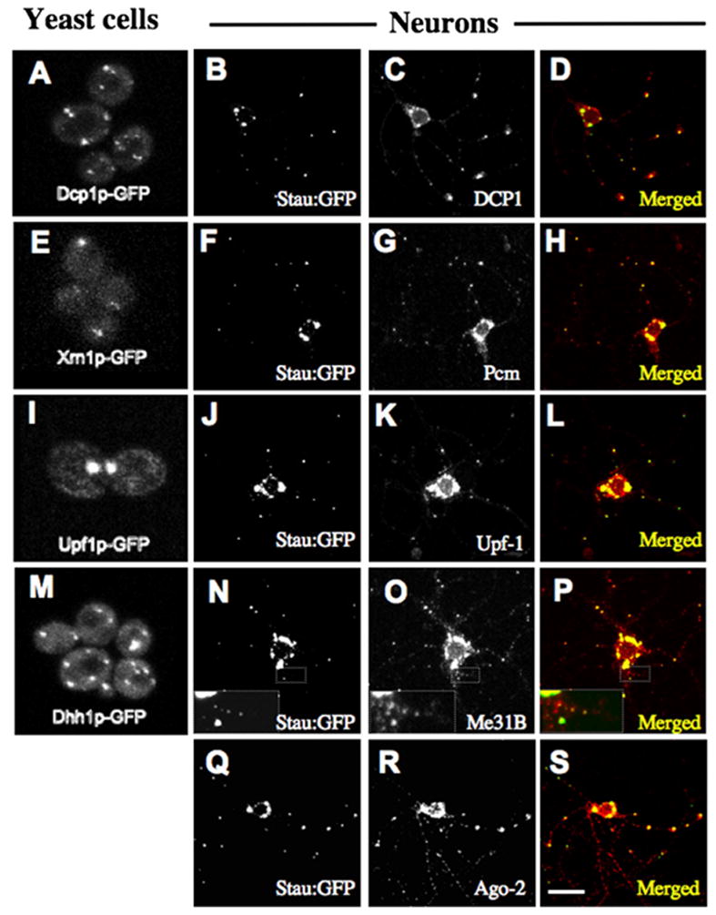
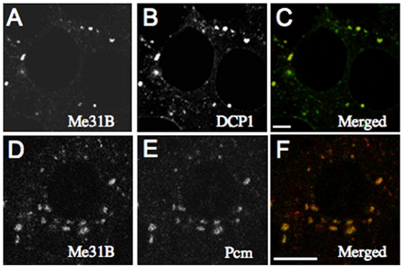
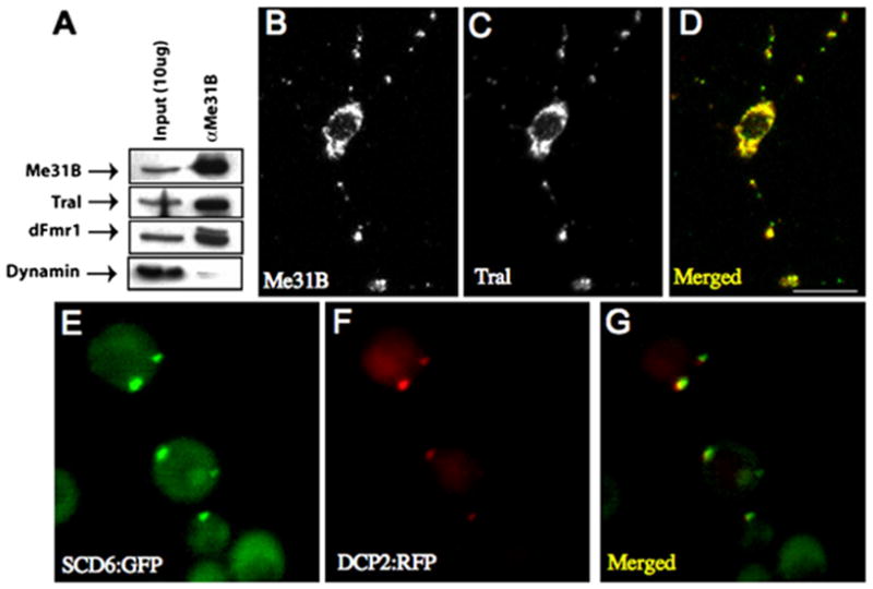
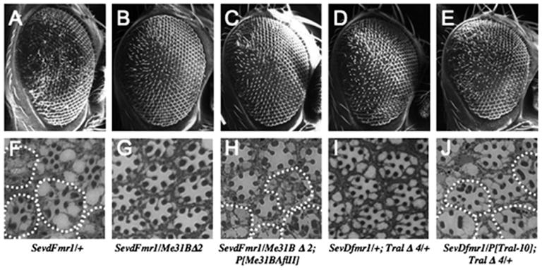
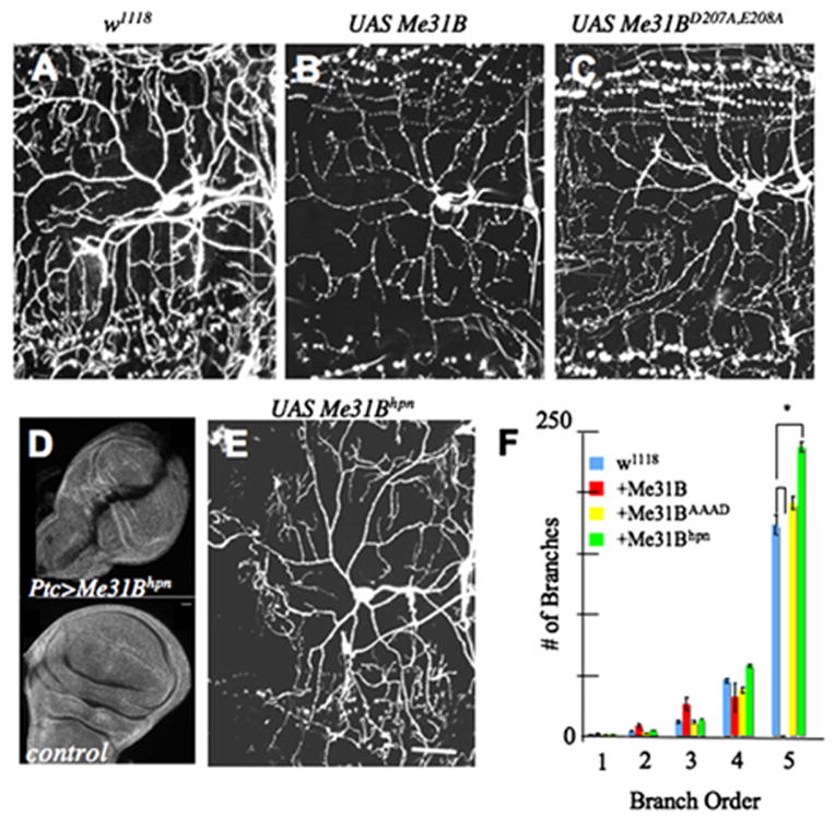
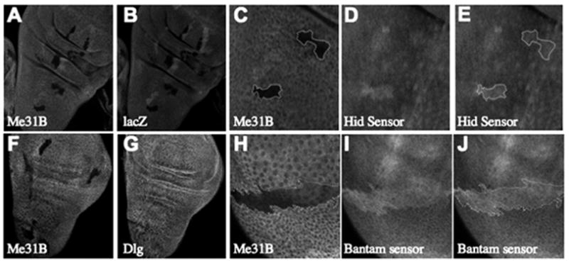
Local control of mRNA translation modulates neuronal development, synaptic plasticity, and memory formation. A poorly understood aspect of this control is the role and composition of ribonucleoprotein (RNP) particles that mediate transport and translation of neuronal RNAs. Here, we show that staufen- and FMRP-containing RNPs in Drosophila neurons contain proteins also present in somatic "P bodies," including the RNA-degradative enzymes Dcp1p and Xrn1p/Pacman and crucial components of miRNA (argonaute), NMD (Upf1p), and general translational repression (Dhh1p/Me31B) pathways. Drosophila Me31B is shown to participate (1) with an FMRP-associated, P body protein (Scd6p/trailer hitch) in FMRP-driven, argonaute-dependent translational repression in developing eye imaginal discs; (2) in dendritic elaboration of larval sensory neurons; and (3) in bantam miRNA-mediated translational repression in wing imaginal discs. These results argue for a conserved mechanism of translational control critical to neuronal function and open up new experimental avenues for understanding the regulation of mRNA function within neurons.


Figures

Figure 1
Figure 1. Drosophila Neurons Have Ribonucleoprotein Particles Containing Stau, dFMR1, Btz, and a Dendritically Targeted mRNA
(A) Stau:GFP (green) in cultured Drosophila motor neurons counterstained with a anti-HRP antibody (red). The inset shows Stau:GFP puncta at the base of small neurite branches (arrows). These puncta show occasional bidirectional movement within neurites (Movies S1 and S2). (B) View of a Drosophila larval ventral ganglion and emerging nerve from an animal expressing Stau:GFP in the nervous system. (C–E) Confocal image pair and merged image of a cultured motor neuron labeled for Stau:GFP (C) and endogenous dFMR1 (D). Dashed boxes show regions optimized for displaying faint spots: the yellow arrowheads show that particles appearing red on the merged image (E) in fact contain Stau:GFP (green). (F–H) Cell double labeled with Stau:GFP (F) and endogenous Btz (G). (I–K) Drosophila CaMKII mRNA (I) visualized by ms2-tagged CamKII mRNA combined with MCP:GFP detection (Ashraf et al., 2006) is present on dFMR1-positive particles (J). (L) In FRAP experiments in live cultured motor neurons expressing Stau:GFP, images of a staufen granule were recorded “before,” immediately after bleaching (“0 sec”), and once every 30 s during the course of recovery. (M) For each time point, fluorescence intensity within a small region of interest (ROI) was measured and plotted on the graph after normalization to a paired “unbleached” spot. From the data set (n = 6 cells; 11 spots), a fluorescence recovery curve was calculated using nonlinear regression. Rectangles frame the bleached particle; ROIs, not shown, were smaller and closer to spot dimensions. Scale Bar, 10 μm.
Figure 2
Figure 2. Neuronal Staufen Granules Contain P Body Components Mediating Translation Repression and RNA Decay
Yeast P-body proteins tagged with GFP in S. cerevisiae cells (left column) with their Drosophila orthologs localized relative to Stau:GFP in cultured Stau:GFP-expressing motor neurons. (A–D) Dcp1p/DCP1; (E–H) Xrn1p/Pcm; (I–L) Upf1p/UFP1; (M–P) Dhh1p/Me31B; (Q–S) and Ago-2 are also present on neuronal staufen granules. The inset with a magnified view of small Me31B particles in a neurite show that these also contain Stau:GFP. Scale bar, 10 μm for neurons.
Figure 3
Figure 3. RNA Decapping and Degradative Enzymes Are Present on Maternal RNP Granules
(A–C) DCP1 and (D–F) Pcm colocalize with Me31B in cytoplasmic foci in nurse cells (stage 8 is shown). (C and F) Merged images. Scale bar, 10 μm.
Figure 4
Figure 4. Tral Is an Me31B/dFMR1-Associated Protein Present on Staufen RNPs with a Conserved Homolog, Scd6p, in Yeast P Bodies
(A) Western blot of Me31B coimmunoprecipitates probed with antibodies against Me31B, Tral, dFMR1, and dynamin. (B–D) Me31B (B) and Tral (C) colocalize in neuronal granules of dFMR1 expressing cultured motor neurons (similar results in w1118 cells are shown in Figures S5D–S5I). (E–G) Yeast cells expressing Scd6p:GFP (E) and Dcp2p:RFP (F) showing colocalization of Scd6p:GFP to P bodies. Scale bar, 10 μm.
Figure 5
Figure 5. Me31B and Tral Are Required for dFMR1-Induced Defects in the Drosophila Eye
(A–E) SEMs of adult compound eyes with paired retinal sections (F–J). Magnification of SEMs is 150×. Tangential sections of each genotype are at approximately the same depth.
Figure 6
Figure 6. Me31B Regulates Dendritic Growth in Sensory Neurons
(A) Control class IV ddaC neuron expressing UAS-mCD8:GFP alone. (B) Class IV ddaC neurons overexpressing Me31B and UAS-mCD8: GFP showing a reduction in higher-order dendrite arborization. (C) The same neurons overexpressing a mutant Me31B incapable of translational repression (Me31BD207A, E208A) show normal dendritic branching. (D) Transgenic RNAi dramatically reduces Me31B protein levels. Anti-Me31B staining of third-instar imaginal discs shows that UAS-Me31Bhpn expressed in the patched domain of wing imaginal discs reduces Me31B levels along the anterior-posterior border (top panel) compared to control wing imaginal discs (lower panel). (E) Class IV ddaC neurons overexpressing a Me31B RNA hairpin (UAS-Me31Bhpn) exhibit abnormal dendrite morphology and increased high-order branching. (F) Numbers of dendritic branches in each order, as revealed by reversed Strahler analysis (see Supplemental Data). Number of neurons analyzed for each genotype are: UAS-mCD8:GFP control (n = 15), UAS-Me31B (n = 10), UAS-Me31BD207A, E208A (n = 11), and UAS-Me31Bhpn (n = 13). Values are mean ± standard error. A star (*) indicates a significant reduction in fifth-order dendrite branching following Me31B overexpression compared to the control (p < 0.001) and a significant increase in fifth-order dendrite branching following Me31B RNAi (p < 0.001). Scale bar, 20 μm.
Figure 7
Figure 7. Me31B Is Required for the In Vivo Function of an Endogenous MicroRNA
Heat-shock-induced clones in wing imaginal discs of hs-FLP1; FRT40A, arm-lacZ/FRT40A, me31BΔ1; reporter flies show dark me31B/me31B clones revealed either by staining for LacZ (B) or Me31B (A and C). Anti-GFP staining shows that the hid reporter (D and E) is upregulated in cell clones lacking Me31B (C). Similar analyses (F and G) show that a control protein Dlg (G) is not upregulated in me31B/me31B clones (F). However, consistent with a Me31B requirement in miRNA/RNAi, the bantam reporter expression is upregulated in me31B/me31B-lacking cells (H–J).

Similar articles

Cited by

References

    1. Andrei MA, Ingelfinger D, Heintzmann R, Achsel T, Rivera-Pomar R, Luhrmann R. A role for eIF4E and eIF4Etransporter in targeting mRNPs to mammalian processing bodies. RNA. 2005;11:717–727. - PMC - PubMed
    1. Antar LN, Dictenberg JB, Plociniak M, Afroz R, Bassell GJ. Localization of FMRP-associated mRNA granules and requirement of microtubules for activity-dependent trafficking in hippocampal neurons. Genes Brain Behav. 2005;4:350–359. - PubMed
    1. Ashraf SI, McLoon AL, Sclarsic SM, Kunes S. Synaptic protein synthesis associated with memory is regulated by the RISC pathway in Drosophila. Cell. 2006;124:191–205. - PubMed
    1. Audhya A, Hyndman F, McLeod IX, Maddox AS, Yates JR, 3rd, Desai A, Oegema K. A complex containing the Sm protein CAR-1 and the RNA helicase CGH-1 is required for embryonic cytokinesis in Caenorhabditis elegans. J Cell Biol. 2005;171:267–279. - PMC - PubMed
    1. Bhattacharyya SN, Habermacher R, Martine U, Closs EI, Fillipowicz W. Relief of microRNA-mediated translational repression in human cells subjected to stress. Cell. 2006;125:1111–1124. - PubMed

Publication types

MeSH terms