. 2007 Mar;75(3):1484-92.
doi: 10.1128/IAI.01315-06. Epub 2006 Dec 18.

Cellular pyrin domain-only protein 2 is a candidate regulator of inflammasome activation

Affiliations

Cellular pyrin domain-only protein 2 is a candidate regulator of inflammasome activation

Andrea Dorfleutner et al. Infect Immun. 2007 Mar.

Abstract

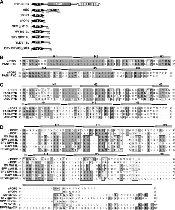
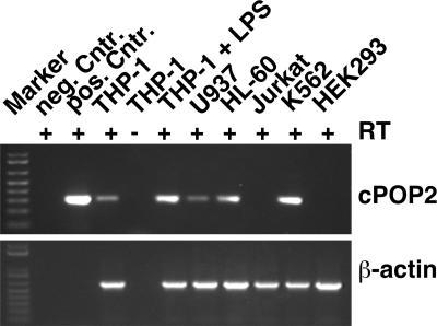
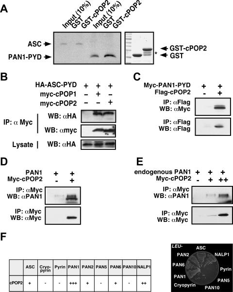
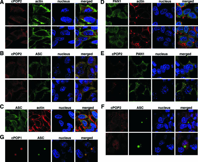
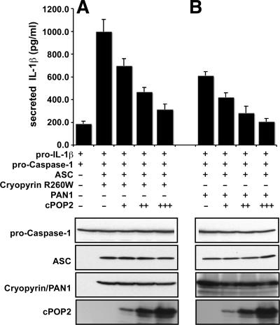
Pyrin domain (PYD) proteins have recently emerged as important signaling molecules involved in the development of innate immunity against intracellular pathogens through activation of inflammatory mediator pathways. ASC is the central adaptor protein, which links pathogen recognition by PYD-containing pathogen recognition receptors, known as PYD-Nod-like receptors (NLR), PAN, PYPAF, NALP, Nod, and Caterpiller proteins, to the activation of downstream effectors, including activation of caspase-1 and NF-kappaB. Activation of these effectors occurs when specific protein complexes, known as inflammasomes, are formed. PYD signal transduction leads to inflammasome assembly and activation of specific effector proteins. It is modulated by a cellular PYD-only protein (cPOP1), which binds to ASC and interferes with the recruitment of ASC to activated PYD-NLRs. Here we describe the identification and characterization of a second cellular POP (cPOP2), which shows highest homology to the PYD of PAN1. cPOP2 binds to ASC and PAN1, thereby blocking formation of cryopyrin and PAN1-containing inflammasomes, activation of caspase-1, and subsequent processing and secretion of bioactive interleukin-1beta. Existence of a second cPOP provides additional insights into inflammasome formation and suggests that POPs might be a common regulatory mechanism to "fine-tune" the activity of specific PYD-NLR family protein-containing inflammasomes.


Figures

FIG. 1.
FIG. 1.
The human genome encodes a second cellular PYD-only protein. (A) Schematic domain representation of cPOP2 and other known viral and cellular POPs, ASC, and PYD-NLR family proteins. Myxoma virus (MV; M013L), rabbit Shope fibroma virus (SFV; gp013L), swinepox virus (SPV; SPV14L), Yaba-like disease virus (YLDV; 18L), mule deer poxvirus (DPV; DPV83gp024). (B) Clustal W alignment of cPOP2 and the most similar PYD of PAN1 (PYPAF2, NALP2, NBS1, CLR19.9). (C) cPOP2 and the PYDs of the three most similar PYD-containing proteins, PAN1, PAN 7 (PYPAF3, NALP7, NOD12, CLR19.4), and ASC. (D) cPOP2 with other known POPs. Dark gray and light gray shading indicates identical and similar (conserved) amino acid residues, respectively. The α-helices, as determined for the PYD of ASC, are marked above the sequences (e.g., H1, H2, etc.) (19).
FIG. 2.
FIG. 2.
cPOP2 is expressed in human cell lines. cPOP2-specific transcripts were detected in several human cell lines by RT-PCR, which was performed with cPOP2-specific primers and primers specific for β-actin. RT-PCR was also performed using pooled RNA from THP-1 cells treated with 600 ng/ml LPS (E. coli serotype O111:B4) for 2, 4, 6, 12, and 24 h (THP-1 + LPS). A cPOP1 expression plasmid (pcDNA3-myc-cPOP1) was included as a negative control, and a cPOP2 expression plasmid (pcDNA3-myc-cPOP2) was used as a positive control. Pooled RNA not incubated with reverse transcriptase during the RT-PCR step was also used as a negative control.
FIG. 3.
FIG. 3.
cPOP2 associates with ASC and PYD-NLR family proteins. (A) In vitro binding between cPOP2, ASC, and PAN1. ASC and the PYD of PAN1 were in vitro translated, labeled with biotin, and subjected to in vitro GST pull-down assays using GST-POP2 and a GST control immobilized on GSH-Sepharose, as indicated. Protein complexes were separated by SDS-PAGE and transferred onto polyvinylidene difluoride (PVDF) membranes, and bound proteins were visualized by immunoblotting with streptavidin-HRP and ECL-Plus (Amersham Pharmacia Biotech) detection. The recombinant GST-cPOP2 and GST proteins are visualized by Coomassie blue staining. The asterisk denotes several degradation products of cPOP2, which did not affect this assay. (B) In vivo binding between cPOP2 and ASC. HEK293T cells were transiently transfected with the HA-tagged PYD of ASC (ASC-PYD), myc-tagged cPOP1, and myc-tagged cPOP2, as indicated. At 36 h posttransfection, clarified and normalized cell lysates were subjected to coimmunoprecipitation, using immobilized anti-myc antibodies (Santa Cruz Biotechnology). Immune complexes were separated by SDS-PAGE, transferred onto PVDF membranes, and probed with anti-HA antibodies directly conjugated to HRP and detected with ECL (Amersham Pharmacia Biotech). Membranes were stripped and reprobed with anti-myc-HRP antibodies. Five percent of the total lysate was also immunoblotted with HA-HRP antibodies to control for ASC-PYD expression. IP, immunoprecipitation; WB, Western blot. (C, D, E) In vivo binding between cPOP2 and PAN1. HEK293T cells were transiently transfected with the myc-tagged PYD of PAN1 (PAN1-PYD) and Flag-tagged cPOP2 (C) or full-length, untagged PAN1 and myc-tagged cPOP2 (D), as indicated. At 36 h posttransfection, clarified and normalized cell lysates were subjected to coimmunoprecipitation, using immobilized anti-Flag antibodies (Sigma) (C) or immobilized anti-myc antibodies (Santa Cruz Biotechnology) (D). Immune complexes were separated by SDS-PAGE, transferred onto PVDF membranes, and probed with anti-myc antibodies directly conjugated to HRP (Santa Cruz Biotechnology) (C) or with anti-PAN1 antibodies (Imgenex) and secondary anti-rabbit antibodies conjugated to HRP (Amersham Pharmacia Biotech) and detected with ECL and ECL-Plus (Amersham Pharmacia Biotech) (D). Membranes were stripped and reprobed with anti-Flag-HRP antibodies (Sigma) (C) or anti-myc HRP antibodies (D). Jurkat cells were transiently transfected with small (+) and large (++) amounts of myc-tagged cPOP2, and cleared lysates were coimmunoprecipitated at 36 h posttransfection, using immobilized anti-myc antibodies (Santa Cruz Biotechnology) (E). Immune complexes were separated by SDS-PAGE, transferred onto PVDF membranes and probed with anti-PAN1 antibodies (Imgenex) and secondary anti-rabbit HRP antibodies (Amersham Pharmacia), and detected with ECL-Plus (Amersham Pharmacia Biotech). Membranes were stripped and reprobed with anti-myc-HRP antibodies (Santa Cruz Biotechnology). (F) Direct interaction screen in yeast between cPOP2, ASC, and PYD-NLR family proteins. Yeast two-hybrid assays were performed, scoring for activation of reporter genes encoding leucine (LEU2) and β-galactosidase (left panel). Plasmid combinations that resulted in growth on leucine-deficient media within 4 days were scored as positive (right panel). The β-galactosidase activity of each colony was also tested by filter assay and scored according to the time required to yield a blue color. cPOP2 was fused to the DNA binding domain of LexA, and the PYD family proteins were fused to the activation domain of B42. The scoring was obtained by considering also the reciprocal experiments, where cPOP2 was fused to the activation domain of B4, and the PYD family proteins were fused to the DNA binding domain of LexA. In all cases, there was good agreement between data obtained by both experiments and also between the leucine and the β-galactosidase assays. Note that all plasmids contained only the PYD. Results for PAN2 (PYPAF4, NALP4, CLR19.5), PAN5 (NALP10, PYNOD, NOD8, CLR11.1), PAN6 (PYPAF7, NALP12, Monarch-1, CLR19.3), PAN10 (PYPAF6, NALP11, NOD17, CLR19.6), NAC (CARD7, DEFCAP, NALP1, CLR17.1), and cryopyrin (CIAS1, PYPAF1, NALP3, CLR1.1) are shown.
FIG. 4.
FIG. 4.
cPOP2 colocalizes with ASC and PAN1. Localization of epitope-tagged proteins was analyzed following transient transfection into HEK293T cells. (A) Myc-tagged cPOP2 was immunostained with a rabbit polyclonal anti-myc antibody and visualized with an Alexa Fluor 546-conjugated anti-rabbit antibody. Actin was visualized with Alexa Fluor 488-conjugated phalloidin, and the nucleus was stained with ToPro-3. Shown from left to right are myc-tagged cPOP2 (red), actin (green), the nucleus (blue), and a merged image. (B) Myc-tagged cPOP2 and Flag-tagged ASC and were immunostained with rabbit polyclonal anti-myc and mouse monoclonal anti-Flag antibodies and visualized with Alexa Fluor 546- and 488-conjugated anti-rabbit and anti-mouse antibodies, respectively. The nucleus was stained with ToPro-3. Shown from left to right are myc-tagged cPOP2 (red), Flag-tagged ASC (green), the nucleus (blue), and a merged image. (C) Flag-tagged ASC was immunostained with a mouse monoclonal anti-Flag antibody and visualized with Alexa Fluor 488-conjugated anti-mouse antibodies. The nucleus was stained with ToPro-3. Shown from left to right are Flag-tagged ASC (green), actin (red), the nucleus (blue), and a merged image. (D) HA-tagged PAN1 was immunostained with a rat polyclonal anti-HA antibody and visualized with Alexa Fluor 488-conjugated anti-rat antibodies. The nucleus was stained with ToPro-3. Shown from left to right are HA-tagged PAN1 (green), actin (red), the nucleus (blue), and a merged image. (E) Myc-tagged cPOP2 and HA-tagged PAN1 were immunostained with rabbit polyclonal anti-myc and rat polyclonal anti-HA antibodies and visualized with Alexa Fluor 546- and 488-conjugated anti-rabbit and anti-rat antibodies, respectively. The nucleus was stained with ToPro-3. Shown from left to right are myc-tagged cPOP2 (red), HA-tagged PAN1 (green), the nucleus (blue), and a merged image. (F) Myc-tagged cPOP2 and Flag-tagged ASC were immunostained as described for panel B, and a representative cell that showed an ASC-formed speck was imaged. Shown from left to right are myc-tagged cPOP2 (red), Flag-tagged ASC (green), the nucleus (blue), and a merged image. (G) Myc-tagged cPOP1 and Flag-tagged ASC were immunostained as described above, and a representative cell that showed an ASC-formed speck was imaged. Shown from left to right are myc-tagged cPOP1 (red), Flag-tagged ASC (green), the nucleus (blue), and a merged image.
FIG. 5.
FIG. 5.
cPOP2 inhibits caspase-1-mediated processing of pro-IL-1β. HEK293N cells were transiently transfected in triplicate with expression constructs for pro-caspase-1, murine pro-IL-1β, ASC, cPOP2, and cryopyrin (R260W) (A) or PAN1 (B) (small dose, +; medium dose, ++; and large dose, +++), as indicated. At 36 h posttransfection, secreted IL-1β was measured by ELISA (BD Pharmingen) from normalized culture supernatants using a standard curve generated with recombinant IL-1β. Data are presented as picograms per milliliter of secreted IL-1β (mean ± standard deviation; n = 3). Transfected cells were also directly lysed in Laemmli buffer and analyzed by SDS-PAGE immunoblotting for the expression of all constructs, except pro-IL-1β, which was measured by ELISA.

References

    1. Agostini, L., F. Martinon, K. Burns, M. F. McDermott, P. N. Hawkins, and J. Tschopp. 2004. NALP3 forms an IL-1beta-processing inflammasome with increased activity in Muckle-Wells autoinflammatory disorder. Immunity 20:319-325. - PubMed
    1. Bruey, J. M., N. Bruey-Sedano, R. Newman, S. Chandler, C. Stehlik, and J. C. Reed. 2004. PAN1/NALP2/PYPAF2, an inducible inflammatory mediator that regulates NF-kappaB and caspase-1 activation in macrophages. J. Biol. Chem. 279:51897-51907. - PubMed
    1. Brydges, S., and D. L. Kastner. 2006. The systemic autoinflammatory diseases: inborn errors of the innate immune system. Curr. Top. Microbiol. Immunol. 305:127-160. - PubMed
    1. Condorelli, G., G. Vigliotta, A. Cafieri, A. Trencia, P. Andalo, F. Oriente, C. Miele, M. Caruso, P. Formisano, and F. Beguinot. 1999. PED/PEA-15: an anti-apoptotic molecule that regulates FAS/TNFR1-induced apoptosis. Oncogene 18:4409-4415. - PubMed
    1. Conway, K. E., B. B. McConnell, C. E. Bowring, C. D. Donald, S. T. Warren, and P. M. Vertino. 2000. TMS1, a novel proapoptotic caspase recruitment domain protein, is a target of methylation-induced gene silencing in human breast cancers. Cancer Res. 60:6236-6242. - PubMed

Publication types

MeSH terms