

doi: 10.1128/IAI.01316-06.
Epub 2006 Dec 18.
c-Fos-deficient mice are susceptible to Salmonella enterica serovar Typhimurium infection
Affiliations
- PMID: 17178788
- PMCID: PMC1828558
- DOI: 10.1128/IAI.01316-06
Item in Clipboard
c-Fos-deficient mice are susceptible to Salmonella enterica serovar Typhimurium infection
Infect Immun.
2007 Mar.
Abstract
c-Fos is a component of transcription factor AP-1. We show that macrophages lacking c-Fos exhibit enhanced production of proinflammatory cytokines, potentiated NF-kappaB phosphorylation, and increased cell death following Salmonella enterica serovar Typhimurium infection. Furthermore, mice lacking c-Fos are highly susceptible to infection, suggesting that c-Fos confers resistance to Salmonella infection in mice.
Figures

Proinflammatory cytokine production by MDMs after Salmonella infection. MDMs were generated by culturing nonadherent cells in the presence of 10 ng/ml M-CSF (R&D) for 4 days as described previously (8). Adherent cells were replated at 1 × 10⁵ cells per well in 48-well plates. MDMs from wild-type (WT) and Fos−/− (KO) mice were infected with Salmonella enterica serovar Typhimurium strain χ3306 at an MOI of 10 for 30 min. Infected cells were washed and fed with fresh medium containing 25 μg/ml gentamicin to kill extracellular bacteria. Cellular mRNA (left panels) and supernatant protein levels (right panels) of TNF-α, IL-12 p40, and IL-6 were measured by quantitative reverse transcription-PCR (RT-PCR) and enzyme-linked immunosorbent assay, respectively. Bars represent means ± standard deviations (SD) (n = 3 for each time point).
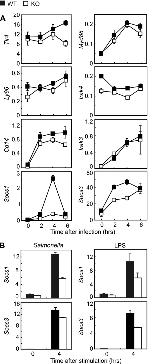
Induction of Socs1 and Socs3 mRNA is suppressed in Fos−/− macrophages in response to Salmonella infection. (A) TLR signaling-associated transcripts in MDMs. MDMs were infected with Salmonella as described for Fig. 1. Cellular mRNA levels of Tlr4, Ly96 (Md2), Cd14, Myd88, Irak4, Irak3, Socs1, and Socs3 were measured by quantitative RT-PCR from triplicate cultures. The transcripts are relative to Gapdh. Bars represent means ± standard errors of the means. WT, wild type; KO, Fos−/−. (B) Socs1 and Socs3 transcripts in PMs. Peritoneal cells were plated at 1 × 10⁵ cells per well in 48-well plates and cultured overnight, and adherent cells were used as PMs (duplicate cultures for each genotype). Cells were infected with Salmonella as described for Fig. 1 or stimulated with 100 ng/ml LPS (S. enterica serovar Minnesota Re595; Sigma) for 4 h, and Socs1 and Socs3 mRNA levels were measured by quantitative RT-PCR from duplicate cultures. The transcripts are relative to Gapdh. Bars represent means ± standard errors of the means.

Western blotting of NF-κB p65 and Mal after Salmonella infection. MDMs prepared from wild-type (WT) or Fos−/− (KO) mice in the presence of 30 ng/ml M-CSF were plated at 4 × 10⁶ cells per 6-cm dish and infected with Salmonella as described for Fig. 1. Total cell lysates were harvested, and Western blotting was performed as previously described (8) using mouse monoclonal anti-phospho (Ser536) NF-κB p65 (phospho-p65) (7F1; Cell Signaling), rabbit polyclonal anti-NF-κB p65 (F-6; Santa Cruz), rabbit polyclonal anti-TIRAP (Mal/TIRAP) (Abcam), and anti-actin (I-19; Santa Cruz).
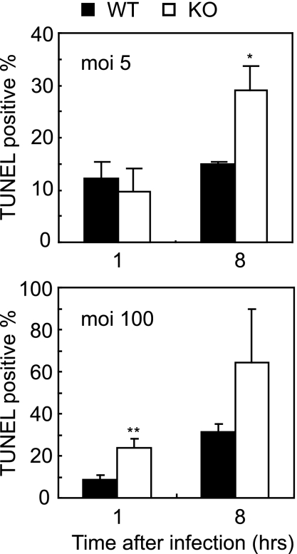
Induction of apoptosis by Salmonella infection. MDMs prepared as described for Fig. 3 were infected at 3 × 10⁵ cells per 24 wells as in Fig. 1 at an MOI of 5 (upper panel) or 100 (lower panel) for 1 h. After an additional 1 or 8 h of incubation, cells were stained by TUNEL (Promega), and the percentages of TUNEL-positive cells relative to total DAPI (4′,6′-diamidino-2-phenylindole)-positive nuclei were calculated. Error bars represent means ± SD (n = 3 each).  , P < 0.05 versus wild-type control; , P < 0.01 versus wild-type control.
, P < 0.05 versus wild-type control; , P < 0.01 versus wild-type control.
Fos−/− mice are susceptible to Salmonella infection. (A and B) Wild-type (WT) and Fos−/− (KO) mice (n = 4 for each genotype) were inoculated orally with 3.3 × 10⁷ CFU/g body weight of Salmonella. Serum cytokine levels (A) and CFU in blood, liver, and spleen (B) were determined 3 days after infection. Error bars represent means ± SD (n = 3 each). , P < 0.05 versus wild-type control; , P < 0.01 versus wild-type control. Cont., control; Infec., infected. (C) Survival of mice was monitored for 1 week after infection as described for panel A. n = 4 for each genotype.
, P < 0.05 versus wild-type control; , P < 0.01 versus wild-type control. Cont., control; Infec., infected. (C) Survival of mice was monitored for 1 week after infection as described for panel A. n = 4 for each genotype.References
-
- Aderem, A., and R. J. Ulevitch. 2000. Toll-like receptors in the induction of the innate immune response. Nature 406:782-787. - PubMed
-
- Dillon, S., A. Agrawal, T. Van Dyke, G. Landreth, L. McCauley, A. Koh, C. Maliszewski, S. Akira, and B. Pulendran. 2004. A Toll-like receptor 2 ligand stimulates Th2 responses in vivo, via induction of extracellular signal-regulated kinase mitogen-activated protein kinase and c-Fos in dendritic cells. J. Immunol. 172:4733-4743. - PubMed
-
- Grigoriadis, A. E., Z. Q. Wang, M. G. Cecchini, W. Hofstetter, R. Felix, H. A. Fleisch, and E. F. Wagner. 1994. c-Fos: a key regulator of osteoclast-macrophage lineage determination and bone remodeling. Science 266:443-448. - PubMed
-
- Hueffer, K., and J. E. Galán. 2004. Salmonella-induced macrophage death: multiple mechanisms, different outcomes. Cell. Microbiol. 6:1019-1025. - PubMed
MeSH terms
Substances
LinkOut - more resources
Full Text Sources
Medical
Molecular Biology Databases
