. 2007 Mar;27(5):1558-67.
doi: 10.1128/MCB.01495-06. Epub 2006 Dec 18.

Fission yeast Cut8 is required for the repair of DNA double-strand breaks, ribosomal DNA maintenance, and cell survival in the absence of Rqh1 helicase

Affiliations

Fission yeast Cut8 is required for the repair of DNA double-strand breaks, ribosomal DNA maintenance, and cell survival in the absence of Rqh1 helicase

Stephen E Kearsey et al. Mol Cell Biol. 2007 Mar.

Abstract

Schizosaccharomyces pombe Rqh1 is a member of the RecQ DNA helicase family. Members of this protein family are mutated in cancer predisposition diseases, causing Bloom's, Werner, and Rothmund-Thomson syndromes. Rqh1 forms a complex with topoisomerase III and is proposed to process or disrupt aberrant recombination structures that arise during S phase to allow proper chromosome segregation during mitosis. Intriguingly, in the absence of Rqh1, processing of these structures appears to be dependent on Rad3 (human ATR) in a manner that is distinct from its role in checkpoint control. Here, we show that rad3 rqh1 mutants are normally committed to a lethal pathway of DNA repair requiring homologous recombination, but blocking this pathway by Rhp51 inactivation restores viability. Remarkably, viability is also restored by overexpression of Cut8, a nuclear envelope protein involved in tethering and proper function of the proteasome. In keeping with a recently described function of the proteasome in the repair of DNA double-strand breaks, we found that Cut8 is also required for DNA double-strand break repair and is essential for proper chromosome segregation in the absence of Rqh1, suggesting that these proteins might function in a common pathway in homologous recombination repair to ensure accurate nuclear division in S. pombe.


Figures

FIG. 1.
FIG. 1.
Rad3 is required for the survival of rqh1Δ cells. (A) Five tetrads derived from diploid h+/h rqh1::kanr/rqh1+ rad3::ura4+/rad3+ (left) and rqh1::kanr/rqh1+ cds1::ura4+/cds1::ura4+ chk1::ura4+/chk1::ura4+ (right) strains were microdissected onto yeast extract (YE) agar, and the resulting colonies were photographed after 5 days of growth at 30°C. The genotypes of the segregants were determined by replica plating and are indicated schematically (W, rqh1+ rad3+; R, rad3Δ; Q, rqh1Δ; S, cds1Δ; K, chk1Δ). Boxes indicate the positions of rqh1 rad3 double (left) and rqh1 cds1 chk1 triple (right) mutants. (B) The strains, as indicated, were streaked in parallel onto YE agar plates and photographed after 3 days of incubation at 26°C or 36°C, as indicated. (C) The strains, as indicated, were grown in liquid culture to mid-logarithmic phase at 26°C and shifted to 36°C, the restrictive temperature. Samples of 500 cells taken at the indicated times after the shift to 36°C were plated in duplicate onto YE agar and incubated at 26°C. After 5 days of growth, viability was scored as a percentage of the number of colonies formed by the sample taken at time zero. Samples taken at the same time points were fixed, DAPI stained, and examined by fluorescence microscopy. The percentage of each sample exhibiting aberrant mitosis was scored as a total of at least 200 cells for each time point. (D) Fluorescence micrographs show representative fields of DAPI-stained cells of the indicated strains grown at 26°C (right panels) or 9 h after the shift to 36°C (left panels). Cells exhibiting aberrant mitosis are indicated (arrowheads). Bar, 10 μm.
FIG. 2.
FIG. 2.
Inactivation of HR suppresses rad3 rqh1 synthetic lethality. (A) Fluorescence micrographs showing Rad22-cyan fluorescent protein (CFP) localization in rad3ts and rqh1Δ rad3ts cells grown at 26°C or 6 h after the shift to 36°C. Bar, 10 μm. (B) The percentages of cells with Rad22 foci in each strain are shown. (C) Five tetrads derived from the diploid mat1PΔ17::LEU2/mst0 rqh1::kanr/rqh1+ rhp51::ura4+/rhp51+ rad3::LEU2/rad3+ strain were microdissected onto yeast extract (YE) agar, and the resulting colonies were photographed after 5 days of growth at 30°C. The genotypes of the segregants were determined by replica plating and are indicated schematically (W, rqh1+ rad3+ rhp51+; Q, rqh1Δ; R, rad3Δ; F, rhp51Δ). The positions of rqh1 rad3 double mutants (boxes) and rqh1 rad3 rhp51 triple mutants (circles) are indicated. (D) The strains, as indicated, were streaked in parallel onto YE agar plates. Plates were photographed after 3 days of incubation at 30°C.
FIG. 3.
FIG. 3.
Cut8 is required for the survival of rqh1Δ cells. (A) The strains, as indicated, containing plasmid pcut8 or empty vector were streaked in parallel onto Edinburgh minimal medium agar plates. Plates were photographed after 3 days of incubation at 26°C and 36°C, as indicated. (B) Five tetrads derived from the diploid h+/h rqh1::kanr/rqh1+ cut8::ura4+/cut8+ strain were microdissected onto yeast extract (YE) agar, and the resulting colonies were photographed after 5 days of growth at 26°C. The genotypes of the segregants were determined by replica plating and are indicated schematically (W, rqh1+ cut8+; Q, rqh1Δ; C, cut8Δ). Boxes indicate the position of the rqh1 double mutant with cut8. (C) The strains, as indicated, were streaked in parallel onto YE agar plates and photographed after 3 days of incubation at 26°C. (D) Fluorescence micrographs show representative fields of DAPI-stained cells of the indicated strains grown at 26°C. Cells exhibiting aberrant mitosis are indicated (arrowheads). Bar, 10 μm. w.t., wild type.
FIG. 4.
FIG. 4.
Cut8 is required for DNA repair. (A) Tenfold serial dilutions of the indicated strains spanning the range from 106 to 102 cells were spotted onto yeast extract (YE) agar containing 5 mM HU, 0.005% methyl methanesulfonate (MMS), 5 μg/ml bleomycin, or no drug (control). “UV” cells were exposed to 150 J/m2 UV-C. Plates were photographed after 3 to 5 days of incubation at 26°C. (B) Fluorescence micrographs showing cut8Δ and rqh1Δ cells after incubation at 26°C in the presence of 10 mM HU for 16 h or no drug. Cells exhibiting aberrant mitosis are indicated (arrowheads). Bar, 10 μm. (C) Pulsed-field gel electrophoresis analyses of chromosomes (Ch) from bleomycin-treated cells. Equal numbers of cells were prepared in agarose gel plugs from exponentially growing cultures of the indicated strains following bleomycin treatment (5 μg/ml bleomycin at 26°C for 1 h). Cells were harvested at the time of bleomycin addition (“C”) and at 3-h intervals after the removal of bleomycin for up to 6 h. Pulsed-field gel electrophoresis was carried out as described in Materials and Methods. (D) Fluorescence micrographs showing Rad22-cyan fluorescent protein (CFP) localization in wild-type and cut8Δ cells after incubation at 26°C in the presence of 10 μg/ml bleomycin for 1 h or no drug (control). Bar, 10 μm. (E) Five tetrads derived from the diploid h+/h cut8::ura4+/cut8+ rad3::LEU2/rad3+ strain were microdissected onto YE agar, and the resulting colonies were photographed after 5 days of growth at 26°C. The genotypes of the segregants were determined by replica plating and are indicated schematically (W, cut8+ slx1+; C, cut8Δ; R, rad3Δ). Boxes indicate the position of the cut8 double mutant with rad3. (F) The strains, as indicated, were streaked in parallel onto YE agar plates and photographed after 3 days of incubation at 26°C. (G) Wild-type strains containing the indicated plasmids under the control of the nmt41 promoter were streaked in parallel onto Edinburgh minimal medium (EMM) agar containing 10 mM HU or no drug (control). Plates were photographed after 3 days of incubation at 30°C. (H) The strains, as indicated, containing plasmid pcut1 or empty vector were streaked in parallel onto EMM agar plates. Plates were photographed after 3 days of incubation at 26°C and 36°C, as indicated.
FIG. 5.
FIG. 5.
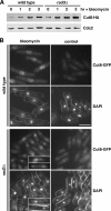
Accumulation of Cut8 in response to DNA damage. (A) Bleomycin (10 μg/ml) was added to asynchronous cultures of wild-type and rad3Δ cells that expressed the chromosomally integrated Cut8-HA gene under the native promoter growing at 26°C in yeast extract medium. Cells harvested at hourly intervals were processed for immunoblotting using antibodies against HA. Antibodies against Cdc2 were used as controls. (B) Fluorescence micrographs showing Cut8-GFP localization in wild-type and rad3Δ cells after incubation at 26°C in the presence of 10 μg/ml bleomycin for 3 h or no drug (control). Bar, 10 μm. Note that unlike wild-type cells, which became highly elongated in response to bleomycin treatment, rad3Δ cells failed to arrest cell cycle progression and displayed the “cut” phenotype, as shown in the insets.
FIG. 6.
FIG. 6.
Cut8 is important for rDNA maintenance. (A) Pulsed-field gel electrophoresis analyses of chromosomes (Ch) from cut8Δ mutants. Equal numbers of cells were prepared in agarose gel plugs from exponentially growing cultures of wild-type or cut8Δ cells (four independent isolates). Pulsed-field gel electrophoresis was carried out as described in Materials and Methods. (B) Five tetrads derived from the diploid h+/h cut8::ura4+/cut8+ slx1::kanr/slx1+ strain were microdissected onto yeast extract (YE) agar, and the resulting colonies were photographed after 5 days of growth at 26°C. The genotypes of the segregants were determined by replica plating and are indicated schematically (W, cut8+ slx1+; C, cut8Δ; S, slx1Δ). Boxes indicate the position of the cut8 double mutant with slx1. (C) The strains, as indicated, were streaked in parallel onto YE agar plates and photographed after 3 days of incubation at 26°C.
FIG. 7.
FIG. 7.
rad3 mutants are defective in nucleolar segregation. (A) Pulsed-field gel electrophoresis analyses of chromosomes (Ch) from rad3Δ mutants. Equal numbers of cells were prepared in agarose gel plugs from exponentially growing cultures of wild-type or rad3Δ cells (two independent isolates). Pulsed-field gel electrophoresis was carried out as described in Materials and Methods. (B) Fluorescence micrographs showing Gar2-GFP and DNA (Hoechst 33342) localization in living rad3Δ and rad9Δ cells. The percentages of binucleated cells displaying aberrant nucleolar structures in each strain are shown (n = 200). Bar, 10 μm. (C) Visualization of lagging Gar2-GFP signal in rad3Δ cells. Individual cells of the indicated strains expressing Gar2-GFP were observed as in panel B, over an 8-minute period, with images collected every minute.

Similar articles

Cited by

References

    1. Bachrati, C. Z., and I. D. Hickson. 2003. RecQ helicases: suppressors of tumorigenesis and premature aging. Biochem. J. 374:577-606. - PMC - PubMed
    1. Barbet, N., W. J. Muriel, and A. M. Carr. 1992. Versatile shuttle vectors and genomic libraries for use with Schizosaccharomyces pombe. Gene 114:59-66. - PubMed
    1. Bennett, R. J., and J. C. Wang. 2001. Association of yeast DNA topoisomerase III and Sgs1 DNA helicase: studies of fusion proteins. Proc. Natl. Acad. Sci. USA 98:11108-11113. - PMC - PubMed
    1. Carr, A. M. 2002. DNA structure dependent checkpoints as regulators of DNA repair. DNA Repair (Amsterdam) 1:983-994. - PubMed
    1. Coulon, S., P. H. Gaillard, C. Chahwan, W. H. McDonald, J. R. Yates III, and P. Russell. 2004. Slx1-Slx4 are subunits of a structure-specific endonuclease that maintains ribosomal DNA in fission yeast. Mol. Biol. Cell 15:71-80. - PMC - PubMed

Publication types

MeSH terms