. 2006 Dec 18;175(6):937-46.
doi: 10.1083/jcb.200604073.

Rac1 and a GTPase-activating protein, MgcRacGAP, are required for nuclear translocation of STAT transcription factors

Affiliations

Rac1 and a GTPase-activating protein, MgcRacGAP, are required for nuclear translocation of STAT transcription factors

Toshiyuki Kawashima et al. J Cell Biol. .

Abstract

STAT transcription factors are tyrosine phosphorylated upon cytokine stimulation and enter the nucleus to activate target genes. We show that Rac1 and a GTPase-activating protein, MgcRacGAP, bind directly to p-STAT5A and are required to promote its nuclear translocation. Using permeabilized cells, we find that nuclear translocation of purified p-STAT5A is dependent on the addition of GTP-bound Rac1, MgcRacGAP, importin alpha, and importin beta. p-STAT3 also enters the nucleus via this transport machinery, and mutant STATs lacking the MgcRacGAP binding site do not enter the nucleus even after phosphorylation. We conclude that GTP-bound Rac1 and MgcRacGAP function as a nuclear transport chaperone for activated STATs.


Figures

Figure 1.
Figure 1.
MgcRacGAP, Rac1, and STAT5 formed a protein complex in IL-3–dependent Ba/F3 cells. (A) STAT5A and MgcRacGAP were coprecipitated with Rac1. The cell lysates of IL-3–dependent Ba/F3 cells were subjected to immunoprecipitation with an anti-Rac1 or control antibody, followed by the immunoblotting with the anti-MgcRacGAP, anti-STAT5A, or anti-Rac1 antibody. (B) IL-3 enhanced association between STAT5A and MgcRacGAP. Ba/F3 cells were incubated in the presence or absence of 5 ng/ml IL-3 for the times indicated, and the cell lysates were subjected to immunoprecipitation with the anti-MgcRacGAP, anti-STAT5A, or control antibody, followed by the immunoblotting with the anti-p-STAT5 (top), anti-STAT5A (middle), or anti-MgcRacGAP antibody (bottom). Each row of images of the immunoprecipitation using the anti-MgcRacGAP and anti-STAT5A antibodies is derived from the same exposure of one gel, and each using the control antibody is derived from a similar exposure of the different gel. (C) The association of STAT5A and MgcRacGAP was enhanced in Ba/F3 cells expressing CA-STAT5A. Ba/F3 cells expressing CA-STAT5A were incubated in the presence or absence of 5 ng/ml IL-3 for 30 min, and cell lysates were subjected to immunoprecipitation with the anti-STAT5A (left) or anti-MgcRacGAP antibody (right), followed by the immunoblotting with the anti-p-STAT5 (top), anti-STAT5A (middle), or anti-MgcRacGAP antibody (bottom). Each row of images is derived from the same exposure of one gel.
Figure 2.
Figure 2.
STAT5A and MgcRacGAP simultaneously entered the nucleus. (A) Stoichiometry of the association between MgcRacGAP and p-STAT5A or total STAT5A in the cytoplasm and nucleus. IL-3–starved Ba/F3 cells were stimulated with IL-3 for the times indicated, and cytosol and nuclear extracts were prepared as described previously (Nakamura et al., 2002). 10 μg of protein for each of the extracts was immunodepleted with the anti-MgcRacGAP, anti-STAT5A, or control antibody, followed by immunoblotting with the anti–p-STAT5 and anti-STAT5A antibodies (a–d). The immunoprecipitates were also examined by Western blotting with the anti–p-STAT5 and anti-STAT5 antibodies (e–h). (B) STAT5A and MgcRacGAP translocated into the nucleus upon ITD-Flt3 stimulation in 293T cells. Cells were transfected with pME/STAT5A-Flag together with pMKIT (MOCK; top) or pMKIT/ITD-Flt3 (bottom). After 36 h, cells were immunostained with the anti-MgcRacGAP and anti-Flag antibodies and viewed using a Fluoview FV300 confocal microscope. Bar, 10 μm.
Figure 3.
Figure 3.
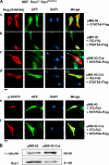
The nuclear translocation of p-STAT5 was not observed in the Rac1-knockout mouse embryonic fibroblasts. (A) The nuclear translocation of ITD-Flt3–induced p-STAT5 was impaired in the Rac2−/−Rac1flox/flox fibroblasts. The Rac2−/−Rac1flox/flox fibroblasts were transduced with a control pMX-IG (a and b) or pMX-IG-Cre (c and d) retrovirus vector. After 3 d, cells were transiently cotransfected with pME/STAT5A-Flag and MOCK (a and c) or pMKIT/ITD-Flt3 (b and d). After 36 h, the cells were fixed and immunostained with the anti-Flag antibody (a–d, left) or anti–p-STAT5 antibody (e and f, left). Bars, 10 μm. (B) Expression of Rac1 was depleted in the Rac2−/−Rac1flox/flox fibroblasts by Cre recombinase. Expression of α-tubulin and Rac1 was examined in the Rac2−/−Rac1flox/flox fibroblasts retovirally transduced with a control pMX-IG or pMX-IG-Cre.
Figure 4.
Figure 4.
Rac1 and MgcRacGAP were required for IL-3–induced nuclear accumulation and transcriptional activation of p-STAT5A. (A) IL-3–induced transcriptional activation of STAT5A was suppressed by knock down of Rac1 or MgcRacGAP. Expression of bcl-xL or GAPDH mRNA was examined in Ba/F3 cells treated with the control siRNA (lane 1), Rac1 siRNA (lane 2), or MgcRacGAP siRNA (lane 3). 24 h after the siRNA treatment, the live cells were collected using Ficoll and subjected to semiquantitative RT-PCR. (B) IL-3–induced nuclear accumulation of p-STAT5A was impaired by knock down of Rac1 or MgcRacGAP. The intracellular distribution of p-STAT5A or total STAT5A in the IL-3–stimulated or unstimulated Ba/F3 cells pretreated with the control, Rac1, or MgcRacGAP siRNA (a and b). Note that Rac1 or MgcRacGAP expression was specifically suppressed by siRNA (c and d). Cytosol and nuclear extracts were prepared as described previously (Nakamura et al., 2002) and validated by Western blot using an anti-HDAC antibody or anti-RhoA antibody (e and f).
Figure 5.
Figure 5.
The mutant of STAT5A, which lacks MgcRacGAP binding site, was not efficiently tyrosine phosphorylated by ITD-Flt3 stimulation and did not enter the nucleus even after tyrosine phosphorylation. (A) The DB2 region of STAT5 directly interacted with MgcRacGAP in vitro. Full-length MgcRacGAP was expressed in Sf-9 cells using the baculovirus vector and was purified from infected Sf-9 cells. The recombinant MgcRacGAP was pulled down by MBP-DB2 or MBP-bound beads and subjected to Western blot analysis with the anti-MgcRacGAP (top) or anti-MBP antibody for the loading control (bottom). (B) The deletion mutant of DB2 did not bind MgcRacGAP, and the STAT5 phosphorylation was considerably impaired by the deletion of DB2. Expression and tyrosine phosphorylation of Flag-tagged STAT5A-dDB2 (top and middle, respectively) were examined in the MOCK or ITD-Flt3–transfected 293T cells. The interactions of MgcRacGAP with the WT-STAT5A or STAT5A-dDB2 were also examined in the MOCK or ITD-Flt3–transfected 293T cells (bottom). Images of the immunoblots using the MOCK or ITD-Flt3–transfected cells are derived from the same exposure of one gel that was cut to remove intervening lanes. (C) The transcriptional activity of STAT5-dDB2 was impaired. Luciferase activities were examined in the lysates of ITD-Flt3–stimulated 293T cells cotransfected with the STAT5-reporter plasmid together with internal control reporter plasmids and the MOCK vector (pME), the expression vector for the Flag-tagged WT-STAT5, or STAT5-dDB2 mutant. The results shown are the mean ± SD of three independent experiments. (D) MgcRacGAP was coprecipitated with JAK2. The cell lysates of 293T cells transfected with the expression vector (pRK5) for JAK2 were subjected to immunoprecipitation with the anti-MgcRacGAP or control antibody, followed by the immunoblotting with the anti-JAK2 (top) or anti-MgcRacGAP antibody (middle). Levels of transfected JAK2 were assayed by blotting with the anti-JAK2 antibody (bottom). (E) STAT5A-dDB2 did not enter the nucleus even after the phosphorylation. The 293T cells were cotransfected with pMKIT/ITD-Flt3 together with the MOCK (left), the expression vector for the Flag-tagged WT-STAT5A (middle), or STAT5A-dDB2 (right). After 24 h, the cells were fixed and immunostained with the anti–p-STAT5 antibody. Bar, 10 μm.
Figure 6.
Figure 6.
Purified p-STAT5A accumulated to the nuclear envelope in the presence of V12Rac1 and MgcRacGAP in the nuclear transport assay. (A) Coomassie blue (CBB) staining of purified STAT5A, p-STAT5A, V12Rac1, N17Rac1, importin α1, importin α5, importin β1, Ran, NTF2, or MgcRacGAP. (B) Western blot analysis of the STAT5A-Flag protein purified from Sf-9 cells with or without coexpression with the kinase domain of JAK using the anti–p-STAT5 antibody. Total cell lysate of GM-CSF–stimulated TF-1 was used as a control. (C) The nuclear transport assay. HeLa cells were permeabilized with 40 μg/ml digitonin. Incubation with 50 μl import mix was done at 37°C for 30 min. Import mix contained TB, an energy regenerating system, and a single or combinations of the following purified proteins as indicated: 1 μM STAT5A, p-STAT5A, V12Rac1, N17Rac1, MgcRacGAP, importin α1, importin β1, Ran, or NTF2. After the import reaction, the cells were fixed. STAT5A protein was detected using the anti-STAT5A antibody. Cells were examined using a Fluoview FV300 confocal microscope. A representative result of three independent experiments is shown. Bar, 10 μm. (D) The direct bindings of both GTP-bound Rac1 and MgcRacGAP facilitated the interaction of p-STAT5A with importin αs. Purified p-STAT5A was incubated with importin αs in the absence or presence of the indicated combinations of V12Rac1, L61Rac1, N17Rac1, or MgcRacGAP in TB containing 5% BSA to block nonspecific bindings. 1 μg of each purified protein was used for each sample. After the incubation for 30 min at RT, STAT5A was immunoprecipitated with anti-STAT5A antibody and washed three times with TB. The immunoprecipitates were subjected to Western blot analysis with the anti–importin α1, anti–importin α5, anti-Rac1, anti-MgcRacGAP, or anti-STAT5A antibody.

References

    1. Adam, S.A., R.S. Marr, and L. Gerace. 1990. Nuclear protein import in permeabilized mammalian cells requires soluble cytoplasmic factors. J. Cell Biol. 111:807–816. - PMC - PubMed
    1. Bild, A.H., J. Turkson, and R. Jove. 2002. Cytoplasmic transport of Stat3 by receptor-mediated endocytosis. EMBO J. 21:3255–3263. - PMC - PubMed
    1. Bishop, A.L., and A. Hall. 2000. Rho GTPases and their effector proteins. Biochem. J. 348:241–255. - PMC - PubMed
    1. Blagoev, B., I. Kratchmarova, S.E. Ong, M. Nielsen, L.J. Foster, and M. Mann. 2003. A proteomics strategy to elucidate functional protein-protein interactions applied to EGF signaling. Nat. Biotechnol. 21:315–318. - PubMed
    1. Bromberg, J.F., M.H. Wrzeszczynska, G. Devgan, Y. Zhao, R.G. Pestell, C. Albanese, and J.E. Darnell Jr. 1999. The JAK-STAT pathway: summary of initial studies and recent advances. Stat3 as an oncogene. Cell. 98:295–303. - PubMed

Publication types