. 2006 Dec 26;103(52):19683-8.
doi: 10.1073/pnas.0609283103. Epub 2006 Dec 18.

Mechanism of allosteric regulation of transglutaminase 2 by GTP

Affiliations

Mechanism of allosteric regulation of transglutaminase 2 by GTP

Gillian E Begg et al. Proc Natl Acad Sci U S A. .

Abstract

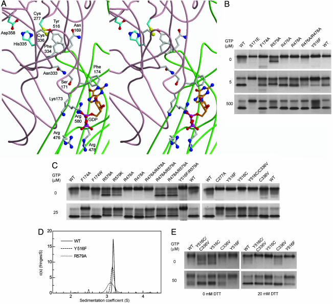
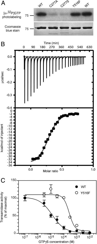
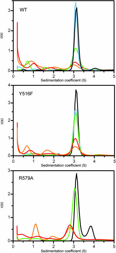
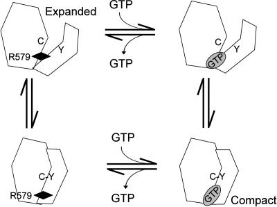
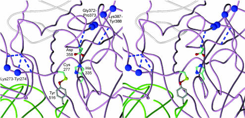
Allosteric regulation is a fundamental mechanism of biological control. Here, we investigated the allosteric mechanism by which GTP inhibits cross-linking activity of transglutaminase 2 (TG2), a multifunctional protein, with postulated roles in receptor signaling, extracellular matrix assembly, and apoptosis. Our findings indicate that at least two components are involved in functionally coupling the allosteric site and active center of TG2, namely (i) GTP binding to mask a conformationally destabilizing switch residue, Arg-579, and to facilitate interdomain interactions that promote adoption of a compact, catalytically inactive conformation and (ii) stabilization of the inactive conformation by an uncommon H bond between a cysteine (Cys-277, an active center residue) and a tyrosine (Tyr-516, a residue located on a loop of the beta-barrel 1 domain that harbors the GTP-binding site). Although not essential for GTP-mediated inhibition of cross-linking, this H bond enhances the rate of formation of the inactive conformer.


Conflict of interest statement

The authors declare no conflict of interest.

Figures

Fig. 1.
Fig. 1.
Mutations of residues involved in GTP-binding or the Cys-277–Tyr-516 H bond affect TG2 conformation. (A) Model of GDP-bound human TG2 showing the transamidase active center (Cys-277, His-335, and Asp-358) and other key residues [(Protein Data Bank ID code 1KV3) (7); core domain backbone, pale pink; β-barrel 1 domain, green]. Carbon atoms are gray, except those of active site residues (cyan), and GDP (orange). Other atoms include oxygen, red; nitrogen, deep blue; sulfur, yellow; phosphorous, bright pink. Arg-580 in human TG2 is equivalent to Arg-579 in rat TG2. (B and C) Samples (3 μg) of WT and mutant TG2 were incubated (3 h at 25°C) in buffer (50 mM Tris·HCl, pH 7.5/50 mM NaCl/0.5 mM EDTA/1.0 mM MgCl2/5 mM DTT/GTP as indicated). nPAGE was performed with 0.5 mM MgCl2, and the indicated GTP concentrations were included in gel solutions and running buffer. Data are representative of two to eight experiments. (D) Sedimentation coefficient distributions, cM(s), for 13 μM WT (solid line), Y516F (dashed line), and R579A (dotted line) in 10 mM Tris·HCl pH 7.5/100 mM NaCl/0.5 mM EDTA/1 mM DTT/500 μM GTP at 4°C. Molar mass was fixed at 78.0 kDa for 2.5–3.8 S species. Data are representative of three to four experiments. (E) Disulfide trapping of Cys-277 with a cysteine substituted for Tyr-516. Samples (3 μg) of WT and mutant TG2 were oxidized (2 h at 25°C) in buffer (50 mM Tris·HCl, pH 7.5/50 mM NaCl/200:800 μM Cu2+:phenanthroline/50 μM GTP), then incubated (1 h at 25°C) in 0.5 mM MgCl2, without or with 20 mM DTT, as indicated. nPAGE was performed as in B. Data are representative of two experiments.
Fig. 2.
Fig. 2.
Y516F mutation does not reduce GTP affinity but weakens GTP inhibition of transamidase activity. (A) WT or mutant TG2 (1.3 μM) were photoaffinity-labeled with [α-32P]GTP and size fractionated by SDS/PAGE. Radiolabeling was detected by autoradiography (Upper), and proteins were visualized with Coomassie blue (Lower). Data are representative of two experiments. (B) Isothermal titration calorimetry of GTPγS binding to Y516F. GTPγS (150 μM) was titrated into solutions of 29 μM Y516F. (B Upper) Raw data from injections. (B Lower) Peaks were integrated to yield injection-associated heat change and buffer control was subtracted. Data are representative of three experiments. (C) GTPγS inhibition of transamidase activity was assayed at ≈20% maximal transamidase activity. Activities as a percentage of maximal were 25% for WT and 22% for Y516F at 50 μM free Ca2+. Data are means of triplicate determinations ± SE and are representative of two experiments.
Fig. 3.
Fig. 3.
GTP-bound TG2 is in a slow reversible equilibrium with multiple GTP-free conformers. Sedimentation coefficient distributions c(s) of WT (Top), Y516F (Middle), and R579A (Bottom) TG2 with equimolar GTPγS in 20 mM Tris·HCl, pH 7.5/100 mM NaCl/0.5 mM EDTA/1 mM MgCl2/2 mM DTT at 4°C: black, undiluted (≈25 μM) TG2/GTP; blue, 3-fold dilution; green, 10-fold dilution; red, 33-fold dilution; orange, 100-fold dilution. Distributions, calculated by using 300 s values between 0.2 and 15 S, are shown normalized for unit area. Data are representative of two experiments.
Fig. 4.
Fig. 4.
Model of allosteric inhibition of TG2 by GTP. Transition from expanded, GTP-free TG2 (upper left) to compact, GTP-bound TG2 (lower right) may occur via an induction mechanism (clockwise) in which GTP binding to the destabilizing Arg-579 (black diamond) triggers a conformational change to the compact form, which is stabilized by Cys-277–Tyr-516 interaction, or a selection mechanism (counterclockwise) in which an unstable conformational intermediate facilitated by Cys-277–Tyr-516 interaction (GTP-free compact, lower left) is stabilized by GTP binding. Removal of the destabilizing Arg-579 by substitution stabilizes the GTP-free compact form in Arg-579 mutants.
Fig. 5.
Fig. 5.
Structural relationship between β-bulges and conserved cis-peptide bonds in TG2. GDP-bound human TG2, colored as in Fig. 1A, with residues linked by cis-peptide bonds shown as purple spheres. The proline cis-peptide bond, Gly-372-Pro-373, is located within a W β-bulge (37), and together with the Lys-387-Tyr-388 cis-peptide bond, clusters with the active site Asp-358. The Lys-273-Tyr-274 cis-peptide bond is part of a PC β-bulge (37) and is N-terminal to the active site Cys-277. Dashed lines indicate β-bulge-associated H bonds.

References

    1. Achyuthan KE, Greenberg CS. J Biol Chem. 1987;262:1901–1906. - PubMed
    1. Lorand L, Graham RM. Nat Rev Mol Cell Biol. 2003;4:140–157. - PubMed
    1. Lee KN, Birckbichler PJ, Patterson MK., Jr Biochem Biophys Res Commun. 1989;162:1370–1375. - PubMed
    1. Nakaoka H, Perez DM, Baek KJ, Das T, Husain A, Misono K, Im MJ, Graham RM. Science. 1994;264:1593–1596. - PubMed
    1. Park ES, Won JH, Han KJ, Suh PG, Ryu SH, Lee HS, Yun HY, Kwon NS, Baek KJ. Biochem J. 1998;331:283–289. - PMC - PubMed

Publication types

MeSH terms

LinkOut - more resources