Comparative Study
. 2007 Mar;175(3):1089-104.
doi: 10.1534/genetics.106.065995. Epub 2006 Dec 18.

Exploring strategies for protein trapping in Drosophila

Affiliations
Comparative Study

Exploring strategies for protein trapping in Drosophila

Ana T Quiñones-Coello et al. Genetics. 2007 Mar.

Abstract

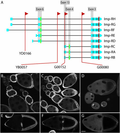
The use of fluorescent protein tags has had a huge impact on cell biological studies in virtually every experimental system. Incorporation of coding sequence for fluorescent proteins such as green fluorescent protein (GFP) into genes at their endogenous chromosomal position is especially useful for generating GFP-fusion proteins that provide accurate cellular and subcellular expression data. We tested modifications of a transposon-based protein trap screening procedure in Drosophila to optimize the rate of recovering useful protein traps and their analysis. Transposons carrying the GFP-coding sequence flanked by splice acceptor and donor sequences were mobilized, and new insertions that resulted in production of GFP were captured using an automated embryo sorter. Individual stocks were established, GFP expression was analyzed during oogenesis, and insertion sites were determined by sequencing genomic DNA flanking the insertions. The resulting collection includes lines with protein traps in which GFP was spliced into mRNAs and embedded within endogenous proteins or enhancer traps in which GFP expression depended on splicing into transposon-derived RNA. We report a total of 335 genes associated with protein or enhancer traps and a web-accessible database for viewing molecular information and expression data for these genes.


Figures

F<sc>igure</sc> 1.—
Figure 1.—
Protein trap expression patterns. (A) Expression of GFP inserted in I'm not dead yet (Indy) localizes to follicle cell membranes (line YC0017). (B) Expression of GFP inserted in Rtnl1 localizes to endoplasmic reticulum (line G00071). (C) Expression of GFP inserted in Calmodulin (Cam) localizes to nurse cell and oocyte membranes and also to nurse cell nuclei (line P00695). (D) Expression of GFP inserted in trailer hitch (tral) localizes germline cytoplasm and is enriched in the oocyte (line G01240). (E) Expression of GFP inserted in CG6416 localizes to muscle sheath (line ZCL0663). (F) Expression of GFP inserted in CG15926 localizes to follicle cell membranes (line G00035). Bar, 50 μm.
F<sc>igure</sc> 2.—
Figure 2.—
Expression and mRNA analyses of enhancer trap and no gene lines. Ovarian expression patterns of GFP in enhancer trap lines (A, C, and G–H) and no gene lines (B and D–F) are shown. (A) GFP localizes to germline nuclei in enhancer trap line YD0184. (B) GFP localizes to follicle cell nuclei in no gene line YD0570. (C) GFP localizes to follicle cell nuclei in enhancer trap line YB0172. (D) GFP localizes to nuclei in no gene line ZCL2825. (E) GFP localizes to follicle cell nuclei in no gene line ZCL2860. (F) GFP localizes to nuclei in no gene line YB0147. (G) GFP localizes to nuclei and is concentrated in the germinal vesicle and puncta in the nurse cell nuclei (arrow) in enhancer trap line YB0011. (H) GFP localizes to the cytoplasm in enhancer trap line ZCL3170. (I) Western analysis with anti-GFP antibody of enhancer trap (Enh) and no gene (NG) lines. w1118 is the control showing several background bands recognized by the antibody. Circled numbers indicate the type of 3′ end seen in P-element insertions (see J). Lines YD0184, YD0570, YB0172, ZCL2825, ZCL2860, and YB0147 show protein products that run at ∼40 kDa (solid arrowhead). Line YB0011 shows a protein product at ∼110 kDa and ZCL3170 shows a protein at ∼75 kDa (open arrowheads). (J) Splicing schematic of P-element lines. In all lines examined, exon 0 of the P-element transposase gene (dark blue), which contains the methionine codon, splices in frame with the GFP. This splice completely removes the white gene that is in the opposite orientation to both the P element and the GFP gene. There are three distinct types of mRNA 3′ of GFP: (1) Readthrough of the splice donor (pink), which adds one amino acid followed by a stop codon and is followed by the P-element poly(A) addition signal 197 nucleotides downstream; (2) splicing from GFP to an exon with a noncanonical SA in the genome adding between 1 and 19 amino acids and a poly(A) addition signal; and (3) splicing from GFP to an annotated exon that contains the start codon of a known gene that is in frame with GFP with no intervening stop codon in the linker sequence. (K) Splicing schematic of PBac lines. All lines investigated showed the exact same splicing pattern. The most 5′ sequence is an annotated noncoding exon (red) that splices into two cryptic exons (light purple) in the 5′ PBac end (dark purple). The second PBac “exon” contains the start codon (Met) and the first 91 codons of the PBac transposase. Splicing from the second PBac exon to the GFP exon maintains the open reading frame. Splicing 3′ of GFP is to a cryptic exon that lies in the yellow gene. The cryptic exon is upstream of, and in the opposite orientation to, the yellow coding sequence. Six additional codons are in the cryptic exon, followed by a poly(A) addition signal.
F<sc>igure</sc> 3.—
Figure 3.—
Multiple insertions in Imp reveal isoform-specific expression patterns. (A) The IGF-II mRNA-binding protein (Imp) gene in FlyBase (FBgn0030235) has eight annotated transcripts, designated Imp-RA through Imp-RH. Insertion sites of the GFP protein traps in YD0166, YB0057, G00152, and G00080 are marked with red lines. All lines produced mRNAs with splicing of the GFP exon to Imp exon 3, which is contained in all the mature transcripts. Splicing 5′ of the GFP exon in G00080 incorporated exons 13 and 4 of the Imp-RC transcript. Splicing 5′ of the GFP exon in G00152 and YB0057 incorporated exon 6, but none of the transcript-specific exons upstream of exon 6 were detected in our RT–PCR analysis. (B–F) Confocal micrographs illustrating the expression patterns for the Imp insert lines YB0057 (B and E), G00152 (C and F), and G00080 (D and G). YB0057 had strong GFP expression in the somatic follicle cells, especially during stages 5–7, and less expression in the germline, with very little oocyte enrichment. G00152 expression was strong in follicle cells throughout oogenesis and showed some oocyte enrichment. G00080 expressed GFP in the germline throughout oogenesis, with strong oocyte enrichment and cortical and posterior localization in late oocytes, while follicle cell expression was reduced in comparison with the other insertions.
F<sc>igure</sc> 4.—
Figure 4.—
Comparison of GFP expression from P-element and PBac Protein traps. Confocal projection images of P-element and piggyBac protein traps inserted in the same gene are shown. (A and B) Protein traps inserted in Fasciclin3 (Fas3). Prominent expression of GFP is seen in follicle cell subsets including the cells that envelop germline cysts in the germarium (brackets), the stalk cells (arrowheads), and the polar cells (arrows). GFP fluorescence was much weaker in the pBac trap, as B was imaged with increased gain and laser power relative to the P-element trap in A (inset in B shows part of the pBac ovariole imaged under the same conditions as A). (C and D) Protein traps in shaggy (sgg) captured using identical imaging conditions to allow for an approximate comparison of GFP-fusion protein levels. Uniform cytoplasmic fluorescence is seen along with enrichment on plasma membranes. (E and F) Protein traps in Ornithine decarboxylase antizyme (Oda). The same pattern of nuclear enrichment is seen with both types of traps, although gain and laser power had to be increased when imaging the PBac trap. (G and H) Protein traps in Protein disulfide isomerase (Pdi). Pdi exhibits a characteristic ER subcellular localization pattern: nuclear envelope enrichment and a fenestrated cytoplasmic distribution. Again, GFP fluorescence in the PBac line was significantly weaker, requiring increased PMT gain relative to the P-element line. One notable difference between the two trap types in Pdi was the high level of GFP∷Pdi present in follicle cells relative to germ cells in the P-element insertion; this difference was not apparent in the PBac trap. Bars, 100 μm.

References

    1. Altschul, S. F., T. L. Madden, A. A. Schaffer, J. Zhang, Z. Zhang et al., 1997. Gapped BLAST and PSI-BLAST: a new generation of protein database search programs. Nucleic Acids Res. 25: 3389–3402. - PMC - PubMed
    1. Balciunas, D., A. E. Davidson, S. Sivasubbu, S. B. Hermanson, Z. Welle et al., 2004. Enhancer trapping in zebrafish using the Sleeping Beauty transposon. BMC Genomics 5: 62. - PMC - PubMed
    1. Bellen, H. J., 1999. Ten years of enhancer detection: lessons from the fly. Plant Cell 11: 2271–2281. - PMC - PubMed
    1. Bellen, H. J., R. W. Levis, G. Liao, Y. He, J. W. Carlson et al., 2004. The BDGP gene disruption project: single transposon insertions associated with 40% of Drosophila genes. Genetics 167: 761–781. - PMC - PubMed
    1. Bobinnec, Y., C. Marcaillou, X. Morin and A. Debec, 2003. Dynamics of the endoplasmic reticulum during early development of Drosophila melanogaster. Cell Motil. Cytoskeleton 54: 217–225. - PubMed

Publication types

MeSH terms

LinkOut - more resources