. 2007;35(2):634-47.
doi: 10.1093/nar/gkl686. Epub 2006 Dec 19.

Functional organization of the Rpb5 subunit shared by the three yeast RNA polymerases

Affiliations

Functional organization of the Rpb5 subunit shared by the three yeast RNA polymerases

Cécile Zaros et al. Nucleic Acids Res. 2007.

Abstract

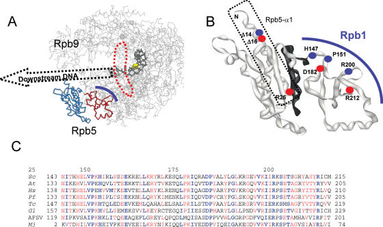
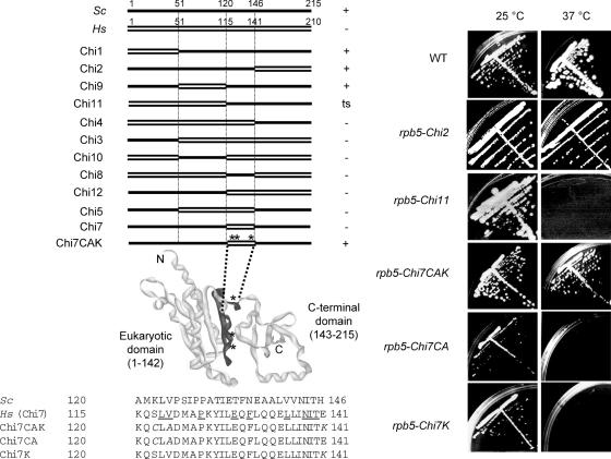
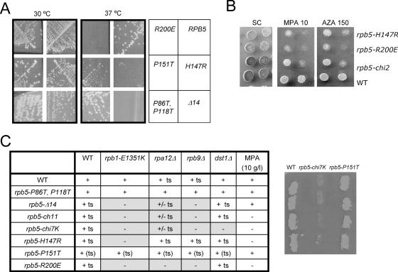
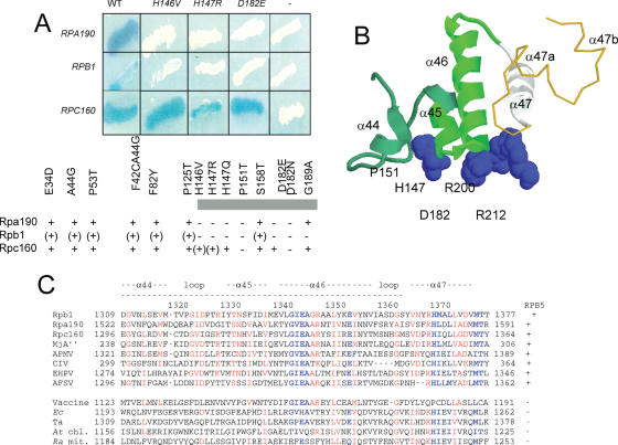
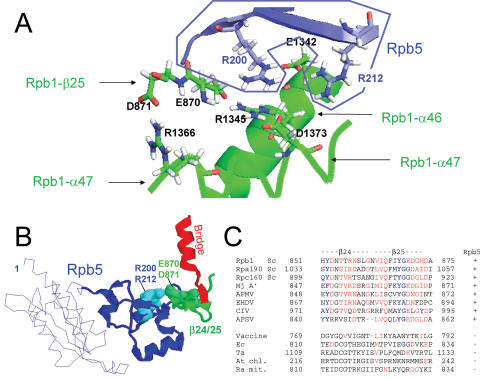
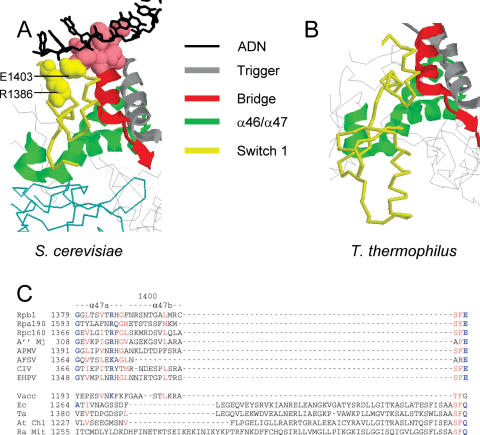
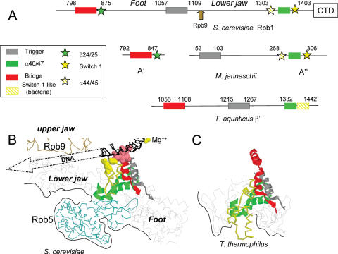
Rpb5, a subunit shared by the three yeast RNA polymerases, combines a eukaryotic N-terminal module with a globular C-end conserved in all non-bacterial enzymes. Conditional and lethal mutants of the moderately conserved eukaryotic module showed that its large N-terminal helix and a short motif at the end of the module are critical in vivo. Lethal or conditional mutants of the C-terminal globe altered the binding of Rpb5 to Rpb1-beta25/26 (prolonging the Bridge helix) and Rpb1-alpha44/47 (ahead of the Switch 1 loop and binding Rpb5 in a two-hybrid assay). The large intervening segment of Rpb1 is held across the DNA Cleft by Rpb9, consistent with the synergy observed for rpb5 mutants and rpb9Delta or its RNA polymerase I rpa12Delta counterpart. Rpb1-beta25/26, Rpb1-alpha44/45 and the Switch 1 loop were only found in Rpb5-containing polymerases, but the Bridge and Rpb1-alpha46/47 helix bundle were universally conserved. We conclude that the main function of the dual Rpb5-Rpb1 binding and the Rpb9-Rpb1 interaction is to hold the Bridge helix, the Rpb1-alpha44/47 helix bundle and the Switch 1 loop into a closely packed DNA-binding fold around the transcription bubble, in an organization shared by the two other nuclear RNA polymerases and by the archaeal and viral enzymes.


Figures

Figure 1
Figure 1
Spatial organization and sequence conservation of Rpb5. (A) Spatial organization of yeast RNA polymerase II. The ‘upper’ view using PDB co-ordinates 1I6H from (45) was drawn with Rasmol (). The RNA polymerase II backbone (without Rpb4/Rpb7) is in grey. The eukaryotic part of Rpb5 (positions 1–142) is shown in blue, and the C-terminal part (positions 143–215) in red. A thick blue line symbolizes the dual binding of Rpb5 to Rpb1. DNA is indicated by a black wire-frame (template strand only) and its hypothetical trajectory downstream of position +4 (where +1 marks the beginning of the DNA–RNA hybrid) is suggested by a dotted arrow. The catalytic Mg(A) atom is shown as a yellow sphere. A doted red line surrounds the invariant Bridge helix. (B) Close-up of the Rpb5 (ribbon structure). Dots indicate the distribution of lethal (red) and conditional (blue) mutants. Positions 121–146, (see Figure 2) are shown in black. A dotted line surrounds the hydrophilic helix Rpb5-α1. (C) Sequence conservation of the C-terminal globe. The amino acid conservation groups considered were: AG, ST, CS, DN, DE, EQ, MILV, KR, FWY, when present in at least half of the compared sequences (red letters). Highly conserved positions present in all amino acid sequences are in blue. Species symbols: S.cerevisiae (Sc), Homo sapiens (Hs), Arabidopsis thaliana (At), Plasmodium falciparum (Pf), Trypanosoma cruzi (Tc) and Giardia lamblia (Gl). These Unikonts (Fungi and Animals, represented here by S.cerevisiae and H.sapiens), Plants (A.thaliana), Alveolates (P.falciparum) or Excavates (T.cruzi) correspond to four of the five main eukaryotic phyla recognized by recent phylogenies (58). One viral (African Swine Fever Virus = AFSV) and one archaeal (Mj = M.jannaschii) subunit are shown for comparison.
Figure 2
Figure 2
In vivo complementation by the human subunit (A) Hybrid constructions based on domain swapping. Black and white boxes correspond to the yeast and human Rpb5, respectively. Symbols on the right denote the mutant growth pattern on complete YPD medium. +: wild- type like growth at 25, 30 and 37°C; ts: no growth at 37°C; −: no growth at all three temperatures. (B) Three viable mutants derived from random mutagenesis of the human domain in the lethal rpb5-chi7 allele (see Materials and Methods for a description of the mutagenesis procedure). A local sequence alignment is shown for the corresponding S.cerevisiae and H.sapiens domain. Amino acid identities (relatively to the S.cerevisiae sequence) are underlined. The amino acids mutated in rpb5-chi7CAK are italicised. The panel above provides a spatial view of Rpb5, with the mutagenized region in black. (C) Growth of the viable rpb5-chi2, rpb5-chi11, rpb5-chi7CAK, rpb5-chi7CA and rpb5-chi7K mutants at 25 and 37°C. Cells were streaked on YPD and examined after five days of incubation.
Figure 3
Figure 3
Conditional mutants of RPB5. (A) Growth pattern of rpb5 mutants. Strains YFN13 (RPB5), YFN6 (rpb5-H147R), YFN5 (rpb5-P151T), YFN49 (rpb5-R200E), YFN60 (rpb5-Δ14) and YFN46 (rpb5-P86T,P118T) were streaked on YPD and tested for growth after three days of incubation at 30 and 37°C. Note the wild-type growth of the rpb5-P86T,P118T double mutant, despite the hypothetical DNA-interacting properties assigned to the corresponding Prolines rings (18,45). (B) Sensitivity to NTP-depleting inhibitors (6-azauracail and mycophenolic acid). Serial dilutions of strains YFN13 (RPB5), YFN6 (rpb5-H147R), YFN49 (rpb5-R200E) and YFN42 (rpb5-chi2) were dropped on SC medium with or without mycophenolate (MPA) or 6-azauracil at the concentration indicated and incubated for three days at 30°C. (C) Summary of the growth defect and synthetic lethality of rpb5 mutants. Strains YFN13 (rpb5Δ/2μ URA3 RPB5), D471-13B (rpb5Δ rpb1-E1351K/2μ URA3 RPB5), YCZ44-5B (rpb5Δ rpb9Δ/2μ URA3 RPB5), D481-1B (rpb5Δ rpa12Δ/2μ URA3 RPB5) and D487-17C (rpb5Δ dst1Δ/2μ URA3 RPB5) were transformed with plasmids of the pGEN-rpb5 and pGEN-Chi series bearing the rpb5-Δ14, rpb5-chi11, rpb5-chi7K, rpb5-H147R, rpb5-P151T or rpb5-R200E alleles, using pGEN-RPB5, pGEN-RPB9 and pGEN-DST1 as positive controls and the void pGEN plasmid as negative control. Four independent transformed clones were re-isolated on selective medium, grown on YPD, replica plated on FOA medium to chase the 2 μ URA3 RPB5 host plasmid and incubated for three days at 30°C. A lack of growth indicated that the transforming plasmid harboured a non-functional rpb5 allele, unable to complement rpb5Δ in the host strain considered. Results are summarized for all four tester strains. The panel on the right illustrates the lethality of rpb5-chi7K and the viability of rpb5-P151T in an rpb9Δ background, as measured after transformation in the YCZ44-5B (rpb5Δ rpb9Δ/2 μ URA3 RPB5) host strain.
Figure 4
Figure 4
Binding of Rpb5 to the Rpb1-α44/47-fold (A) Two-hybrid interactions between Rpb5 and Rpa190, Rpb1 and Rpc160. Wild-type and mutant forms of Rpb5 fused to Gal4BD in plasmid pAS2Δ were tested against plasmids pACT2-RPA190(1615), pACT2-RPB1(1545) and pACT2-RPC160(1594) listed in table 1 and containing the partner regions of Rpa190, Rpb1 and Rpc160 fused to the Gal4AD domain of pACT2. β-Galactosidase was tested in an overlay assay (29), as shown for the RPB5, rpb5-H146V, rpb5-H147R and rpb5-D182E constructs and as summarized below for the other mutants. +, (+) and − denote positive, reduced and negative responses in the β-galactosidase plate assay, respectively. (B) Spatial organization of H147, P151, D182, R200 and R212 relatively to the Rpb1α44/47-fold in the elongating RNA polymerase II. H147, P151, D182, R200 and R212 positions of Rpb5 are space-filled. The Rpb1-α44/46-fold (positions 1309–1362, corresponding to the minimal two-hybrid domain of Figure 4A is shown in green ribbons. The Rpb1-α47 helix (positions 1363–1377), not comprised in the minimal two-hybrid domain but binding Rpb5, notably through position R212) is shown in white ribbons. A gold thin line corresponding to Switch 1 backbone (positions 1378–1403). (C). Sequence conservation of the Rpb1-α44/47 domains. Highly conserved domain are blue. Species symbols: Sc (S.cerevisiae); Mj (M.jannaschii), Ec (Escherichia coli); Ta (T.aquaticus); At (A.thaliana, chloroplastic), Ra (R.americana, mitochondrial). Viral species: Acanthamoeba Polyphaga Mimivirus (APMV); Chilo Iridescent Virus (CIV); Emiliana Huxleyi PhycoDNA Virus (EHPV); African Swine Fever Virus (AFSV) and Vaccine.
Figure 5
Figure 5
Dual binding of Rpb5 to Rpb1 (A) View of the salt bridge system involving R200 and R212 (Rpb5) and six Rpb1 amino acids: E870, D871 (Rpb1-β25), E1342 and R1345 (Rpb1-α46), R1366 and D1373 (Rpb1-α47). Drawing based on the PDB crystallographic coordinates 1I6H (41), with Hydrogen atoms recalculated by the PDB-viewer programme. The Rpb5 domain is in blue, and the two Rpb1 parts in green. (B) Spatial organization of Rpb5, the Rpb1-β24/25-fold and the Bridge helix (Rpb1-α25), using the same orientation as in Figure 1. Rpb5 is shown in blue, with the eukaryotic backbone domain shown as a thin line. R200 and R212 are space-filled. Rpb1-β24/25 and Rpb1-α25 are shown in green and red, respectively. (C) Conservation of the Rpb1-β24/25 sequence. Species symbols as in Figure 4. Conserved amino acids are in red.
Figure 6
Figure 6
The switch 1 loop in RNA polymerase II. (A) Spatial organization of the Switch 1 loop (1378–1403), Bridge helix (C-end, positions 804–835), trigger helix (positions 1057–1089) and Rpb1-α46/47 domain (1338–1377) in RNA polymerase II, based on the PDB crystallographic coordinates 1I6H (41). (B) Spatial organization of the corresponding bacterial domain, based on the PDB crystallographic coordinates 1IW7 (47). Sequence conservation of the switch 1 loop domain. Species symbols as in Figure 4. (C) Positions R1386 and E1403 are shown in bold characters.
Figure 7
Figure 7
Spatial organization of Rpb5, Rpb9 and their interacting domains. (A) Distribution of the Bridge, Rpb1-b24/25, Trigger, Rpb1-a44/47 and Switch 1 domains on Rpb1(S.cerevisiae), A′ and A′ (M.jannaschii) and β′ (T.aquaticus). (B) View of the same domains and of Rpb5 and Rpb9 in the elongating RNA polymerase II, based on the PDB crystallographic coordinates 1I6H (45). This view corresponds to the ‘upper’ representation of Figure 1A. The Bridge C-end (positions 831–851), Trigger (1057–1088), α46/47 (1340–1379) and Switch 1 (1380–1406) modules are shown as ribbon structures. DNA-binding amino acids of the Bridge (positions 831–836) and Switch 1 modules (R1386 and E1403) are space-filled. The template strand of DNA (black wire-frame) and the catalytic Mg (yellow sphere) are also indicated. The Rpb9 and Rpb5 backbones are shown in brown and blue, respectively. Black lines correspond to the outer surface of the crystal structure. (C) Equivalent view of the Thermus thermophilus holoenzyme, based on the PDB crystallographic coordinates 1IW7 (47). Note that the amino acid sequences and spatial structures of T.aquaticus and T.thermophilus are practically identical. The T.aquaticus (β′ subunit) equivalent of the yeast bridge (β′F), trigger (β′G) and α46/47 modules correspond to positions 1089–1109, 1215–1246 and 1341–1380, respectively. The yellow backbone (positions 1381–1443 of the β′ subunit) is no related to the Switch 1-fold in sequence (except for its last three positions, see Supplementary Data S3).
Figure 8
Figure 8
Transcriptional activation of GAL1 in RNA polymerase II defective mutants. Strains YFN68 (wild-type), YFN6 (rpb5-H147R), YFN10 (rpb5-chi7K) and YVV9 (rpb9Δ) were grown on raffinose (−) and galactose (Gal). GAL1 and PEP4 mRNAs were assayed by quantitative reverse transcribed PCR (A) as described in Materials and Methods, using the oligonucleotide probes listed in Supplementary Data S1. The same measures are shown for strain YFN13 (pTET::RPB5), YFN50 (pTET::RPB10), YFN27 (pTET::RPB11), YFN25 (pTET::RPA43) and YMLF2 (pTET::RPC17) exponentially grown on Gal medium in the presence of increasing concentrations of doxycycline (B and C). In each case, the higher concentration of doxycycline used reduced growth rate by a factor of three. Note that the depletion of the Rpa43 subunit of RNA polymerase I (YFN25) or the Rpc17 subunit of RNA polymerase III (YMLF2) has no effect on the accumulation of GAL1 mRNA.
Figure 9
Figure 9
RNA polymerase I and III defects of rpb5 mutants (A) RNA polymerase I and III activity in vivo. The level of 20S pre-rRNA (compared to the mature 18S rRNA product) and of the pre-tRNALeu3 transcript (compared to the mature tRNA) was measured by northern hybridization using the oligonucleotide probes listed in Supplementary Data S1. Cells were exponentially grown on YPD at 30°C until they reached an O.D. of 0.5 and were then shifted to 37°C for 3, 5 and 7 h. (B) In vitro transcription of 35S rRNA, 5S rRNA and tRNATyr templates. Transcription was assayed on cell-free extract prepared from strains YFN13 (wild-type), YFN6 (rpb5-H147R) and YFN10 (rpb5-chi7K). The templates used were pRS316-SUP4 (tRNATyr) for RNA polymerase III activity (34) and pSIRT (5S rRNA and 35S rRNA) for RNA polymerase I (35). Transcription assays are described in the Materials and Methods section. (C) Subunit composition of the purified RNA polymerase I of strains YFN13, YFN6 and YFN10, using samples with similar specific activities when tested on poly(dA–dT) templates (36). The first two panels, corresponding to standard MW markers and to an RNA polymerase I sample (kindly given by Christophe Carles and Michel Riva) were used to identify individual RNA polymerase I subunits. The star denotes a sub-stoichiometric polypeptide band that was identified by mass spectrometry as being the Rpc53 subunit of RNA polymerase III. However, there is currently no evidence that Rpc53 could be a bona fide subunit of RNA polymerase I.

Similar articles

Cited by

References

    1. Werner F., Weinzierl R.O. A recombinant RNA polymerase II-like enzyme capable of promoter- specific transcription. Mol. Cell. 2002;10:635–646. - PubMed
    1. Langer D., Hain J., Thuriaux P., Zillig W. Transcription in Archaea: similarity to that in Eucarya. Proc. Natl Acad. Sci. USA. 1995;92:5768–5772. - PMC - PubMed
    1. Briand J.F., Navarro F., Rematier P., Boschiero C., Labarre S., Werner M., Shpakovski G.V., Thuriaux P. Partners of Rpb8p, a small subunit shared by yeast RNA polymerases I, II and III. Mol. Cell. Biol. 2001;21:6056–6065. - PMC - PubMed
    1. Pontier D., Yahubyan G., Vega D., Bulski A., Saez-Vasquez J., Hakimi M.A., Lerbs-Mache S., Colot V., Lagrange T. Reinforcement of silencing at transposons and highly repeated sequences requires the concerted action of two distinct RNA polymerases IV in Arabidopsis. Genes Dev. 2005;19:2030–2040. - PMC - PubMed
    1. Wery M., Shematorova E., Van Driessche B., Vandenhaute J., Thuriaux P., Van Mullem V. Members of the SAGA and Mediator complexes are partners of the transcription elongation factor TFIIS. EMB0 J. 2004;23:42132–44242. - PMC - PubMed

Publication types

MeSH terms