. 2007 Apr 15;109(8):3325-32.
doi: 10.1182/blood-2006-04-017863. Epub 2006 Dec 19.

Nonmalignant T cells stimulate growth of T-cell lymphoma cells in the presence of bacterial toxins

Affiliations

Nonmalignant T cells stimulate growth of T-cell lymphoma cells in the presence of bacterial toxins

Anders Woetmann et al. Blood. .

Abstract

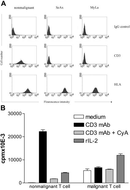
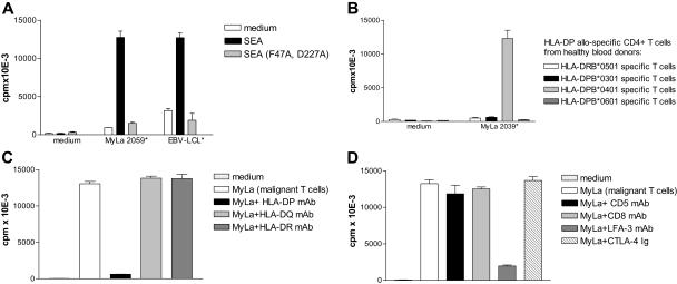
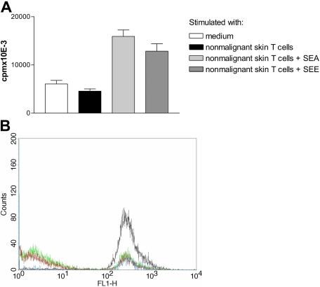
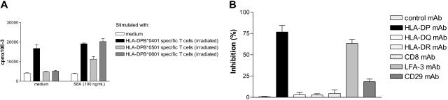
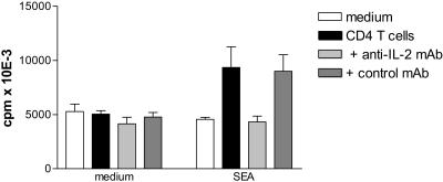
Bacterial toxins including staphylococcal enterotoxins (SEs) have been implicated in the pathogenesis of cutaneous T-cell lymphomas (CTCLs). Here, we investigate SE-mediated interactions between nonmalignant T cells and malignant T-cell lines established from skin and blood of CTCL patients. The malignant CTCL cells express MHC class II molecules that are high-affinity receptors for SE. Although treatment with SE has no direct effect on the growth of the malignant CTCL cells, the SE-treated CTCL cells induce vigorous proliferation of the SE-responsive nonmalignant T cells. In turn, the nonmalignant T cells enhance proliferation of the malignant cells in an SE- and MHC class II-dependent manner. Furthermore, SE and, in addition, alloantigen presentation by malignant CTCL cells to irradiated nonmalignant CD4(+) T-cell lines also enhance proliferation of the malignant cells. The growth-promoting effect depends on direct cell-cell contact and soluble factors such as interleukin-2. In conclusion, we demonstrate that SE triggers a bidirectional cross talk between nonmalignant T cells and malignant CTCL cells that promotes growth of the malignant cells. This represents a novel mechanism by which infections with SE-producing bacteria may contribute to pathogenesis of CTCL.


Figures

Figure 1
Figure 1
Malignant T cells have deficient expression and function of the TCR/CD3 complex. (A) Nonmalignant (MyLa 1885) and malignant (MyLa 2039) T-cell lines from skin from a patient with tumor-stage MF and malignant T cells from blood of a patient with Sezary syndrome (SeAx) were stained with anti-CD3 mAb, anti–MHC class I (HLA-A, HLA-B, and HLA-C) mAb, or isotype IgG control. Flow cytometry results are shown for viable cells gated from Fsc/Ssc-scatter plots. The data are representative of 3 independent experiments. (B) Nonmalignant (P1183) and malignant (MyLa 2039) T cells were grown in microtiter plates coated with or without anti-CD3 mAb or isotype control mAb and with or without cyclosporine A (10 nM final concentration) or with rIL-2 (100 U/mL) for 48 hours at 37°C in a humidified 5% CO2 atmosphere. Twelve hours prior to harvest, 3H-thymidine (1 μCi [0.037 MBq]/well) was added, the cells were harvested onto glass fiber filters, and 3H-thymidine incorporation was measured. The proliferation was expressed as mean counts per minute (+ SD) of triplicate cultures. The data are representative of 5 independent experiments with 3 malignant and 3 nonmalignant T-cell lines.
Figure 2
Figure 2
Malignant T cells function as superantigen- and alloantigen-presenting cells. (A) SE presentation by malignant to nonmalignant T cells is MHC class II dependent. Nonmalignant T cells (P1119) from healthy donors were grown in microtiter plates with or without malignant T cells (MyLa 2059) or EBV-LCLs and with or without SEA (wt) or SEA (F47A, D227A) with mutations in the HLA class II–binding sites. Malignant T cells and EBV-LCLs were irradiated (22 Gy, as indicated by *) to avoid proliferation. Twelve hours prior to harvest, 3H-thymidine (1 μCi [0.037 MBq]/well) was added, the cells were harvested onto glass fiber filters, and 3H-thymidine incorporation was measured. The proliferation was expressed as mean counts per minute of triplicate cultures. The data are representative of 3 independent experiments. (B) Malignant T cells present HLA-DP allospecific T cells. HLA-DPB*0301–, HLA-DPB*0401–, HLA-DPB*0501–, and HLA-DPB*0601–specific CD4+ T-cell lines from healthy donors were grown in microtiter plates with or without HLA-DPB0401–positive irradiated (to avoid proliferation) malignant T cells (MyLa 2039). 3H-thymidine uptake was performed and measured. The data are representative of 3 independent experiments. (C) HLA-DP presentation is blocked by HLA-DP mAb. HLA-DPB*0401–positive malignant T cells (MyLa 2039) were irradiated and incubated with or without HLA-DP, HLA-DQ, or HLA-DR framework mAbs (10 μg/mL) prior to coculture for 48 hours with HLA-DPB*0401–specific nonmalignant T cells in microtiter plates. The data are representative of 3 independent experiments. (D) Alloantigen stimulation by malignant T cells is inhibited by LFA-3 mAb. HLA-DPB*04.01–positive malignant T cells (MyLa 2039) were irradiated and incubated with or without CD5, CD8, LFA-3 mAb (10 μg/mL), or CTLA4-Ig (1 μg/mL) prior to coculture for 48 hours with HLA-DPB*0401–specific nonmalignant T cells in microtiter plates. The data are representative of 3 independent experiments. Error bars represent SD.
Figure 3
Figure 3
Nonmalignant T cells stimulate growth of malignant T cells in the presence of SEA. (A) Malignant T cells (SeAx) were grown with or without irradiated (to be devoid of the proliferative capacity) nonmalignant T cells (P1675) and/or SEA (100 ng/mL) in microtiter plates. Twelve hours prior to harvest, 3H-thymidine (1 μCi [0.037 MBq]/well) was added, the cells were harvested onto glass fiber filters, and 3H-thymidine incorporation was measured. The proliferation was expressed as mean counts per minute of triplicate cultures. The data are representative of 3 independent experiments. (B) Malignant T cells (MyLa 2039) and nonmalignant T cells were grown with or without irradiated nonmalignant T cells (MyLa 2355) and/or SEA (100 ng/mL) in microtiter plates. The data are representative of 4 independent experiments. (C) Malignant T cells (MyLa 1929) were grown with or without irradiated nonmalignant T cells (MyLa 1850) and/or SEA (100 ng/mL) in microtiter plates. Error bars represent SD.
Figure 4
Figure 4
Nonmalignant T cells stimulate growth of malignant T cells in the presence of SEE. (A) Malignant T cells (MyLa 2059) were grown with or without irradiated nonmalignant T cells (MyLa 3241) with or without SEA (100 ng/mL) or SEE (100 ng/mL) for 48 hours in microtiter plates. The data are representative of 3 independent experiments. Error bars represent SD. (B) Nonmalignant T cells stimulate growth of primary SS T cells in the presence of SEA. Flow cytometric analyses of CFSE expression in malignant cells grown for 3 days in medium (blue line), or with SEA (100 ng/mL) (black line), nonmalignant, blood-derived T cells (MyLa 1885) (green line), or SEA and nonmalignant T cells (red line). The following MFI values were obtained: MFI = 264 (blue line); MFI = 339 (black line); MFI = 90 (green line); and MFI = 70 (red line). The data are representative of 2 independent experiments.
Figure 5
Figure 5
Alloantigen presentation by malignant T cells to nonmalignant T cells stimulates growth of malignant T cells. (A) HLA-DPB*0401–positive malignant T cells (MyLa 2039) were grown with and without SEA (100 ng/mL) and/or HLA-DP*0401–, HLA-DP*0501–, and HLA-DP*0601–specific nonmalignant T cells (irradiated to avoid proliferation) for 48 hours in microtiter plates. The data are representative of 3 independent experiments. (B) HLA-DPB*0401–positive malignant T cells (MyLa 2039) were grown with or without HLA-DPB*0401–specific T cells (irradiated) with or without HLA-DP, HLA-DQ, HLA-DR, CD8, LFA-3, or CD29 mAb (10 μg/mL) for 48 hours in microtiter plates. The percentage inhibition of growth stimulation by coculture was calculated as described in “Materials and methods.” The data are representative of 3 independent experiments. Error bars indicate SD.
Figure 6
Figure 6
Costimulation of SeAx cells by nonmalignant cells and SEA is blocked by an anti–IL-2 mAb. Malignant T cells (SeAx) were grown with or without irradiated (to avoid proliferation) nonmalignant CD4+ T cells (P1675), and/or SEA (100 ng/mL) with or without control mAb or an IL-2–blocking (25 μg/mL) mAb in microtiter plates. Twelve hours prior to harvest, 3H-thymidine (1 μCi [0.037 MBq]/well) was added, the cells were harvested onto glass fiber filters, and 3H-thymidine incorporation was measured. The proliferation was expressed as mean counts per minute of triplicate cultures. Essentially identical results were obtained in 2 independent experiments. Error bars represent SD.

References

    1. Berger CL, Mariwalla K, Girardi M, Edelson RL. Advances in understanding the immunobiology and immunotherapy of cutaneous T-cell lymphoma. Adv Dermatol. 2004;20:217–235. - PubMed
    1. Dummer R. Future perspectives in the treatment of cutaneous T-cell lymphoma (CTCL). Semin Oncol. 2006;33(suppl 3):33–36. - PubMed
    1. Willemze R, Jaffe ES, Burg G, et al. WHO-EORTC classification for cutaneous lymphomas. Blood. 2005;105:3768–3785. - PubMed
    1. Kim EJ, Hess S, Richardson SK, et al. Immunopathogenesis and therapy of cutaneous T cell lymphoma. J Clin Invest. 2005;115:798–812. - PMC - PubMed
    1. Bagot M, Boumsell L, Bensussan A. Immunopathogenesis of cutaneous T-cell lymphomas. Hematol Oncol Clin North Am. 2003;17:1313–1317. vii. - PubMed

Publication types

MeSH terms