. 2007 Jan 2;104(1):78-82.
doi: 10.1073/pnas.0609973104. Epub 2006 Dec 20.

The 1',4'-iminopyrimidine tautomer of thiamin diphosphate is poised for catalysis in asymmetric active centers on enzymes

Affiliations

The 1',4'-iminopyrimidine tautomer of thiamin diphosphate is poised for catalysis in asymmetric active centers on enzymes

Natalia Nemeria et al. Proc Natl Acad Sci U S A. .

Abstract

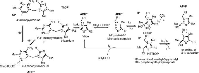
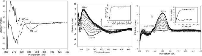
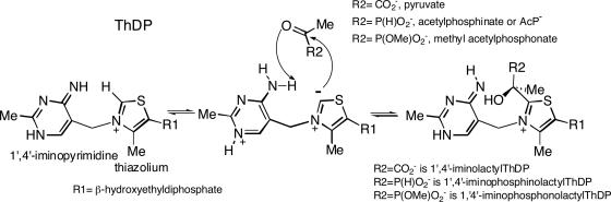
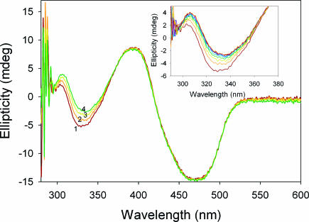
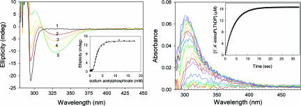
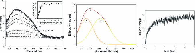
Thiamin diphosphate, a key coenzyme in sugar metabolism, is comprised of the thiazolium and 4'-aminopyrimidine aromatic rings, but only recently has participation of the 4'-aminopyrimidine moiety in catalysis gained wider acceptance. We report the use of electronic spectroscopy to identify the various tautomeric forms of the 4'-aminopyrimidine ring on four thiamin diphosphate enzymes, all of which decarboxylate pyruvate: the E1 component of human pyruvate dehydrogenase complex, the E1 subunit of Escherichia coli pyruvate dehydrogenase complex, yeast pyruvate decarboxylase, and pyruvate oxidase from Lactobacillus plantarum. It is shown that, according to circular dichroism spectroscopy, both the 1',4'-iminopyrimidine and the 4'-aminopyrimidine tautomers coexist on the E1 component of human pyruvate dehydrogenase complex and pyruvate oxidase. Because both tautomers are seen simultaneously, these two enzymes provide excellent evidence for nonidentical active centers (asymmetry) in solution in these multimeric enzymes. Asymmetry of active centers can also be induced upon addition of acetylphosphinate, an excellent electrostatic pyruvate mimic, which participates in an enzyme-catalyzed addition to form a stable adduct, resembling the common predecarboxylation thiamin-bound intermediate, which exists in its 1',4'-iminopyrimidine form. The identification of the 1',4'-iminopyrimidine tautomer on four enzymes is almost certainly applicable to all thiamin diphosphate enzymes: this tautomer is the intramolecular trigger to generate the reactive ylide/carbene at the thiazolium C2 position in the first fundamental step of thiamin catalysis.


Conflict of interest statement

The authors declare no conflict of interest.

Figures

Scheme 1.
Scheme 1.
Mechanism of YPDC.
Fig. 1.
Fig. 1.
Near-UV CD spectra of PDHc-E1h. (Left) Spectrum 1 shows PDHc-E1h (concentration of active centers = 25 μM) in 10 mM KH2PO4 (pH 7.0) with 0.20 mM ThDP and 1 mM MgCl2. Spectrum 2 shows PDHc-E1h in the absence of ThDP. (Center) Near-UV CD spectra of PDHc-E1h titrated with AcP. PDHc-E1h (concentration of active centers = 19.6 μM) in 10 mM KH2PO4 (pH 7.0) containing 0.20 mM ThDP and 1 mM MgCl2 was titrated with AcP (1–150 μM). Inset shows that saturation is reached for a 1:1 molar ratio of [AcP]/[PDHc-E1h active centers]. (Right) The PDHc-E1h (1.0 mg/ml, concentration of active centers = 13.0 μM) was diluted in 50 mM KH2PO4 (pH 7.0) containing 1 mM MgCl2 and then thiamin 2-thiothiazolone diphosphate (1–50 μM) was added. Upon addition of thiamin 2-thiothiazolone diphosphate the positive CD band at 325 nm developed and reached saturation with Kd,ThTTDP = 0.59 μM.
Scheme 2.
Scheme 2.
Formation of 1′,4′-iminophosphinolactyl-ThDP, a stable LThDP analogue.
Fig. 2.
Fig. 2.
Near-UV CD spectra of POX. Spectrum 1 shows POX (concentration of active centers = 2.0 mg/ml or 30 μM) at 30°C in 50 mM KH2PO4 (pH 6.0) containing 0.1 mM ThDP, 1 mM MgCl2, and 6% glycerol. Spectra 2, 3, and 4 were recorded after addition of 5, 10, and 40 μM AcP, respectively, at 30°C and show the presence of 1′,4′-iminophosphinolactyl-ThDP at 308 nm and the Michaelis complex at 336 nm. Inset shows the enlarged spectra in the 290- to 380-nm range upon addition of acetylphosphinate.
Fig. 3.
Fig. 3.
Formation of 1′,4′-iminophosphinolactyl-ThDP on YPDC. (Left) Near-UV CD spectra of YPDC. Spectrum 1 shows YPDC (concentration of active centers is 2.5 mg/ml or 42 μM) in 50 mM KH2PO4 (pH 6.0) containing 0.1 mM ThDP and 2.0 mM MgCl2. Spectra 2, 3, 4, and 5 were recorded after addition of 2, 2.5, 3.5, and 17.5 mM sodium acetylphosphinate, respectively, at 30°C. The spectra show presence of 1′,4′-iminophosphinolactyl-ThDP at 302 nm and of the Michaelis complex at 328 nm. (Inset) Dependence of 1′,4′-iminophosphinolactyl-ThDP formation at 302 nm on concentration of AcP after subtraction of the spectrum of YPDC in the absence of AcP. Strong cooperativity (Hill coefficient ≈ 3.17) probably reflects the presence of an allosteric substrate activation site in addition to the active centers in the functional dimer (1, 2). (Right) Photodiode array stopped-flow UV-VIS difference spectra showing the formation of 1′,4′-iminophosphinolactyl-ThDP at 302 nm in YPDC. YPDC (2 mg/ml) in 50 mM phosphate (pH 6.0) containing 0.1 mM ThDP and 2.0 mM MgCl2 in one syringe was mixed with an equal volume of 40 mM AcP in the same buffer in the second syringe. The reaction was monitored for 33 s, and the spectra were recorded at 2.5-ms interval at 30°C. Inset shows the rate of formation of 1′,4′-iminophosphinolactyl-ThDP at 302 nm (kobs = 0.25 s−1).
Fig. 4.
Fig. 4.
Formation of 1′,4′-iminophosphinolactyl-ThDP on PDHc-E1ec. (Left) Difference CD spectra of the PDHc-E1ec titrated with AcP. PDHc-E1ec (concentration of active centers = 15 μM) in 10 mM KH2PO4 (pH 7.0) containing 2 mM MgCl2 and 0.20 mM ThDP was titrated with AcP (1–100 μM). Difference spectra were obtained upon subtraction of the spectrum of PDHc-E1ec in the absence of AcPI. Inset shows that the CD maximum was reached at a 1:1 molar ratio of [AcP]/[PDHc-E1ec active centers]. (Center) The CD curve resulting from addition of saturating AcP (70 μM) to PDHc-E1ec (2.43 mg/ml; 0.1 mM ThDP/1 mM MgCl2/20 mM potassium phosphate, pH 7.0) was resolved into peaks 2 and 3 by using Peak Fit v 4.12 from Systat Software (San Jose, CA). The λmax for peaks 1, 2, and 3 are 318, 309, and 343 nm, respectively, and the rate constants measured at these wavelengths were identical within experimental error. (Right) Stopped-flow CD determination of the rate of 1′,4′-iminophosphinolactyl-ThDP formation at 30°C. PDHc-E1ec [5 mg/ml or 50 μM active centers dissolved in 20 mM KH2PO4 (pH 7.0) also containing 0.1 mM ThDP and 1 mM MgCl2] in one syringe was mixed with 50 μM AcP in the same buffer. The reaction was monitored for 5 s, 2,000 data points were collected at 2.5-ms intervals, and the trace was fitted to a double exponential equation with rate constants of 4.44 ± 0.34 s−1 and 0.593 ± 0.064 s−1.

References

    1. Jordan F. Nat Prod Rep. 2003;20:184–201. - PubMed
    1. Jordan F, Nemeria NS. Bioorg Chem. 2005;33:190–215. - PMC - PubMed
    1. Schellenberger A. Biochim Biophys Acta. 1998;1385:177–186. - PubMed
    1. Jordan F, Mariam YH. J Am Chem Soc. 1978;100:2534–2540.
    1. Lindquist Y, Schneider G, Ermler U, Sundstroem M. EMBO J. 1992;11:3273–3279. - PMC - PubMed

Publication types

LinkOut - more resources