. 2007 Jan 2;104(1):240-5.
doi: 10.1073/pnas.0603420103. Epub 2006 Dec 20.

Gain-of-function mutations in complement factor B are associated with atypical hemolytic uremic syndrome

Affiliations

Gain-of-function mutations in complement factor B are associated with atypical hemolytic uremic syndrome

Elena Goicoechea de Jorge et al. Proc Natl Acad Sci U S A. .

Erratum in

  • Proc Natl Acad Sci U S A. 2007 Jun 19;104(25):10749

Abstract

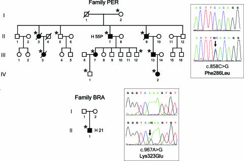
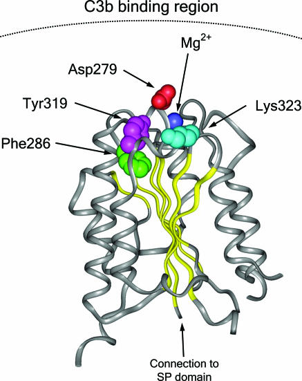
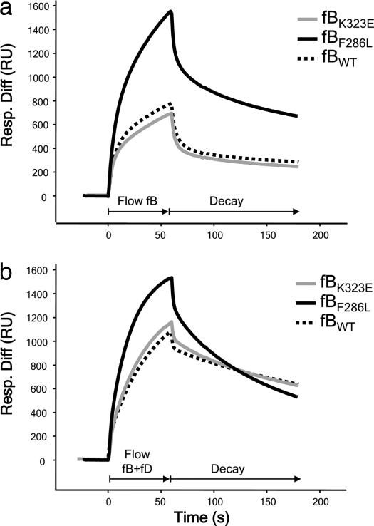
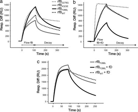
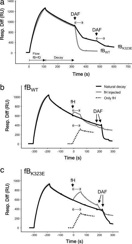
Hemolytic uremic syndrome (HUS) is an important cause of acute renal failure in children. Mutations in one or more genes encoding complement-regulatory proteins have been reported in approximately one-third of nondiarrheal, atypical HUS (aHUS) patients, suggesting a defect in the protection of cell surfaces against complement activation in susceptible individuals. Here, we identified a subgroup of aHUS patients showing persistent activation of the complement alternative pathway and found within this subgroup two families with mutations in the gene encoding factor B (BF), a zymogen that carries the catalytic site of the complement alternative pathway convertase (C3bBb). Functional analyses demonstrated that F286L and K323E aHUS-associated BF mutations are gain-of-function mutations that result in enhanced formation of the C3bBb convertase or increased resistance to inactivation by complement regulators. These data expand our understanding of the genetic factors conferring predisposition to aHUS, demonstrate the critical role of the alternative complement pathway in the pathogenesis of aHUS, and provide support for the use of complement-inhibition therapies to prevent or reduce tissue damage caused by dysregulated complement activation.


Conflict of interest statement

The authors declare no conflict of interest.

Figures

Fig. 1.
Fig. 1.
Mutations in the BF gene in two aHUS pedigrees. Pedigrees of families PER and BRA are described. Individuals are identified by numbers within each generation (in Roman numerals). Affected individuals are indicated with filled symbols. Deceased individuals are crossed. Carriers of BF mutations are indicated by an asterisk. For each BF mutation, the chromatogram corresponding to the DNA sequence surrounding the mutated nucleotide in BF is shown for the appropriate HUS patient and for a control sample. The nucleotide and amino acid numbering are referred to the translation start site (A in ATG is + 1; Met is + 1).
Fig. 2.
Fig. 2.
Structural implications for the HUS-associated BF mutations. Diagram of the von Willebrand type A domain of fB. Insight II (BYOSYM software package; Molecular Simulations, San Diego, CA) was used to draw structure by using PDB files 1Q0P and 1RS0_A. The positions of the residues Phe-286 and Lys-323 that are mutated in the HUS patients are indicated. The position of the Mg2+ ion and that of the Asp-279 and Tyr-319 are also indicated. Notice the edge-to-face stacking of Phe-286 and Tyr-319 residues. Numbering of residues is referred to with the initial methionine as “1” and therefore includes the sequence of the N-terminal signal peptide. Residue Asp-279 was described as Asp-254 by Hourcade et al. (32).
Fig. 3.
Fig. 3.
SPR analysis of fBs purified from plasma. (a) Formation of C3bB complexes. Control fB (fBWT) and fB purified from H21 (fBK323E) formed similar levels of the proenzyme, whereas fB purified from H55P (fBF286L) formed C3bB complexes much more rapidly and to a much higher level. (b) Formation of C3bBb complexes. fBWT and fBK323E formed similar levels of complexes with C3b. fBF286L formed high and similar levels of complexes with C3b in the absence or presence of fD. Resp. Diff, response difference; RU, resonance units.
Fig. 4.
Fig. 4.
SPR analysis of recombinant fB proteins. (a) Formation of C3bB complexes. fBWT and fBK323E formed similar levels of the proenzyme, whereas fBF286L formed C3bB complexes much more rapidly and to a much higher level, similar to those formed by the fBD279G mutant described by Hourcade et al. (20). (b) Formation of C3bBb. At 10 μg/ml, fBWT and fBK323E formed similar levels of complexes with C3b. fBF286L formed lower levels of complexes with C3b in the presence than in the absence of fD as a consequence of accelerated rate of spontaneous decay of the cleaved fB. (c) Formation of C3bBb. At 37 μg/ml, fBF286L formed high levels of proenzyme, but when fD was included in the incubation, levels of activated enzyme formed by either recombinant fBWT or fBF286L were comparable. Resp. Diff, response difference; RU, resonance units.
Fig. 5.
Fig. 5.
fBK323E convertase decay by sDAF or fH monitored by SPR. (a) Decay by sDAF. C3bBb was allowed to decay spontaneously for several minutes before injection of sDAF. Bb from fBWT was removed from the surface by accelerated decay, whereas Bb formed from fBK323E demonstrated a much slower rate of accelerated decay. (b) Decay of fBWT by fH. After a period of spontaneous decay, fH or buffer (“natural decay”) was injected over the surface. Mass of protein bound reflected a balance between fH binding to C3b and Bb release. To visualize the binding of fH to C3b in isolation, fH was injected over the C3b surface (“only fH”). To confirm efficiency of fH decay, DAF was flowed and released no more Bb. (c) Decay of fBK323E by fH. Injects were as described above. Bb from fBK323E was much less efficiently decayed by fH as evidenced by the increased protein mass at the surface during the inject and the presence of residual Bb revealed by DAF-mediated decay. Resp. Diff, response difference; RU, resonance units.

References

    1. Moake JL. N Engl J Med. 2002;347:589–600. - PubMed
    1. Karmali MA. Mol Biotechnol. 2004;26:117–122. - PubMed
    1. Caprioli J, Bettinaglio P, Zipfel PF, Amadei B, Daina E, Gamba S, Skerka C, Marziliano N, Remuzzi G, Noris M. J Am Soc Nephrol. 2001;12:297–307. - PubMed
    1. Manuelian T, Hellwage J, Meri S, Caprioli J, Noris M, Heinen S, Jozsi M, Neumann HPH, Remuzzi G, Zipfel PF. J Clin Invest. 2003;111:1181–1190. - PMC - PubMed
    1. Pérez-Caballero D, González-Rubio C, Gallardo ME, Vera M, López-Trascasa M, Rodríguez de Córdoba S, Sánchez-Corral P. Am J Hum Genet. 2001;68:478–484. - PMC - PubMed

Publication types

MeSH terms