Comparative Study
. 2006 Dec 20;26(51):13167-79.
doi: 10.1523/JNEUROSCI.4238-06.2006.

Deletion of the Ttf1 gene in differentiated neurons disrupts female reproduction without impairing basal ganglia function

Affiliations
Comparative Study

Deletion of the Ttf1 gene in differentiated neurons disrupts female reproduction without impairing basal ganglia function

Claudio Mastronardi et al. J Neurosci. .

Abstract

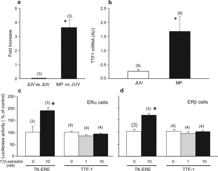
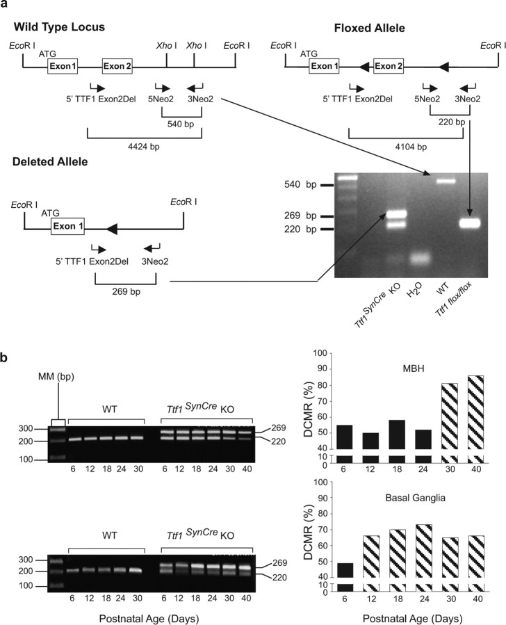
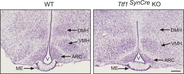
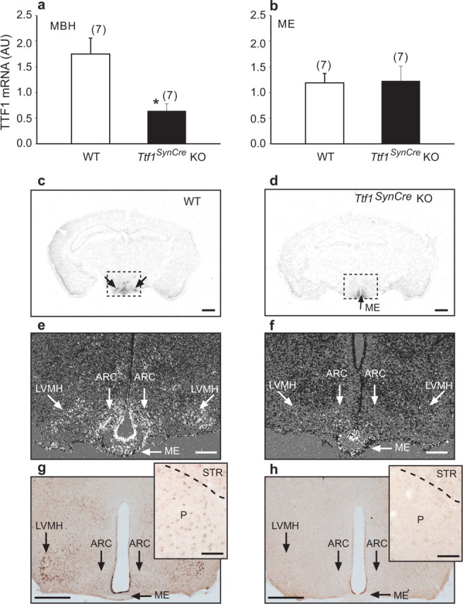
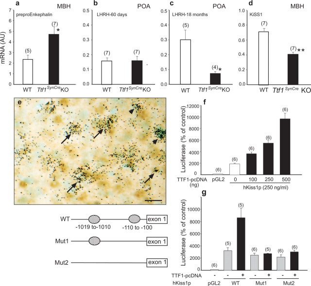
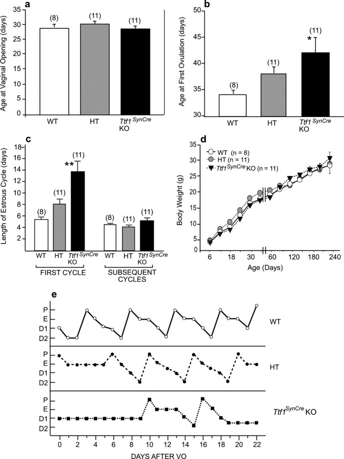
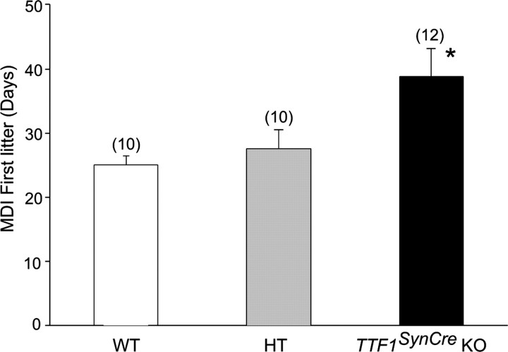
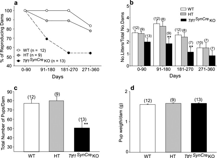
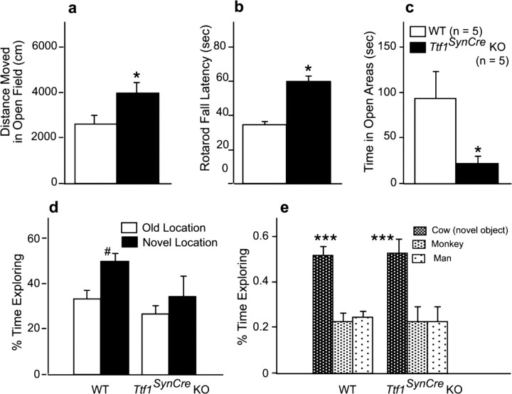
Thyroid transcription factor 1 (TTF1) [also known as Nkx2.1 (related to the NK-2 class of homeobox genes) and T/ebp (thyroid-specific enhancer-binding protein)], a homeodomain gene required for basal forebrain morphogenesis, remains expressed in the hypothalamus after birth, suggesting a role in neuroendocrine function. Here, we show an involvement of TTF1 in the control of mammalian puberty and adult reproductive function. Gene expression profiling of the nonhuman primate hypothalamus revealed that TTF1 expression increases at puberty. Mice in which the Ttf1 gene was ablated from differentiated neurons grew normally and had normal basal ganglia/hypothalamic morphology but exhibited delayed puberty, reduced reproductive capacity, and a short reproductive span. These defects were associated with reduced hypothalamic expression of genes required for sexual development and deregulation of a gene involved in restraining puberty. No extrapyramidal impairments associated with basal ganglia dysfunction were apparent. Thus, although TTF1 appears to fulfill only a morphogenic function in the ventral telencephalon, once this function is satisfied in the hypothalamus, TTF1 remains active as part of the transcriptional machinery controlling female sexual development.


Figures

Figure 1.
Figure 1.
TTF1 mRNA abundance increases in the female monkey hypothalamus at the time of puberty. a, Puberty-related increase in hypothalamic TTF1 mRNA content detected using human cDNA microarrays. Changes in mRNA content are expressed as fold increase over values present in prepubertal juvenile (JUV) animals. MP, Midpuberty. b, Verification of the array results by real-time PCR. *p < 0.05 versus JUV controls. AU, Arbitrary units. c, Lack of effect of 17β-estradiol-dependent stimulation of ERαs on TTF1 promoter activity measured in transcription activity assays using a luciferase reporter gene driven by the rat TTF1 gene 5′ flanking region. d, Lack of effect of ERβ stimulation by estradiol on TTF1 promoter activity. C6 cells stably expressing either ERα or ERβ were exposed to estradiol (1 or 10 nm), and luciferase activity was measured after 48 h of exposure to the steroid. Tk-ERE, Reporter plasmid in which luciferase expression is driven by estrogen-responsive elements fused to the thymidine kinase promoter. *p < 0.05 versus control wells not exposed to estradiol. In this and the following figures, bars represent group means, error bars represent SEM, and numbers in parentheses indicate the number of animals or independent observations per group.
Figure 2.
Figure 2.
PCR strategies used to genotype and calculate the DCMR in brain tissue. a, Three primers (horizontal arrows) were designed to distinguish among three possible TTF1 alleles. The wild-type allele is identified by a 540 bp PCR product amplified by primers 5Neo2 and 3Neo2. The intact floxed allele is detected by a 220 bp PCR product amplified by the same pair of primers. To estimate DCMR in different brain regions, the 5Neo2 and 3Neo2 primers were used in conjunction with a second forward primer (5′TTF1 Exon2Del). When paired with 3Neo2, this primer amplifies a 269 bp band that identifies a deleted Ttf1 allele. Although three exons have been described in the mouse genome (http://www.ensembl.org/Mus_musculus/transview?db=core&transcript=ENSMUST00000001536), we represent in the diagram only two exons, because the exact location of an exon upstream from the exon containing the main ATG (as shown in the figure) is uncertain (Lonigro et al., 1996; Oguchi and Kimura, 1998). It is also unclear whether just one or more upstream exons might be present. b, DCMR in MBH and basal ganglia. The PCR products shown on the left correspond to the floxed allele (220 bp band) and the recombined allele (269 bp band). The bar graphs on the right represent the DCMR calculated to occur in Ttf1SynCre KO mice at the indicated postnatal ages. Each bar is the mean of 2–3 animals. AU, Arbitrary units; MM, molecular marker.
Figure 3.
Figure 3.
Gross morphology of the MBH is similar in WT and Ttf1SynCre KO mice. The images depict the morphological aspect of 30 μm tissue sections stained with 0.1% thionin. DMH, Dorsomedial hypothalamic nucleus; VMH, ventromedial hypothalamic nucleus; ARC, arcuate nucleus; V, third ventricle. Scale bar, 400 μm.
Figure 4.
Figure 4.
SynCre-mediated deletion of the Ttf1 gene reduces TTF1 expression in hypothalamic neurons but not ependymoglial cells, as assessed by real-time PCR (n = 7 per group), in situ hybridization (n = 3 per group), and immunohistochemistry (n = 3 per group) analysis of the brains from 60-d-old female mice. a, b, TTF1 mRNA content decreases in the MBH (a) but not in the ME (b) of Ttf1SynCre KO mice compared with WT animals, as assessed by real-time PCR. c, d, Coronal sections of the brain at the level of the MBH illustrating the presence of TTF1 mRNA in the MBH of WT mice (c; double arrows) and the loss of expression in the MBH but not the ME (d; single arrow) in Ttf1SynCre KO mice, as determined by in situ hybridization using a 35S-UTP-labeled TTF1 cRNA. e, f, Dark-field, higher-magnification in situ hybridization images showing the presence of TTF1 mRNA in specific MBH subregions including the arcuate (ARC), lateroventral medial nuclei (LVMH), and the ME of WT mice (e) and the selective loss of TTF1 mRNA in the same nuclei of Ttf1SynCre KO mice, without a change of expression in the ME (f). g, Detection of TTF1 protein in the LVMH, ARC, and ME of WT mice. Inset, TTF1 protein is also detected in cells of the pallidum (P), although at lower levels. h, Selective loss of the protein in the LVMH and ARC, with persistent expression in the ME of Ttf1SynCre KO mice, as determined by immunohistochemistry. Inset, Loss of TTF1 in the pallidum of Ttf1SynCre KO mice. STR, Striatum. Scale bars: c, d, 400 μm; e, f, 200 μm; g, h, 400 μm; insets, 50 μm. *p < 0.05 versus WT.
Figure 5.
Figure 5.
Hypothalamic expression of TTF1 target genes is altered in 60-d-old Ttf1SynCre KO mice. a, Preproenkephalin mRNA levels are increased in the MBH. Normally, TTF1 represses preproenkephalin gene transcription. b, LHRH mRNA abundance in the POA is similar to WT mice at 60 d. c, However, the age-related increase in LHRH mRNA levels seen in WT mice is abolished in Ttf1SynCre KO animals. Normally, TTF1 transactivates the LHRH promoter. d, KiSS1 mRNA abundance in the MBH is decreased. e, KiSS1 neurons of the arcuate nucleus identified by in situ hybridization (black grains) also express TTF1 protein, identified by immunostaining (brown color). Examples of colocalization are denoted by arrows. Some KiSS1 mRNA-containing cells are TTF1 negative (arrowheads). Scale bar, 20 μm. f, TTF1 transactivates the KiSS1 promoter, as assessed by functional promoter assays using a luciferase reporter gene. g, Deletion of either a single proximal putative TTF1 recognition motif [located at −110 to −100 relative to the presumed transcription start site; mutation 1 (Mut1)] or both this motif and an additional site (located at −1019 to −1010; Mut2) in the 5′ flanking region of the hKiSS1 gene obliterates the transactivating effect of TTF1 on the KiSS1 promoter. *p < 0.05; ***p < 0.001.
Figure 6.
Figure 6.
Neuronal deletion of the Ttf1 gene delays the onset of female puberty. a–c, Although the age at vaginal opening occurred at similar ages in WT, HT, and Ttf1SynCre KO mice (a), the age at first ovulation was significantly delayed in Ttf1SynCre KO animals (b), and the first estrous cycle was longer in Ttf1SynCre KO than WT and HT mice (c). d, These changes were not secondary to differences in body weight gain. e, Representative estrous cycle profiles illustrating the delayed onset of normal cyclicity and the slight lengthening of subsequent estrous cycles in Ttf1SynCre KO mice compared with HT and WT mice. P, Proestrus; E, estrus; D1, diestrous day 1; D2, diestrous day 2; VO, vaginal opening. *p < 0.05, **p < 0.01 versus WT controls.
Figure 7.
Figure 7.
Mating–delivery interval for the first litter born to WT, HT, and Ttf1SynCre KO female mice. The KO mice delivered their first litter significantly later than both the HT and WT groups, indicating delayed first ovulation. *p < 0.05.
Figure 8.
Figure 8.
Reproductive capacity is diminished in Ttf1SynCre KO female mice. a, Fifty percent of Ttf1SynCre KO dams ceased delivering pups by 6–9 months of age, a time during which all WT females and 90% of HT mice were reproducing normally. b, The number of litters produced by each dam every 90 d significantly declined in Ttf1SynCre KO females by 3 months of age. c, Ttf1SynCre KO females produce fewer pups than WT or HT dams in a 1 year period. d, The body weight of the pups at birth was similar in all three experimental groups. **p < 0.01 versus WT controls. No., Number.
Figure 9.
Figure 9.
Functions directly or indirectly related to basal ganglia activity are not negatively impacted in Ttf1SynCre KO mice. a–e, Locomotor activity in the open field is increased (a), rotarod performance is increased (b), anxiety is increased as assessed by the elevated zero maze (c), and novel location recognition is reduced (d), but novel object recognition is not affected (e). The tests were performed on 14-month-old WT and Ttf1SynCre KO mice. n = 5 mice per genotype. *p < 0.05 versus WT; #p < 0.05 versus old location; ***p < 0.001 versus the two other objects.

References

    1. Alvarez-Bolado G, Rosenfeld MG, Swanson LW. Model of forebrain regionalization based on spatiotemporal patterns of POU-III homeobox gene expression, birthdates, and morphological features. J Comp Neurol. 1995;355:237–295. - PubMed
    1. Basso K, Margolin AA, Stolovitzky G, Klein U, Dalla-Favera R, Califano A. Reverse engineering of regulatory networks in human B cells. Nat Genet. 2005;37:382–390. - PubMed
    1. Bohinksi RJ, Di Lauro R, Whitsett JA. The lung-specific surfactant protein B gene promoter is a target for thyroid transcription factor 1 and hepatocyte nuclear factor 3, indicating common factors for organ-specific gene expression along the foregut axis. Mol Cell Biol. 1994;14:5671–5681. - PMC - PubMed
    1. Davidson EH, Rast JP, Oliveri P, Ransick A, Calestani C, Yuh CH, Minokawa T, Amore G, Hinman V, Arenas-Mena C, Otim O, Brown CT, Livi CB, Lee PY, Revilla R, Rust AG, Pan Z, Schilstra MJ, Clarke PJ, Arnone MI, et al. A genomic regulatory network for development. Science. 2002;295:1669–1678. - PubMed
    1. Davis AM, Seney ML, Stallings NR, Zhao L, Parker KL, Tobet SA. Loss of steroidogenic factor 1 alters cellular topography in the mouse ventromedial nucleus of the hypothalamus. J Neurobiol. 2004;60:424–436. - PubMed

Publication types

MeSH terms