Molecular interaction between projection neuron precursors and invading interneurons via stromal-derived factor 1 (CXCL12)/CXCR4 signaling in the cortical subventricular zone/intermediate zone
- PMID: 17182777
- PMCID: PMC6674999
- DOI: 10.1523/JNEUROSCI.4162-06.2006
Molecular interaction between projection neuron precursors and invading interneurons via stromal-derived factor 1 (CXCL12)/CXCR4 signaling in the cortical subventricular zone/intermediate zone
Abstract
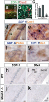
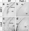
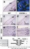
Most cortical interneurons are generated in the subpallial ganglionic eminences and migrate tangentially to their final destinations in the neocortex. Within the cortex, interneurons follow mainly stereotype routes in the subventricular zone/intermediate zone (SVZ/IZ) and in the marginal zone. It has been suggested that interactions between invading interneurons and locally generated projection neurons are implicated in the temporal and spatial regulation of the invasion process. However, so far experimental evidence for such interactions is lacking. We show here that the chemokine stromal-derived factor 1 (SDF-1; CXCL12) is expressed in the main invasion route for cortical interneurons in the SVZ/IZ. Most SDF-1-positive cells are proliferating and express the homeodomain transcription factors Cux1 and Cux2. Using MASH-1 mutant mice in concert with the interneuron marker DLX, we exclude that interneurons themselves produce the chemokine in an autocrine manner. We conclude that the SDF-1-expressing cell population represents the precursors of projection neurons during their transition and amplification in the SVZ/IZ. Using mice lacking the SDF-1 receptor CXCR4 or Pax6, we demonstrate that SDF-1 expression in the cortical SVZ/IZ is essential for recognition of this pathway by interneurons. These results represent the first evidence for a molecular interaction between precursors of projection neurons and invading interneurons during corticogenesis.
Figures


Similar articles
-
Molecules and mechanisms involved in the generation and migration of cortical interneurons.ASN Neuro. 2010 Mar 31;2(2):e00031. doi: 10.1042/AN20090053. ASN Neuro. 2010. PMID: 20360946 Free PMC article. Review.
-
CXCR4 regulates interneuron migration in the developing neocortex.J Neurosci. 2003 Jun 15;23(12):5123-30. doi: 10.1523/JNEUROSCI.23-12-05123.2003. J Neurosci. 2003. PMID: 12832536 Free PMC article.
-
Intermediate Progenitors Facilitate Intracortical Progression of Thalamocortical Axons and Interneurons through CXCL12 Chemokine Signaling.J Neurosci. 2015 Sep 23;35(38):13053-63. doi: 10.1523/JNEUROSCI.1488-15.2015. J Neurosci. 2015. PMID: 26400936 Free PMC article.
-
Dynamics of Cux2 expression suggests that an early pool of SVZ precursors is fated to become upper cortical layer neurons.Cereb Cortex. 2004 Dec;14(12):1408-20. doi: 10.1093/cercor/bhh102. Epub 2004 Jul 6. Cereb Cortex. 2004. PMID: 15238450
-
Control of tangential/non-radial migration of neurons in the developing cerebral cortex.Neurochem Int. 2007 Jul-Sep;51(2-4):121-31. doi: 10.1016/j.neuint.2007.05.006. Epub 2007 May 21. Neurochem Int. 2007. PMID: 17588709 Review.
Cited by
-
The Effect of Pro-Neurogenic Gene Expression on Adult Subventricular Zone Precursor Cell Recruitment and Fate Determination After Excitotoxic Brain Injury.J Stem Cells Regen Med. 2016 May 30;12(1):25-35. doi: 10.46582/jsrm.1201005. eCollection 2016. J Stem Cells Regen Med. 2016. PMID: 27397999 Free PMC article.
-
Molecules and mechanisms involved in the generation and migration of cortical interneurons.ASN Neuro. 2010 Mar 31;2(2):e00031. doi: 10.1042/AN20090053. ASN Neuro. 2010. PMID: 20360946 Free PMC article. Review.
-
Intermediate progenitors and Tbr2 in cortical development.J Anat. 2019 Sep;235(3):616-625. doi: 10.1111/joa.12939. Epub 2019 Jan 24. J Anat. 2019. PMID: 30677129 Free PMC article. Review.
-
Regional distribution of cortical interneurons and development of inhibitory tone are regulated by Cxcl12/Cxcr4 signaling.J Neurosci. 2008 Jan 30;28(5):1085-98. doi: 10.1523/JNEUROSCI.4602-07.2008. J Neurosci. 2008. PMID: 18234887 Free PMC article.
-
Timing of cortical interneuron migration is influenced by the cortical hem.Cereb Cortex. 2011 Apr;21(4):748-55. doi: 10.1093/cercor/bhq142. Epub 2010 Aug 16. Cereb Cortex. 2011. PMID: 20713502 Free PMC article.
References
-
- Casarosa S, Fode C, Guillemot F. Mash1 regulates neurogenesis in the ventral telencephalon. Development. 1999;126:525–534. - PubMed
-
- Daniel D, Rossel M, Seki T, König N. Stromal cell-derived factor-1 (SDF-1) expression in embryonic mouse cerebral cortex starts in the intermediate zone close to the pallial-subpallial boundary and extends progressively towards the cortical hem. Gene Expr Patterns. 2005;5:317–322. - PubMed
-
- Favor J, Peters H, Hermann T, Schmahl W, Chatterjee B, Neuhauser-Klaus A, Sandulache R. Molecular characterization of Pax6(2Neu) through Pax6(10Neu): an extension of the Pax6 allelic series and the identification of two possible hypomorph alleles in the mouse Mus musculus. Genetics. 2001;159:1689–1700. - PMC - PubMed
-
- Guillemot F, Lo LC, Johnson JE, Auerbach A, Anderson DJ, Joyner AL. Mammalian achaete-scute homolog 1 is required for the early development of olfactory and autonomic neurons. Cell. 1993;75:463–476. - PubMed
Publication types
MeSH terms
Substances
LinkOut - more resources
Full Text Sources
Molecular Biology Databases
