

Np95 is implicated in pericentromeric heterochromatin replication and in major satellite silencing
- PMID: 17182844
- PMCID: PMC1805105
- DOI: 10.1091/mbc.e06-09-0874
Np95 is implicated in pericentromeric heterochromatin replication and in major satellite silencing
Abstract
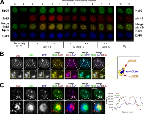
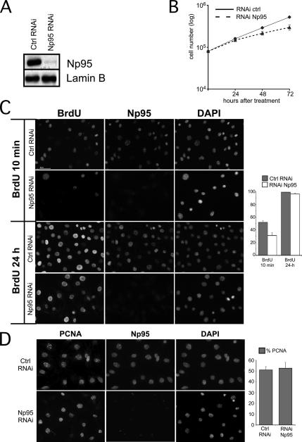
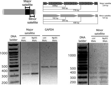
Heterochromatin plays an important role in transcriptional repression, for the correct segregation of chromosomes and in the maintenance of genome stability. Pericentric heterochromatin (PH) replication and formation have been proposed to occur in the pericentric heterochromatin duplication body (pHDB). A central question is how the underacetylated state of heterochromatic histone H4 tail is established and controlled, because it is a key event during PH replication and is essential to maintain the compacted and silenced state of these regions. Np95 is a cell cycle regulated and is a nuclear histone-binding protein that also recruits HDAC-1 to target promoters. It is essential for S phase and for embryonic formation and is implicated in chromosome stability. Here we show that Np95 is part of the pHDB, and its functional ablation causes a strong reduction in PH replication. Depletion of Np95 also causes a hyperacetylation of lysines 8, 12, and 16 of heterochromatin histone H4 and an increase of pericentromeric major satellite transcription, whose RNAs are key players for heterochromatin formation. We propose that Np95 is a new relevant protein involved in heterochromatin replication and formation.
Figures


Similar articles
-
The PHD domain of Np95 (mUHRF1) is involved in large-scale reorganization of pericentromeric heterochromatin.Mol Biol Cell. 2008 Aug;19(8):3554-63. doi: 10.1091/mbc.e07-10-1059. Epub 2008 May 28. Mol Biol Cell. 2008. PMID: 18508923 Free PMC article.
-
Dissecting the role of H3K64me3 in mouse pericentromeric heterochromatin.Nat Commun. 2013;4:2233. doi: 10.1038/ncomms3233. Nat Commun. 2013. PMID: 23903902
-
Silencing markers are retained on pericentric heterochromatin during murine primordial germ cell development.Epigenetics Chromatin. 2017 Mar 11;10:11. doi: 10.1186/s13072-017-0119-3. eCollection 2017. Epigenetics Chromatin. 2017. PMID: 28293300 Free PMC article.
-
Pericentromeric satellite RNAs as flexible protein partners in the regulation of nuclear structure.Wiley Interdiscip Rev RNA. 2024 Jul-Aug;15(4):e1868. doi: 10.1002/wrna.1868. Wiley Interdiscip Rev RNA. 2024. PMID: 38973000 Review.
-
The role of heterochromatin in centromere function.Philos Trans R Soc Lond B Biol Sci. 2005 Mar 29;360(1455):569-79. doi: 10.1098/rstb.2004.1611. Philos Trans R Soc Lond B Biol Sci. 2005. PMID: 15905142 Free PMC article. Review.
Cited by
-
Maintenance of Epigenetic Information.Cold Spring Harb Perspect Biol. 2016 May 2;8(5):a019372. doi: 10.1101/cshperspect.a019372. Cold Spring Harb Perspect Biol. 2016. PMID: 27141050 Free PMC article. Review.
-
UHRF1 is a genome caretaker that facilitates the DNA damage response to gamma-irradiation.Genome Integr. 2010 Jun 8;1(1):7. doi: 10.1186/2041-9414-1-7. Genome Integr. 2010. PMID: 20678257 Free PMC article.
-
UHRF1 is associated with tumor recurrence in non-muscle-invasive bladder cancer.Med Oncol. 2012 Jun;29(2):842-7. doi: 10.1007/s12032-011-9983-z. Epub 2011 May 25. Med Oncol. 2012. PMID: 21611839
-
Three SRA-domain methylcytosine-binding proteins cooperate to maintain global CpG methylation and epigenetic silencing in Arabidopsis.PLoS Genet. 2008 Aug 15;4(8):e1000156. doi: 10.1371/journal.pgen.1000156. PLoS Genet. 2008. PMID: 18704160 Free PMC article.
-
UHRF1 in endothelial cells is essential for angiogenesis and associated with the activation of pro-angiogenic signaling pathways and expression of endothelial genes.Angiogenesis. 2025 Aug 5;28(4):42. doi: 10.1007/s10456-025-09998-0. Angiogenesis. 2025. PMID: 40764462
References
-
- Agalioti T., Chen G., Thanos D. Deciphering the transcriptional histone acetylation code for a human gene. Cell. 2002;111:381–392. - PubMed
-
- Bailis J. M., Forsburg S. L. It's all in the timing: linking S phase to chromatin structure and chromosome dynamics. Cell Cycle. 2003;2:303–306. - PubMed
-
- Bernard P., Maure J. F., Partridge J. F., Genier S., Javerzat J. P., Allshire R. C. Requirement of heterochromatin for cohesion at centromeres. Science. 2001;294:2539–2542. - PubMed
Publication types
MeSH terms
Substances
LinkOut - more resources
Full Text Sources
Other Literature Sources
Molecular Biology Databases
Miscellaneous
