. 2007 Mar;18(3):850-63.
doi: 10.1091/mbc.e06-07-0587. Epub 2006 Dec 20.

Mad2-independent spindle assembly checkpoint activation and controlled metaphase-anaphase transition in Drosophila S2 cells

Affiliations

Mad2-independent spindle assembly checkpoint activation and controlled metaphase-anaphase transition in Drosophila S2 cells

Bernardo Orr et al. Mol Biol Cell. 2007 Mar.

Abstract

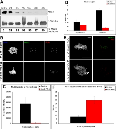
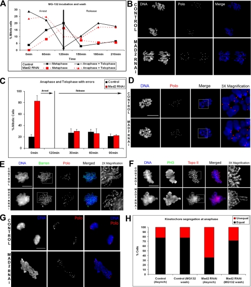
The spindle assembly checkpoint is essential to maintain genomic stability during cell division. We analyzed the role of the putative Drosophila Mad2 homologue in the spindle assembly checkpoint and mitotic progression. Depletion of Mad2 by RNAi from S2 cells shows that it is essential to prevent mitotic exit after spindle damage, demonstrating its conserved role. Mad2-depleted cells also show accelerated transit through prometaphase and premature sister chromatid separation, fail to form metaphases, and exit mitosis soon after nuclear envelope breakdown with extensive chromatin bridges that result in severe aneuploidy. Interestingly, preventing Mad2-depleted cells from exiting mitosis by a checkpoint-independent arrest allows congression of normally condensed chromosomes. More importantly, a transient mitotic arrest is sufficient for Mad2-depleted cells to exit mitosis with normal patterns of chromosome segregation, suggesting that all the associated phenotypes result from a highly accelerated exit from mitosis. Surprisingly, if Mad2-depleted cells are blocked transiently in mitosis and then released into a media containing a microtubule poison, they arrest with high levels of kinetochore-associated BubR1, properly localized cohesin complex and fail to exit mitosis revealing normal spindle assembly checkpoint activity. This behavior is specific for Mad2 because BubR1-depleted cells fail to arrest in mitosis under these experimental conditions. Taken together our results strongly suggest that Mad2 is exclusively required to delay progression through early stages of prometaphase so that cells have time to fully engage the spindle assembly checkpoint, allowing a controlled metaphase-anaphase transition and normal patterns of chromosome segregation.


Figures

Figure 1.
Figure 1.
Depletion of Mad2 by RNAi in S2 cells, (A) Western blot analysis shows Mad2 depletion at different times after addition of Mad2 dsRNA. Below α-tubulin was used as a loading control. At 72 h Mad2 depletion is more than 90%. (B) Immunolocalization of Mad2 RNAi in control and dsRNAi-treated cells at 72 h shows DNA (blue), Mad2 (green), and Polo (red). After 72 h RNAi treated cells do not contain Mad2 at their kinetochores, whereas in control cells Mad2 staining is clearly visible. (C) Quantification of Mad2 levels in control and RNAi-treated prometaphase cells. At least 15 cells were analyzed in each case. (D) Control and RNAi-treated cells were incubated with 30 μM colchicine, and the mitotic index was recorded. Cells lacking Mad2 fail to accumulate in mitosis. (E) These cells were also immunostained to reveal the cohesin subunit DRAD21 to determine whether they exit mitosis prematurely. Note that there is a significant increase in the frequency of DRAD21 negative cells after colchicine incubation (mitotic population only). (F) Immunolocalization of the cohesin subunit DRAD21 in control and Mad2 RNAi-treated cells at 72 h followed by colchicine incubation to depolymerize microtubules revealed a threefold increase in PSCS. DNA (blue), DRAD21 (green), and Polo (red) are shown. Note that after 72 h RNAi treatment most prometaphase cells do not contain centromere-associated DRAD21, whereas in control cells the staining is clearly visible. Bar, (B and E) 5 μm.
Figure 2.
Figure 2.
Mitotic progression after depletion of Mad2. (A–C) Cells were treated with dsRNAi for 72 h before fixation and immunostaining to reveal DNA (blue), phospho-histone H3 (green), and Polo (red). (A) Quantification of mitotic progression revealed that cells lacking Mad2 show a significant reduction in the frequency of metaphases among mitotic cells. (B) During anaphase and telophase, cells show extensive PH3-positive chromatin bridges. Bar, 5 μm. (C) Quantification shows that after depletion of Mad2 most cells show chromatin bridges during anaphase or telophase. Analysis of DNA content in control or (D) Mad2-depleted (E) cells at different times. Note that after 72 h Mad2-depleted cells fail to show a well-defined 4N peak, which becomes very broad, indicating extensive aneuploidy. (F) Quantification of kinetochore numbers in control and Mad2-depleted cells at prometaphase. Cells containing between 20 and 26 kinetochores were considered to be normal. C, control; M, Mad2 RNAi; n = 30 cells at each time point. Note the progressive shift in ploidy along time.
Figure 3.
Figure 3.
Chromosome congression after checkpoint-independent mitotic arrest. (A and C) Cells were incubated with the proteasome inhibitor MG132 for 90 min before fixation and immunostained to reveal DNA (blue), kinetochores (green), and tubulin (red). (B) Quantification shows that when Mad2-depleted cells are incubated with MG132, the frequency of metaphases is similar to controls. (C) To determine the frequency of proper attachment of kinetochore pairs, control and Mad2-depleted cells were treated with Ca2+ to reveal only kinetochore bundles, fixed, and immunostained as before. Kinetochore pairing was determined by following individual chromosomes through different optical layers. (D) Quantification of different kinetochore attachments in control and Mad2-depleted cells after incubation with MG132 to prevent exit from mitosis. Note that in the absence of Mad2 most kinetochore pairs appear to attach properly, and only ∼5% of kinetochore pairs could not be assigned to any particular type of attachment (n = 300 kinetochore pairs). (E) Interkinetochore distance measured in metaphase cells arrested with 20 μM MG132 and in asynchronous cells in culture. When treated with MG132, both control and Mad2-depleted cells are under tension, and the interkinetochore distance is increased. (F) Types of attachment used for quantification shown in D; CID (green) and tubulin (red). Bar, 5 μm.
Figure 4.
Figure 4.
Reversion of the phenotypes caused by depletion of Mad2. To determine whether the Mad2-associated phenotypes could be reverted by providing additional time in prometaphase/metaphase, S2 cells previously treated with dsRNA against Mad2 for 72 h were incubated for 120 min with a low dose of MG132 (2 μM) and then released from the block by performing a threefold dilution on the cell culture media with fresh media. Samples were then collected every 30 min for immunofluorescence analysis. (A) Quantitative analysis of mitotic progression in control and Mad2-depleted cells before and after reversion. From 0 to 120 min both control and Mad2-depleted cells show a strong decrease in anaphases and telophases and a marked increase in the number of metaphases. After washing MG132 (120 min), the number of metaphases begins to decrease, and the number of anaphase and telophase figures increases. (B) Immunofluorescence shows that 180 min after washing the drug, most anaphases in Mad2-depleted cells do not show chromatin bridges. (C) Quantitative analysis of the anaphase and telophase figures before and after the MG132 treatment shows a complete reversal of the phenotype. (D) Normal chromosomes can be obtained in Mad2-depleted cells if cells are incubated with MG132 to prevent exit from mitosis. Before fixation cells were also treated with colchicine to depolymerize microtubules and induce a better chromosome spread. DNA is shown in blue and the kinetochore marker Polo in red. (E and F) Immunolocalization of essential components for the organization and structure of mitotic chromosomes. Barren and Topoisomerase II in control and Mad2-depleted prometaphases are properly localized to a well-defined sister chromatid axis, in asynchronous cell culture. (G) Analysis of kinetochore segregation in control and Mad2-depleted cells after 72 h of RNAi incubation was carried out on anaphase cells after immunostaining for DNA with anti-Polo antibody. (H) Quantification of chromosome segregation at anaphase shows that in control cells almost 80% of cells segregate kinetochores equally. After depletion of Mad2 nearly 70% of cells also show unequal kinetochore segregation. Providing extra time in prometaphase/metaphase to control cells by incubation in MG132 and then washing out the drug does not alter the frequency of unequal kinetochore segregation. However, a similar treatment in Mad2-depleted cells reduces to almost control levels the frequency of unequal kinetochore segregation. Bar, (D–G) 5 μm. A total of 40 cells were analyzed.
Figure 5.
Figure 5.
Maintenance of SAC activity in Mad2-depleted cells but not in cells lacking BubR1. To determine the status of the SAC Mad2-depleted cells were subjected to a transient mitotic arrest by incubation in MG132 for 2 h and then released either into normal media or media containing colchicine and the frequency of prometaphase and metaphases (A) or sister chromatid separation (B) determined by DRad21 immunostaining (mitotic population only). Note that Mad2-depleted cells when released into normal media rapidly exit mitosis, however, if released into media containing colchicine, accumulate in a prometaphase-like state similarly to control cells. In contrast, BubR1-depleted cells fail to accumulate at prometaphase when released into colchicine, suggesting an inactive SAC. (C) Quantification of Mad2 kinetochore signal by mean pixel intensity in cells treated with colchicine or MG132 and in cells released from the MG132 arrest into colchicine (180 min). Although almost no Mad2 was detected in any sample treated with Mad2 RNAi, in control cells there is a strong Mad2 accumulation in both cells treated with colchicine and those that were transiently arrested in mitosis with MG132 and then released into colchicine.
Figure 6.
Figure 6.
Maintenance of centromere-associated DRad21 in Mad2-depleted cells. All cells were collected, fixed, and stained to reveal DNA (blue), Polo (green), and the cohesin subunit DRad21 (red). Cells from control and cells treated with dsRNA against Mad2 or BubR1 were analyzed without colchicine (A), after colchicine treatment (B), and in cells transiently arrested with MG132 (C) and then released into media containing colchicine (180 min). Note that Mad2-depleted cells released into this media are unable to degrade cohesion, whereas BubR1-depleted cells fail to maintain sister chromatid cohesion in the presence of colchicine, suggesting an inactive SAC response (see also higher magnification inserts). Bar, 5 μm.
Figure 7.
Figure 7.
Immunolocalization of BubR1 in cells lacking Mad2. Control and Mad2-depleted cells where either (A) incubated in MG132 for 2 h or (B) released from the arrest into media containing colchicine (180 min). These cells were fixed and stained to reveal DNA (blue), BubR1 (green), and CID (red). (C) Quantification of the BubR1 kinetochore signal in control and Mad2-depleted cells shows an identical twofold increase in cells released into the colchicine. Note that cells lacking Mad2, similarly to control cells, accumulate BubR1 at kinetochores strongly when released into colchicine. Bar, 5 μm.

Similar articles

Cited by

References

    1. Basu J., Bousbaa H., Logarinho E., Li Z., Williams B. C., Lopes C., Sunkel C. E., Goldberg M. L. Mutations in the essential spindle checkpoint gene bub1 cause chromosome missegregation and fail to block apoptosis in Drosophila. J. Cell Biol. 1999;146:13–28. - PMC - PubMed
    1. Bharadwaj R., Yu H. The spindle checkpoint, aneuploidy, and cancer. Oncogene. 2004;23:2016–2027. - PubMed
    1. Bhat M. A., Philp A. V., Glover D. M., Bellen H. J. Chromatid segregation at anaphase requires the barren product, a novel chromosome-associated protein that interacts with Topoisomerase II. Cell. 1996;87:1103–1114. - PubMed
    1. Blower M. D., Karpen G. H. The role of Drosophila CID in kinetochore formation, cell-cycle progression and heterochromatin interactions. Nat. Cell Biol. 2001;3:730–739. - PMC - PubMed
    1. Buffin E., Lefebvre C., Huang J., Gagou M. E., Karess R. E. Recruitment of Mad2 to the kinetochore requires the Rod/Zw10 complex. Curr. Biol. 2005;15:856–861. - PubMed

Publication types

LinkOut - more resources