. 2006 Dec 15;20(24):3475-86.
doi: 10.1101/gad.403406.

The establishment of neuronal properties is controlled by Sox4 and Sox11

Affiliations

The establishment of neuronal properties is controlled by Sox4 and Sox11

Maria Bergsland et al. Genes Dev. .

Abstract

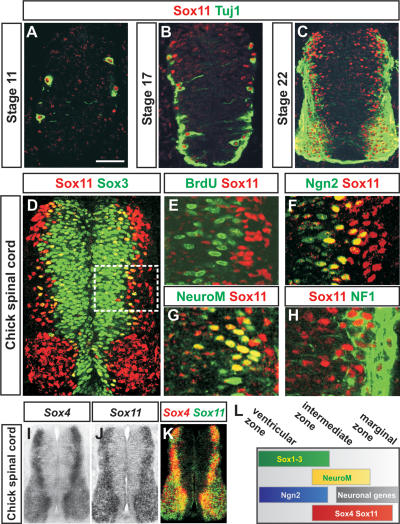
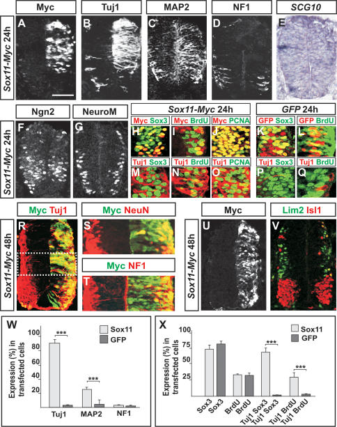
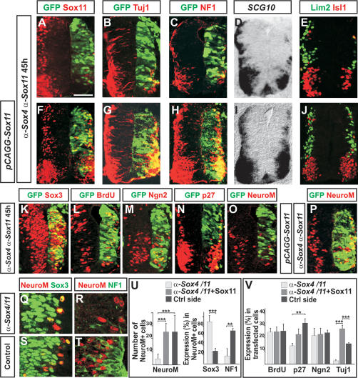
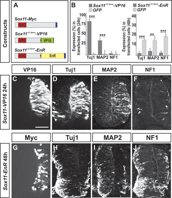
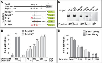
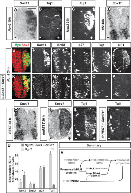
The progression of neurogenesis relies on proneural basic helix-loop-helix (bHLH) transcription factors. These factors operate in undifferentiated neural stem cells and induce cell cycle exit and the initiation of a neurogenic program. However, the transient expression of proneural bHLH proteins in neural progenitors indicates that expression of neuronal traits must rely on previously unexplored mechanisms operating downstream from proneural bHLH proteins. Here we show that the HMG-box transcription factors Sox4 and Sox11 are of critical importance, downstream from proneural bHLH proteins, for the establishment of pan-neuronal protein expression. Examination of a neuronal gene promoter reveals that Sox4 and Sox11 exert their functions as transcriptional activators. Interestingly, the capacity of Sox4 and Sox11 to induce the expression of neuronal traits is independent of mechanisms regulating the exit of neural progenitors from the cell cycle. The transcriptional repressor protein REST/NRSF has been demonstrated to block neuronal gene expression in undifferentiated neural cells. We now show that REST/NRSF restricts the expression of Sox4 and Sox11, explaining how REST/NRSF can prevent precocious expression of neuronal proteins. Together, these findings demonstrate a central regulatory role of Sox4 and Sox11 during neuronal maturation and mechanistically separate cell cycle withdrawal from the establishment of neuronal properties.


Figures

Figure 1.
Figure 1.
Expression of Sox4 and Sox11 in the developing chick spinal cord. (AC) Expression of Sox11 and Tuj1 protein in spinal cords from HH stages 11 to 22. (DH) Expression of Sox11 in comparison with Sox3+ (D), BrdU+ (E), Ngn2+ (F), NeuroM/Math3+ (G), and NF1+ (H) cells. The white box in D indicates the analyzed area in EH. (IK) Expression of Sox4 (I) and Sox11 (J) mRNA. (K) The image represents an overlay of images I and J. (L) The summary figure indicates the distribution of Sox1–3, Ngn2, NeuroM/Math3, Sox4, and Sox11 and neuronal proteins in the developing neural tube. Bars: A,B, 40 μm; C,D, 60 μm;, EH), 15 μm; IK, 100 μm.
Figure 2.
Figure 2.
Sox4 and Sox11 can induce the expression of neuronal proteins. (AE,W) Misexpression of Sox11 for 24 h (A) increased the number of Tuj1+ (B,W) and MAP2+ cells (C,W), whereas no change in the expression levels of NF1 (D,W) or SCG10 (E) could be detected (n ≥ 6 embryos; [*] p < 0.05; [**] p < 0.01; [***] p < 0.001). (F,G) Electroporation of Sox11 did not alter the expression of the proneural bHLH protein Ngn2 (F) or the bHLH protein NeuroM/Math3 (G). (HL,X) Many Sox11-transfected cells expressed the progenitor marker Sox3 (H,X), and the number of Sox11-transfected cells that incorporated BrdU (I,X) or expressed the cell cycle marker PCNA (J) was comparable with cells electroporated with a GFP control vector (K,L,X) (n ≥ 6 embryos; [***] p < 0.001). (MQ,X) Most of the ectopic Tuj1+ cells coexpressed Sox3 (M,X), and several of these also incorporated BrdU (N,X) and expressed PCNA (O). No or very few Tuj1+/Sox3+-incorporating cells (P,X) or Tuj1+/BrdU-incorporating cells (Q,X) could be detected in embryos electroporated with a GFP control vector. (RT) Forty-eight hours after Sox11 transfection (R), many of the electroporated cells were post-mitotic and also expressed NeuN (S) and NF1 (T). (U,V) Misexpression of Sox11 for 48 h (U) did not disrupt the expression pattern of the subtype-specific neuronal markers Lim2 and Isl1 (V). Results are represented as mean ± SD. Bars: AE,HQ,R,U,V, 60 μm; F,G, 75 μm; S,T, 40 μm.
Figure 3.
Figure 3.
Decreased levels of Sox4 and Sox11 block the establishment of a neuronal phenotype. (AE,V) Forty-five hours after siRNA transfection, the expression of Sox11 (A), Tuj1 (B,V), NF1 (C), SCG10 (D), and Lim2 and Isl1 (E) was considerably reduced compared with the nontransfected control side (n ≥ 6 embryos; [**] p < 0.01; [***] p < 0.001). (FJ,V) The generation of Tuj1+ (G,V), NF1+ (H), SCG10+ (I), and Lim2+ and Isl1+ (J) neurons could be rescued in α-Sox4- and α-Sox11-electroporated neural tubes by the presence of Sox11 expression vectors (F). (KM,V) Transfection of α-Sox4 and α-Sox11 siRNAs did not alter the expression of Sox3 (K), the rate of BrdU incorporation (L,V), or the expression of Ngn2 (M,V). (NP,U,V) α-Sox4 and α-Sox11 siRNAs reduced the number of p27Kip1+ and NeuroM/Math3+ cells (N,O,U,V), a reduction that could be rescued by the presence of cotransfected Sox11 expression vectors (P,U,V) (n ≥ 6 embryos; [**] p < 0.01; [***] p < 0.001). (QU) In α-Sox4 and α-Sox11 siRNA-transfected neural tubes, most NeuroM/Math3+ cells coexpressed the progenitor marker Sox3 (Q,U) but had failed to up-regulate NF1 expression (R,U). In contrast, in the nonelectroporated control side, 27% of the NeuroM/Math3+ cells coexpressed Sox3 (S,U) and >70% coexpressed NF1 (T,U). Results are represented as mean ± SEM. Bars: AJ, 60 μm; KP, 90 μm; QT, 120 μm.
Figure 4.
Figure 4.
Active repression of Sox11 target genes prevents the expression of pan-neuronal properties. (A) The constructs used were as follows: The C-terminal part of Sox11 was replaced with the transactivation domain of VP16 (Sox11ΔC-term–VP16), or with the repressor domain of the Drosophila Engrailed gene (Sox11ΔC-term–EnR). (BF) Sox11ΔC-term–VP16 (C) behaved like full-length Sox11 and induced the expression of Tuj1 (B,D) and MAP2 (B,E) but not that of NF1 (B,F). (B,GJ) Forced expression of Sox11ΔC-term–EnR (G) prevented neuronal protein expression, including Tuj1 (B,H), MAP2 (B,I), and NF1 (B,J) (n ≥ 6 embryos; [**] p < 0.01; [***] p < 0.001). Results are represented as mean ± SD. Bars: CF, 60 μm; GJ, 80 μm.
Figure 5.
Figure 5.
Sox4 and Sox11 interact with promoter elements of the neuronal gene Tubb3. (A) Tubb3 promoter constructs used in transactivation studies. S1–S3 indicate potential Sox4- and Sox11-binding sites, S1 (TTCTATTGTCCCC), S2, and S3 (CCGCATTGTGCGG). X marked in red indicates mutated sites. (B) Transactivation of the Tubb3166-LacZ and Tubb3622-LacZ reporters by Sox4 or Sox11. (C) Recombinant Sox4 and Sox11 proteins were able to bind to S1, S2, and S3 (S23) in a DNA-binding gel shift assay. No binding could be detected when these sites were mutated: S1mut (TTCTCCCGTCCCC) and S23mut (CCGCGGGGTGCGG). (D) The ability of Sox4 and Sox11 to transactivate the Tubb3166-LacZ reporter construct was reduced more than sixfold when S1 and S23 were mutated (S1M and S23M). The error bars in B and D indicate the standard deviation (SD) of three independent transfections. Each experiment was repeated six times. (**) p < 0.01; (***) p < 0.001.
Figure 6.
Figure 6.
The expression of Sox4 and Sox11 is controlled by REST and proneural proteins. (AC) Electroporation of Ngn2 induced high levels of ectopic Sox11 expression already 10 h after transfection (A), whereas the expression of Tuj1 first could be readily detected 24 h after electroporation (B,C). (D) Electroporation of Id2 for 48 h reduced the expression of Sox11. (EJ,U) Overexpression of Ngn2 for 24 h promoted cells to suppress progenitor characters (E,G,U) and up-regulate the expression of Sox11 (F), p27Kip1 (H,U), and neuronal proteins (I,J,U) (n ≥ 6 embryos; [*] p < 0.05; [***] p < 0.001). (KP,U) Cells cotransfected with Ngn2 and α-Sox4 and α-Sox11 siRNAs for 24 h expressed reduced levels of Sox3 (K,U) and had exited the cell cycle (M,U) and up-regulated p27Kip1 (N,U). Cotransfected cells did not up-regulate the expression of Sox11 (L) or neuronal markers (O,P,U). (QT) Misexpression of REST for 48 h decreased the expression of Sox11 (Q) (48% ± 11% reduction; [***] p < 0.001), whereas a dominant-negative version of REST, dnREST, 20 h after electroporation had induced high levels of ectopic Sox11 (R) and Tuj1 expression (S). (T) dnREST-transfected cells could not up-regulate the expression of Tuj1 when the expression of Sox4 and Sox11 was prevented by siRNAs. Results are represented as mean ± SD. Bars: AC,RT, 60 μm; EP, 80 μm; D,Q, 50 μm. (V) Proposed molecular network regulating the establishment of pan-neuronal properties. Proneural bHLH proteins drive the initial steps of neurogenesis and direct the exit of neural cells from the division cycle. Proneural proteins also induce the expression of Sox4 and Sox11, which in turn activate neuronal gene expression. According to this model, proneural proteins induce the expression of an additional genetic program (designated X) that together with Sox4 and Sox11 activates a complete neuronal phenotype in differentiating neurons. REST/NRSF prevents precocious expression of neuronal proteins in undifferentiated neural cells both by a direct repression of neuronal genes and by restricting the expression of Sox4 and Sox11 to neural cells that have exited the cell cycle.

References

    1. Aarnisalo, P., Kim, C.H., Lee, J.W., Perlmann, T. Defining requirements for heterodimerization between the retinoid X receptor and the orphan nuclear receptor Nurr1. J. Biol. Chem. 2002;277:35118–35123. - PubMed
    1. Berk, A.J., Boyer, T.G., Kapanidis, A.N., Ebright, R.H., Kobayashi, N.N., Horn, P.J., Sullivan, S.M., Koop, R., Surby, M.A., Triezenberg, S.J. Mechanisms of viral activators. Cold Spring Harb. Symp. Quant. Biol. 1998;63:243–252. - PubMed
    1. Bertrand, N., Castro, D.S., Guillemot, F. Proneural genes and the specification of neural cell types. Nat. Rev. Neurosci. 2002;3:517–530. - PubMed
    1. Briscoe, J., Pierani, A., Jessell, T.M., Ericson, J. A homeodomain code specifies progenitor cell identity and neuronal fate in the ventral neural tube. Cell. 2000;101:435–445. - PubMed
    1. Bylund, M., Andersson, E., Novitch, B.G., Muhr, J. Vertebrate neurogenesis is counteracted by Sox1-3 activity. Nat. Neurosci. 2003;6:1162–1168. - PubMed

Publication types

Substances

LinkOut - more resources