. 2007 Feb 20;500(6):1007-33.
doi: 10.1002/cne.21229.

Chemokine receptor expression by neural progenitor cells in neurogenic regions of mouse brain

Affiliations

Chemokine receptor expression by neural progenitor cells in neurogenic regions of mouse brain

Phuong B Tran et al. J Comp Neurol. .

Abstract

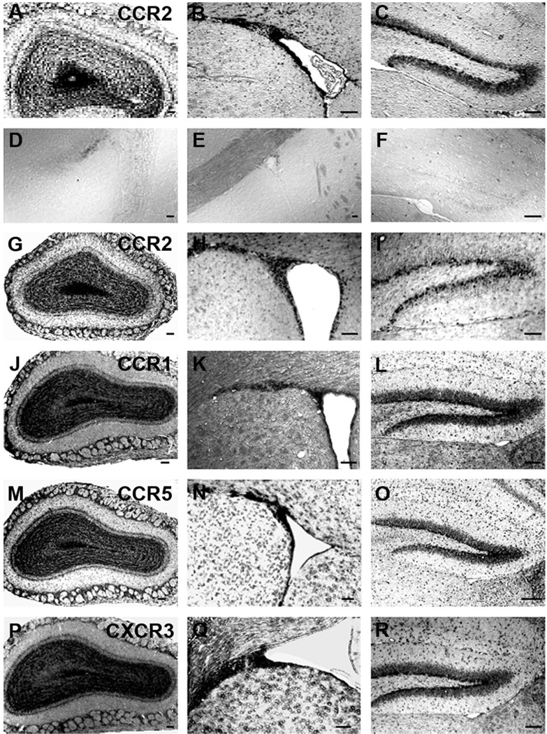
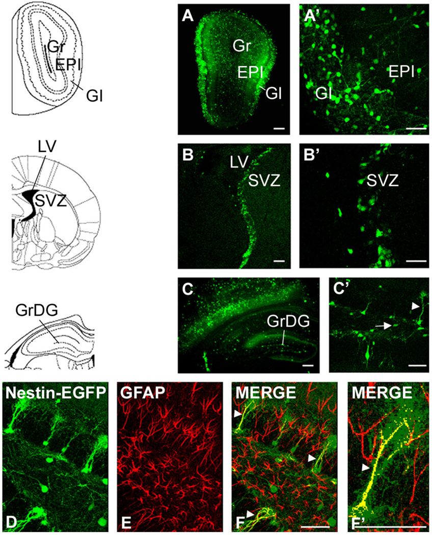
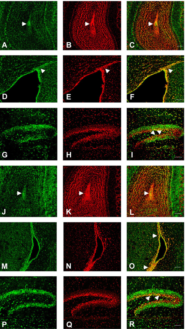
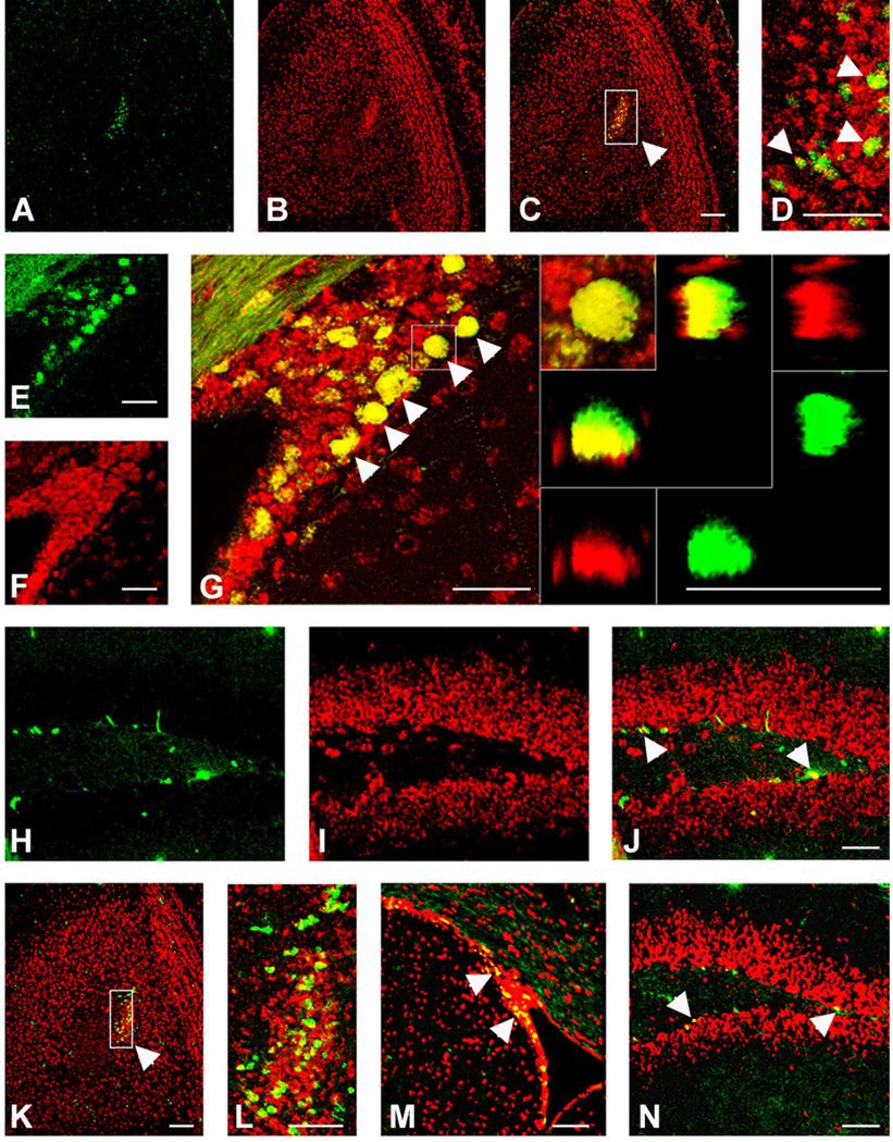
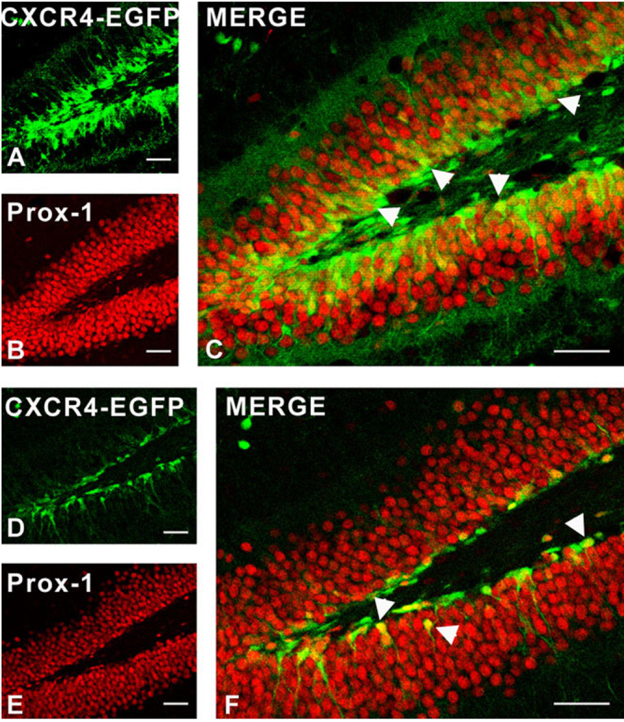
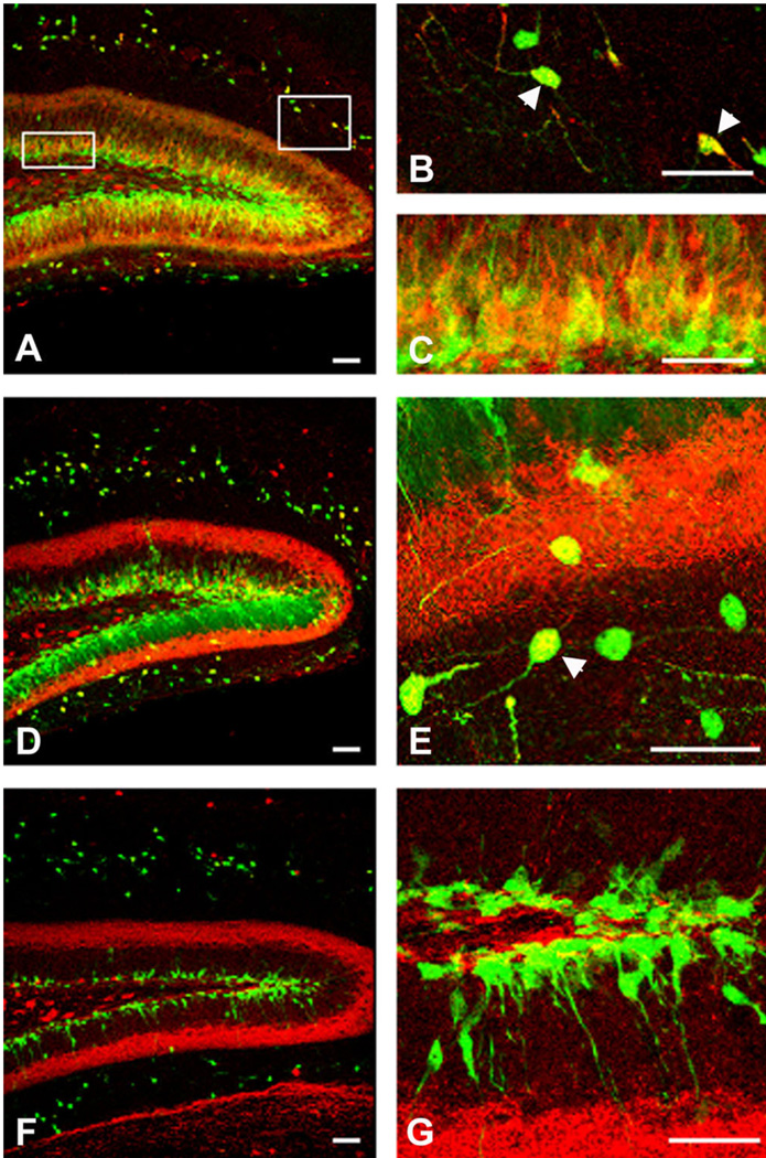
We previously demonstrated that chemokine receptors are expressed by neural progenitors grown as cultured neurospheres. To examine the significance of these findings for neural progenitor function in vivo, we investigated whether chemokine receptors were expressed by cells having the characteristics of neural progenitors in neurogenic regions of the postnatal brain. Using in situ hybridization we demonstrated the expression of CCR1, CCR2, CCR5, CXCR3, and CXCR4 chemokine receptors by cells in the dentate gyrus (DG), subventricular zone of the lateral ventricle, and olfactory bulb. The pattern of expression for all of these receptors was similar, including regions where neural progenitors normally reside. In addition, we attempted to colocalize chemokine receptors with markers for neural progenitors. In order to do this we used nestin-EGFP and TLX-LacZ transgenic mice, as well as labeling for Ki67, a marker for dividing cells. In all three areas of the brain we demonstrated colocalization of chemokine receptors with these three markers in populations of cells. Expression of chemokine receptors by neural progenitors was further confirmed using CXCR4-EGFP BAC transgenic mice. Expression of CXCR4 in the DG included cells that expressed nestin and GFAP as well as cells that appeared to be immature granule neurons expressing PSA-NCAM, calretinin, and Prox-1. CXCR4-expressing cells in the DG were found in close proximity to immature granule neurons that expressed the chemokine SDF-1/CXCL12. Cells expressing CXCR4 frequently coexpressed CCR2 receptors. These data support the hypothesis that chemokine receptors are important in regulating the migration of progenitor cells in postnatal brain.


Figures

Fig. 1
Fig. 1
Specificity of probes used for in situ hybridization experiments. HEK293 cells were transfected with a mouse CCR2 expressing vector and subjected to Northern blot analysis. A strong signal was detected when the CCR2 in situ probe was used (upper panel). No significant signal was detected with the CCR1, CXCR3, and CXCR4 probes. The CCR5 probe generated an appreciable signal. Ribosomal RNAs were used as an internal control for equal loading (lower panel). U, untransfected; C, CCR2-transfected.
Fig. 2
Fig. 2
Expression pattern for CXCR4 mRNA expression in neurogenic regions of mouse brain. In situ hybridization was performed with a probe specific for CXCR4, using digoxygenin (DIG) labeling (A–F) and fluorescence in situ hybridization (FISH) (G–I) methods. CXCR4 mRNA was expressed in the olfactory bulb (OB), subventricular zone (SVZ), and dentate gyrus (DG) of 5-week-old mouse brain. A: In the OB, CXCR4 mRNA was expressed in the rostral extent of the olfactory ventricle, as well as in the granule, periglomerular, and mitral cell layers. B: In the SVZ, CXCR4 mRNA expression was observed in cells surrounding the ventricle and in the dorsolateral extension of the SVZ. C: In the DG, CXCR4 mRNA was expressed in the granule cell layer including the subgranular zone (SGZ). D–F: In situ hybridization performed using a CXCR4 sense probe to test the specificity of the probe. G–I: CXCR4 mRNA expression patterns obtained by FISH. Comparison of the two methods shows that in each case the expression pattern was the same, thereby validating this fluorescent method of detection. Scale bars = 100 µm.
Fig. 3
Fig. 3
Expression patterns for chemokine receptors in neurogenic regions of mouse brain. A–C: The expression of CCR2 mRNA in the OB, SVZ, and DG, respectively, obtained by in situ hybridization using a probe for CCR2. D–F: In situ hybridization performed using a CCR2 sense probe to test the specificity of the probe. Because of potential crossreactivity of the CCR2 probe with CCR5 (see Fig. 1), we performed additional in situ hybridization experiments using a probe for CCR2 taken from the 3′ UTR. In situ hybridization using this probe showed the same expression pattern (G–I). In situ hybridization was also performed using antisense probes for CCR1 (J–L), CCR5 (M–O), and CXCR3 (P–R) chemokine receptors. Experiments were carried out on sections obtained from the OB, SVZ, and DG using antisense probes for each chemokine receptor. Scale bars = 100 µm.
Fig. 4
Fig. 4
Expression pattern for CCR2 receptors in neurogenic regions of the mouse brain. Immunohistochemistry was carried out on free-floating sections obtained from adult (5-week-old) mouse brain using a CCR2 antibody. A: In the olfactory bulb (OB), CCR2- immunoreactive cells were detected in the core as well as the glomerular layer (GI), mitral cell layer (Mi), and granular cell layer (Gr). B: In the subventricular zone (SVZ), CCR2 labeling was observed in cells surrounding the lateral ventricle and in the dorsolateral extension of the SVZ. C: In the dentate gyrus (DG), CCR2-immunopositive cells were localized in the granule cell layer (Gr) as well as in the hilus. D: Lack of CCR2 staining in sections from the dentate gyrus of CCR2 KO mice demonstrated the specificity of the CCR2 antibody. Scale bars = 100 µm.
Fig. 5
Fig. 5
Distribution pattern of EGFP-expressing cells in neurogenic regions of a 5-week-old nestin-EGFP transgenic mouse brain. EGFP is expressed under a neurally specific enhancer region of the nestin promoter (Yaworsky and Kappen, 1999). A,A’: Expression of nestin-EGFP transgene in the olfactory bulb (OB). Nestin-EGFP is expressed primarily in the rostral extent of the olfactory ventricle as well as in the granule (Gr) and periglomerular (GI) cell layers. B,B’: Expression of nestin-EGFP in the subventricular zone (SVZ). EGFP is expressed by ependymal cells and cells in the dorsal-lateral extension of the SVZ. C,C’: Expression of nestin-EGFP in the dentate gyrus (DG). EGFP is expressed in a class of cells (type 1, arrowhead in C’) whose cell bodies are localized in the subgranular cell layer and they extend their radial projections out toward the molecular layer as well as other cells, which extend one or two processes perpendicular to the granule cell layer (type 2 cells, arrow in C’). D–F: Nestin-EGFP type 1 cells (green) express the astrocytic marker GFAP (red) (arrowheads). F’: The colocalization of nestin-EGFP and GFAP at higher magnification. Scale bars = 100 µm in A–C; 50 µm in A’–C’,F,F’.
Fig. 6
Fig. 6
Colocalization of CXCR4 and EGFP in neurogenic regions of 5-week-old nestin-EGFP transgenic mouse brain. FISH was performed using a CXCR4 antisense probe in conjunction with immunostaining with a GFP antibody. A–C: In the olfactory bulb (OB), most of the EGFP-expressing cells also expressed CXCR4. D–F: In the subventricular zone (SVZ), virtually all the nestin-expressing cells coexpressed CXCR4. G–I: In the dentate gyrus (DG), both populations of nestin-expressing cells (types 1 and 2) expressed CXCR4. Insert in I shows higher magnification of CXCR4 and nestin-EGFP colocalization in the DG. Scale bars = 100 µm.
Fig. 7
Fig. 7
Colocalization of CXCR4 and GAD65 in the olfactory bulb (OB) of 5-week-old GAD65-EGFP transgenic mouse brain. FISH was performed using a CXCR4 antisense probe in conjunction with immunostaining with a GFP antibody. I: An xy-projection of a z-stack, GAD65 (E) in green and CXCR4 (J) in red, in cells of the periglomerular layer of the olfactory bulb. Colocalization of CXCR4 and GAD65 is highlighted by a rectangle and was confirmed by 3D reconstitution (A). The top and left panels display the reconstructed view in the z-dimension. Left panels (F–H) illustrate xz plane and top panels (B–D) show the yz plane. Both xz- and yz-reconstructions are also displayed in split channel mode to allow further assessment of the double labeling. In addition, there were also periglomerular cells that expressed CXCR4 but did not express GAD65. Scale bar = 100 µm.
Fig. 8
Fig. 8
Colocalization of CCR2 and EGFP in neurogenic regions of 5-week-old nestin-EGFP transgenic mouse brain. A–I: FISH was performed using a CCR2 antisense probe in conjunction with immuno-staining with a GFP antibody. A–C: In the olfactory bulb (OB), most of the EGFP-expressing cells also expressed CCR2. D–F: In the subventricular zone (SVZ), virtually all the EGFP-expressing cells coexpressed CCR2. G–I: In the dentate gyrus (DG), EGFP-expressing cells expressed CCR2, CCR2 expression being more apparent in nonradial astrocyte-like cells. J–M: Immunohistochemistry was carried out on free-floating sections obtained from nestin-EGFP mouse brain using CCR2 antibody. In the DG, CCR2-immunoreactive cells (red) colocalized with type 2 nestin-EGFP cells (green). M: The CCR2 expression by nestin-EGFP cells at higher magnification (arrowhead). Scale bars = 100 µm in A–I; 50 µm in J–M.
Fig. 9
Fig. 9
Colocalization of CXCR4 or CCR2 with TLX expression in neurogenic regions of TLX-LacZ transgenic mouse brain (16 days). FISH was performed using CXCR4 or CCR2 antisense probes in conjunction with immunostaining with β-gal antibody. Both CXCR4 and CCR2/CCR5 expression colocalized with TLX in the olfactory ventricle (arrowheads in C and L, respectively) of the olfactory bulb (OB) (CXCR4: A–C, CCR2: J–L), in the subventricular zone (SVZ) (arrowheads in F and O) (CXCR4: D–F, CCR2: M–O), and in the subgranular zone (arrowheads in Iand R) of the dentate gyrus (DG) (CXCR4: G–I, CCR2: P–R). Scale bars = 100 µm.
Fig. 10
Fig. 10
Colocalization of CXCR4 (A–J) or CCR2 (K–N) expression with Ki67 in neurogenic regions of 5-week-old mouse brain. FISH was performed using CXCR4 or CCR2 antisense probes in conjunction with immunostaining with a Ki67 antibody. Both CXCR4 and CCR2/ CCR5 colocalized with Ki67 in the olfactory ventricle (arrowheads in picture obtained at higher magnification, D: CXCR4, L: CCR2) of the olfactory bulb (OB), in the subventricular zone (SVZ) (arrowheads in G: CXCR4 and M: CCR2), and in the subgranular zone (arrowheads in J: CXCR4 and N: CCR2) of the dentate gyrus (DG). The right panels inserted in G shows the colocalization of CXCR4 and Ki67 at higher magnification in the SVZ. The panel at the left corner shows a xy-projection of a z-stack, Ki67 in green and CXCR4 in red. Colocalization of CXCR4 and Ki67 was confirmed by 3D reconstitution. Left and bottom panels illustrate the reconstructed view in xz plane and top and right panels show the yz plane. Both xz- and yz-reconstructions are also displayed in split channel mode to allow further assessment of the double labeling. Scale bars = 100 µm.
Fig. 11
Fig. 11
Heterogeneity of EGFP-expressing cells in the dentate gyrus of CXCR4-EGFP BAC transgenic mice during postnatal development. EGFP is expressed in the DG as well as in a population of cells with the position and morphology of Cajal-Retzius cells. At 1 (A) and 2 (B) weeks, CXCR4 is expressed throughout the entire DG, more numerous cells being observed in the inner layer of the granular layer as well as in the SGZ. During development, the concentration of EGFP-expressing cells decreased in the outermost parts of the granule cell layer. At 3–5 weeks (C–F), CXCR4-EGFP-expressing cells are more localized to the SGZ and internal aspects of the granule cell layer. F: The expression pattern of CXCR4-EGFP cells in Cajal- Retzius cells. At 6 weeks (G,H), CXCR4 is expressed mainly in immature granule cells. In addition, it is also expressed in some neural progenitors (type 2 cells, insert in H) and radial astrocyte-like cells (type 1 cells) localized in the SGZ and extending long processes into the granular cell layer, as shown at higher magnification in H. At 3 months (I), the expression of CXCR4 is more restricted. Scale bars = 200 µm in A,B; 100 µm in C,D; 50 µm in E,F,G,I; 20 µm in H.
Fig. 12
Fig. 12
Colocalization of GFAP and CXCR4-EGFP-expressing cells in the dentate gyrus. Immunohistochemistry was carried out on free-floating sections using a GFAP antibody. The overlap of CXCR4-EGFP cells (green) and Alexa 633-labeled GFAP cells (red) shows that at 2 (A–D), 3 (E–H), and 4 (I–L) weeks, some of the CXCR4-EGFP cells expressed the astrocytic marker GFAP (arrowheads in D,H,L). Coexpression was more frequent at earlier times but even at 4 weeks several cells in the SGZ that had radial-astrocyte-like morphology express both CXCR4 (green) and GFAP (red) (L). Arrows illustrate cells that express CXCR4 that do not colocalize with GFAP (L). Scale bars = 50 µm.
Fig. 13
Fig. 13
Colocalization of nestin and CXCR4-EGFP-expressing cells in the dentate gyrus. Immunohistochemistry was carried out on free-floating sections using a nestin antibody. At 2 weeks (A–D), numerous cells that expressed CXCR4 (green, A) also expressed nestin (red, B) in their long radial-astrocyte-like processes (arrowheads in D). The overlap of CXCR4-EGFP cells (green) and nestin-positive cells (red) shows that at 3 (E–H) and 4 (I–L) weeks, the coexpression decreased but is still observed in some cases (arrowheads in H,L). Scale bars = 50 µm.
Fig. 14
Fig. 14
Colocalization of CXCR4 and the granule cell marker Prox-1 in the dentate gyrus (DG) of CXCR4-EGFP transgenic mice at different ages. Immunostaining was performed on free-floating sections using an antibody against Prox-1. At 2 weeks (A–C), many cells in the SGZ coexpressed CXCR4 (green, A) and Prox-1 (red, B) (arrowheads in C). At 4 weeks (D–F) a smaller number of CXCR4-EGFP cells also expressed Prox-1 (arrowheads in F). Scale bars = 50 µm.
Fig. 15
Fig. 15
Colocalization of CXCR4 and calretinin in the dentate gyrus (DG) of CXCR4-EGFP transgenic mice at different ages. Immunostaining was carried out on free-floating sections using an antibody against calretinin. At 2 (A–C) and 3 (D,E) weeks, some cells in the granule cell layer as well as many Cajal-Retzius cells (arrowheads in B,E) coexpressed CXCR4 (green) and calretinin (red). Coexpression was mainly observed in the cell bodies and, to a lesser extent, in proximal processes. At 4 weeks there was very little coexpression of CXCR4 and calretinin, as shown by the merged pictures (F,G). Scale bars = 50 µm.
Fig. 16
Fig. 16
Colocalization of CXCR4 and doublecortin (DCX) and lack of colocalization of CXCR4 and calbindin in the dentate gyrus of CXCR4-EGFP transgenic mice at different ages. Immunostaining was carried out on free-floating sections using calbindin and DCX antibodies. Calbindin expression was observed extensively in the granule cell layer, particularly its outer aspect (red), while CXCR4 (green) was expressed by cells in the subgranular zone and primarily in the interior part of the granule cell layer representing immature granule cells. Thus, as shown by the overlap, at 2 (A), 3 (D), and 4 (G) weeks, there was little coexpression of CXCR4 and calbindin at any stage. At 2 (B,C) and 3 (E,F) weeks, some cells resembling neuroblasts coexpressed CXCR4 (green) and DCX (red) (arrowheads in C,F), although at 3 weeks there were more cells that showed this colocalization. At 4 weeks (H,I), fewer cells showed colocalization between CXCR4 and DCX (arrowhead in I). Scale bars = 50 µm.
Fig. 17
Fig. 17
Colocalization of SDF-1 and PSA-NCAM in the dentate gyrus of SDF-1-EGFP transgenic mice at different ages. Immunostaining was carried out on free-floating sections with an antibody against PSA-NCAM. At 2 weeks (A–D), only a few SDF-1-EGFP cells (green) colocalized with the early neuronal marker PSA-NCAM (red) in the dentate gyrus (DG). C: The overlap of SDF-1-EGFP expression and PSA-NCAM labeling. Arrowheads in D show cells that coexpress SDF-1-EGFP and PSA-NCAM. At 3 (E–H) and 4 (I–L) weeks, numerous cells showed coexpression of SDF-1 and PSA-NCAM (arrowheads in H,L). As shown at higher magnification in inserts in H and L, SDF-1-EGFP cells expressed PSA-NCAM at the cell periphery. Scale bars = 50 µm.
Fig. 18
Fig. 18
Colocalization of CXCR4 and PSA-NCAM in the dentate gyrus (DG) of CXCR4-EGFP transgenic mice at different ages. Immunostaining was performed on free-floating sections using an antibody against PSA-NCAM. At 2 weeks (A–D), there was little colocalization of CXCR4 (green) and Alexa 633-labeled PSA-NCAM (red) as shown by the merged picture (C) and picture obtained at higher magnification (D). At 3 weeks (E–H), some cells in the subgranular zone (SGZ) show coexpression of CXCR4 and PSA-NCAM (arrowhead in H). Insert in H shows the cellular localization of CXCR4-EGFP and PSANCAM in cells of the DG. As shown in this picture obtained at higher magnification, PSA-NCAM is expressed at the periphery of CXCR4- EGFP-expressing cell. This coexpression increased over time; thus, at 4 weeks (I–L), more cells coexpressed CXCR4-EGFP and PSA-NCAM (arrowheads in L). As shown at higher magnification in the insert in L, CXCR4-EGFP cells expressed PSA-NCAM at the periphery of the cell. Scale bars = 50 µm.
Fig. 19
Fig. 19
Colocalization of CXCR4 and CCR2 in the subgranular zone (SGZ) of the dentate gyrus. Immunohistochemistry was performed on free-floating sections of CXCR4-EGFP mouse brain using CCR2 antibody. CXCR4-EGFP (A) colocalized with CCR2 (B) in cells with the morphology of neural progenitor cells as shown by the overlap (C). Inserts in C show the colocalization of CXCR4 and CCR2 at higher magnification. Scale bars = 50 µm.
Fig. 20
Fig. 20
Chemoattractant effects of different chemokines on neural progenitor cells isolated from postnatal mouse brain. Chemotaxis assays were performed using Dunn chambers (see Materials and Methods). A: SDF-1, RANTES, MCP-1, MCP-2, and IP-10 exhibited time-dependent chemoattractant effects on postnatal progenitor cells. B: Graph shows the average responses to the chemokines tested (t-test, *P < 0.05, n = 7).

References

    1. Aarum J, Sandberg K, Haeberlein SL, Persson MA. Migration and differentiation of neural precursor cells can be directed by microglia. Proc Natl Acad Sci U S A. 2003;100:15983–15988. - PMC - PubMed
    1. Aboody KS, Brown A, Rainov NG, Bower KA, Liu S, Yang W, Small JE, Herrlinger U, Ourednik V, Black PM, Breakefield XO, Snyder EY. Neural stem cells display extensive tropism for pathology in adult brain: evidence from intracranial gliomas. Proc Natl Acad Sci U S A. 2000;97:12846–12851. - PMC - PubMed
    1. Abrous DN, Koehl M, Le Moal M. Adult neurogenesis: from precursors to network and physiology. Physiol Rev. 2005;85:523–569. - PubMed
    1. Altman J, Das GD. Autoradiographic and histological evidence of postnatal hippocampal neurogenesis in rats. J Comp Neurol. 1965;124:319–336. - PubMed
    1. Alvarez-Buylla A, Lim DA. For the long run: maintaining germinal niches in the adult brain. Neuron. 2004;41:683–686. - PubMed

Publication types

MeSH terms