. 2006 Dec 20;1(1):e23.
doi: 10.1371/journal.pone.0000023.

Development of a tumor-selective approach to treat metastatic cancer

Affiliations

Development of a tumor-selective approach to treat metastatic cancer

Karen S Aboody et al. PLoS One. .

Abstract

Background: Patients diagnosed with metastatic cancer have almost uniformly poor prognoses. The treatments available for patients with disseminated disease are usually not curative and have side effects that limit the therapy that can be given. A treatment that is selectively toxic to tumors would maximize the beneficial effects of therapy and minimize side effects, potentially enabling effective treatment to be administered.

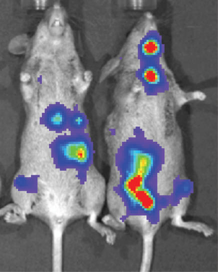
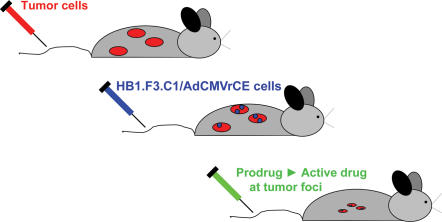
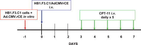
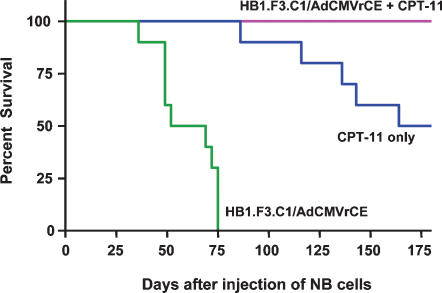
Methods and findings: We postulated that the tumor-tropic property of stem cells or progenitor cells could be exploited to selectively deliver a therapeutic gene to metastatic solid tumors, and that expression of an appropriate transgene at tumor loci might mediate cures of metastatic disease. To test this hypothesis, we injected HB1.F3.C1 cells transduced to express an enzyme that efficiently activates the anti-cancer prodrug CPT-11 intravenously into mice bearing disseminated neuroblastoma tumors. The HB1.F3.C1 cells migrated selectively to tumor sites regardless of the size or anatomical location of the tumors. Mice were then treated systemically with CPT-11, and the efficacy of treatment was monitored. Mice treated with the combination of HB1.F3.C1 cells expressing the CPT-11-activating enzyme and this prodrug produced tumor-free survival of 100% of the mice for >6 months (P<0.001 compared to control groups).

Conclusions: The novel and significant finding of this study is that it may be possible to exploit the tumor-tropic property of stem or progenitor cells to mediate effective, tumor-selective therapy for metastatic tumors, for which no tolerated curative treatments are currently available.


Conflict of interest statement

Competing Interests: The authors have declared that no competing interests exist.

Figures

Figure 1
Figure 1. Es1e SCID mice injected intravenously with neuroblastoma cells develop multiple disseminated tumors.
SK-N-AS cells (5×10⁵) transduced to express luciferase were injected into tail veins. One month following injection of tumor cells, mice were injected intraperitoneally with luciferin and imaged using a Xenogen IVIS imaging system, according the directions of the manufacturer. Multiple tumors were present in 100% of mice. Two representative mice are shown.
Figure 2
Figure 2. Schematic diagram of the protocol for NDEPT.
Human neuroblastoma tumor cells are injected intravenously to produce disseminated tumors. At an appropriate time after injection of neuroblastoma cells, neural stem cells or neural progenitor cells transduced with adenovirus to express a prodrug-activating enzyme (in this study, a secreted form of rabbit carboxylesterase [rCE]) are injected intravenously. Following migration of stem cells or progenitor cells to tumor foci and a delay of 3–4 days to allow relatively high level expression of the prodrug-activating enzyme into the extracellular milieu at the tumor sites, mice are treated with the prodrug (in this study, CPT-11). The prodrug is activated selectively at tumor foci, to increase the therapeutic index of the prodrug.
Figure 3
Figure 3. HB1.F3.C1 cells injected intravenously localize to micrometastatic human NB-1643 neuroblastoma tumors in the liver of a mouse.
(A) Dissected liver from a representative animal with hepatic metastases; the animal was sacrificed two days following injection of HB1.F3.C1 cells into the tail vein. (B) Low- and (C) high-power magnification of a section of tumor-involved liver, stained with hematoxylin and eosin. Tumor cells appear dark purple (black arrows); normal tissue appears pink. (D) Liver section stained with an anti-human mitochondrial protein antibody and counterstained with hematoxylin. Tumor micrometastases stain dark brown (red arrows); normal liver tissue is purple. (E) Immunofluorescence microscopy of liver section. HB1.F3.C1 cells were CM-DiI-labeled prior to injection and are evident as red cells. The liver section was stained with DAPI; tumor foci are identified by areas of densely-packed DAPI-stained tumor cell nuclei. The red arrows show extravasated HB1.F3.C1 cells proximal to a hepatic vein (v). Inset is high magnification of a DiI-labeled HB1.F3.C1 cell within the tumor. (F–H) Liver section from a tumor-bearing animal that received CM-DiI-labeled HB1.F3.C1 cells was stained with FITC-conjugated human specific mitochondrial antibody. (F) Red CM-DiI-labeled HB1.F3.C1 cells. (G) The same section showing FITC-labeled (green) human tumor cells and HB1.F3.C1 cells. (H) Overlay of F (red CM-DiI HB1.F3.C1 cells) and G (green FITC HB1.F3.C1 and tumor cells). HB1.F3.C1 cells (orange/yellow cells indicated by white arrows) migrated to hepatic micrometastases (green cells) and infiltrated the tumor parenchyma in the proximity of a blood vessel (bv, white dotted lines). Scale bars: 1 cm (A), 2 mm (B), 500 µm (C), 200 µm (F, G), 100 µm (D, E, H), 10 µm (E inset).
Figure 4
Figure 4. HB1.F3.C1 cells target macroscopic metastatic neuroblastoma in the bone marrow.
X-ray image of hind limb of a mouse with advanced stage neuroblastoma (Day 82; left panel, scale bar: 1 cm). Confirmation of the tumor mass as human SK-N-AS neuroblastoma cells was performed by immunohistochemistry using anti-human mitochondria antibody (not shown). The CM-DiI-labeled HB1.F3.C1 cells (red cells, injected into the tail vein 3 days prior to sacrifice) localized to tumor in the marrow (right panel; scale bar: 200 µm).
Figure 5
Figure 5. HB1.F3.C1 cells injected intravenously localized to microscopic bone marrow disease.
Concordant detection of v-myc (HB1.F3.C1 cells) by PCR and TH expression (neuroblastoma cells) by RT-PCR in bone marrow specimens. Bone marrow samples isolated from animals injected with HB1.F3.C1 cells were analyzed for the presence of v-myc (HB1.F3.C1 cells) or the expression of TH (NB-1643 cells). HB1.F3.C1 cells were present in the bone marrow only when tumor cells were also present. HB1.F3.C1 cells were not detected in the bone marrow of non-tumor-bearing animals. The positive controls (+) were DNA extracted from HB1.F3.C1 cells for v-myc, and RNA extracted from NB-1691 cells for TH. The negative controls (−) contained no DNA or RNA template, respectively.
Figure 6
Figure 6. Mice bearing microscopic NB-1643 tumors were injected intravenously with 2 million HB1.F3.C1 cells pre-labeled with CM-DiI Red Cell Tracker.
Normal and tumor-bearing organs were harvested, sectioned, and stained with DAPI. HB1.F3.C1 cells were not detected in normal (A) brain, (B) kidney, (C) heart, (D) intestine, or (E) skin tissue. Rare, single, HB1.F3.C1 cells (white arrows) were seen in (F) lung, (G) liver and (H) spleen. Scale bars: 200 µm (A–E), 100 µm (F–H).
Figure 7
Figure 7. HB1.F3.C1 cells transduced with an adenoviral multiplicity of infection of 5, 10, or 20 retain their tumor-tropism toward conditioned medium from SK-N-AS neuroblastoma cells.
Bars represent the average number of migrated cells±SEM of triplicate wells in modified Boyden chamber assays.
Figure 8
Figure 8. Treatment protocol of HB1.F3.C1/AdCMVrCE/CPT-11 NDEPT.
One day prior to being used in treatments, cells were transduced with the AdCMVrCE construct (see text). The treatment schedule was based on the time-course of expression of the secreted form of rCE, following adenoviral transduction of HB1.F3.C1 cells (Danks, unpublished observation) and on a schedule of administration of CPT-11 that has been shown to be relatively effective for neuroblastoma patients .
Figure 9
Figure 9. Therapeutic efficacy of rCE/CPT-11 NDEPT.
Mice (10/group) received one of the following: 1) 2 million HB1.F3.C1 cells transduced with AdCMVrCE (MOI = 20) encoding a secreted form of rCE; 2) CPT-11 (7.5 mg/kg) alone; 3) transduced HB1.F3.C1 cells and CPT-11 (7.5 mg/kg). Animals that received HB1.F3.C1/AdCMVrCE cells with CPT-11 survived significantly longer than mice receiving only HB1.F3.C1/AdCMVrCE cells (P<0.0001) or only CPT-11 (P<0.001), suggesting an enhanced tumor targeting and tumor cell-killing effect of CPT-11.

References

    1. Matthay KK, Seeger RC, Reynolds CP, Stram DO, O'Leary MC, et al. Allogeneic versus autologous purged bone marrow transplantation for neuroblastoma: a report from the Children's Cancer Group. J Clin Oncol. 1994;12:2382–2389. - PubMed
    1. Brodeur GM. Neuroblastoma: biological insights into a clinical enigma. Nat Rev Cancer. 2003;3:203–216. - PubMed
    1. Evans AE, August CS, Kamani N, Bunin N, Goldwein J, et al. Bone marrow transplantation for high risk neuroblastoma at the Children's Hospital of Philadelphia: an update. Med Pediatr Oncol. 1994;23:323–327. - PubMed
    1. Aboody KS, Brown A, Rainov NG, Bower KA, Liu S, et al. Neural stem cells display extensive tropism for pathology in adult brain: evidence from intracranial gliomas. Proc Natl Acad Sci U S A. 2000;97:12846–12851. - PMC - PubMed
    1. Ourednik V, Ourednik J, Park KI, Teng YD, Aboody KS, et al. Neural stem cells are uniquely suited for cell replacement and gene therapy in the CNS. Novartis Found Symp. 2000;231:242–262. Discussion 262–249, 302–246. - PubMed

Publication types

MeSH terms