. 2006 Dec 20;1(1):e33.
doi: 10.1371/journal.pone.0000033.

A weakened transcriptional enhancer yields variegated gene expression

Affiliations

A weakened transcriptional enhancer yields variegated gene expression

Cathy Collins et al. PLoS One. .

Abstract

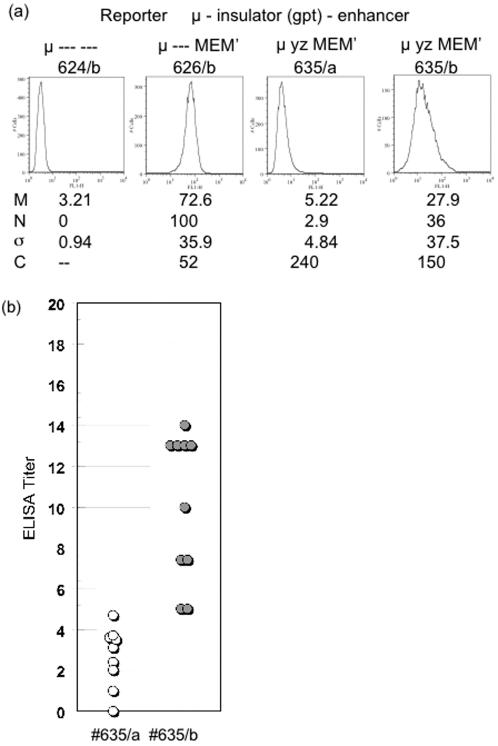
Identical genes in the same cellular environment are sometimes expressed differently. In some cases, including the immunoglobulin heavy chain (IgH) locus, this type of differential gene expression has been related to the absence of a transcriptional enhancer. To gain additional information on the role of the IgH enhancer, we examined expression driven by enhancers that were merely weakened, rather than fully deleted, using both mutations and insulators to impair enhancer activity. For this purpose we used a LoxP/Cre system to place a reporter gene at the same genomic site of a stable cell line. Whereas expression of the reporter gene was uniformly high in the presence of the normal, uninsulated enhancer and undetectable in its absence, weakened enhancers yielded variegated expression of the reporter gene; i.e., the average level of expression of the same gene differed in different clones, and expression varied significantly among cells within individual clones. These results indicate that the weakened enhancer allows the reporter gene to exist in at least two states. Subtle aspects of the variegation suggest that the IgH enhancer decreases the average duration (half-life) of the silent state. This analysis has also tested the conventional wisdom that enhancer activity is independent of distance and orientation. Thus, our analysis of mutant (truncated) forms of the IgH enhancer revealed that the 250 bp core enhancer was active in its normal position, approximately 1.4 kb 3' of the promoter, but inactive approximately 6 kb 3', indicating that the activity of the core enhancer was distance-dependent. A longer segment--the core enhancer plus approximately 1 kb of 3' flanking material, including the 3' matrix attachment region--was active, and the activity of this longer segment was orientation-dependent. Our data suggest that this 3' flank includes binding sites for at least two activators.


Conflict of interest statement

Competing Interests: The authors have declared that no competing interests exist.

Figures

Figure 1
Figure 1. Structure of the reporter cassettes
a) The endogenous μ gene. The boxes labeled V and C represent the exons encoding the variable (V) and constant (Cμ) regions of the immunoglobulin μ heavy chain gene. The relative positions of the intronic enhancer (Eμ) and switch (Sμ) regions in the V-C intron are shown. The Eμ enhancer is depicted with three components: the core enhancer (E) flanked by matrix attachment regions, M and M′. b) Recombination-mediated cassette exchange. The upper panel depicts a DNA segment in which (inverted) LoxP sites (1L and L1) flank a gene encoding the HyTK fusion protein (hygromycin-resistance and thymidine kinase [gancyclovir sensitivity]). As described previously, this DNA segment was inserted in the genome of the recipient hybridoma cell line . The HyTK and μ cassettes are represented as thick lines, with major exons as rectangles, the LoxP sites as triangles (L1 in the “forward” orientation, 1L in the “reverse” orientation). The three-stranded line represents the chromosomal DNA. The residual backbone of the vector that is shared between the target and the replacement is represented as a thin line; the remainder of the reporter cassette is represented as a dotted line. The middle panel depicts the structure of the replacement vector designed to substitute a modified μ gene for the Hy-TK gene via Cre-mediated recombination of the LoxP sites. This vector lacks the switch (Sμ) region and was constructed by joining two segments of the μ gene of the Sp6 hybridoma: The 2.0 kb V-bearing segment includes the DNA between the PacI site (∼850 bp 5′ of the initiator ATG) and the NgoMIV site 3′ of J4. The 4.6 kb Cμ-bearing segment includes the DNA between the SnaI site 5′ of Cμ and the SphI site 3′ of Cμ. DNA segments were inserted either in the intron at the NgoMIV site (denoted i), 1.2 kb 3′ of the initiator ATG, or 3′ of Cμ at the SphI site (denoted 3′), 5.9 kb 3′ of the ATG. The lower panel depicts the structure after the μ reporter cassette has replaced the HyTK gene. To distinguish replacements from random insertions we made use of the HinDIII (H) and NheI (N) sites that distinguish the DNA that flanks the HyTK and μ genes. The notations (i) and (3′) indicate the two sites where enhancer-derived segments were inserted. c) Structure of the reporter gene used for assaying insulator segments. d) The enhancer-derived segments. The “full” enhancer corresponds to the 2034 bp DNA segment bounded by the NgoMIV and Bst1107I sites, which are denoted as nucleotides 1 and 2034, respectively. The indicated subsegments were prepared by PCR, and nucleotide positions of their endpoints, numbered from the first nucleotide of the NgoMIV site are as follows: M, 1-782; E, 783-1035; M′, 1036-2034; p, 604-782; p′, 1036-1295; q′, 1296-1342; r′, 1343-1654; s′, 1655-1976. The XbaI sites that are often used to delimit the MARs are at 448 and 1441. The Bright binding sites are P1, 624-648; P2, 733-767; P4 1183-1202; P4, 1237-1276. e) Subsegments of the gpt cassette. The full gpt expression cassette includes the SV40 promoter (S), the gpt structural gene (gpt) and the SV40 polyA site (T). The gpt structural gene was divided into three subsegments, denoted x, y z. The nucleotide positions are measured from the first nucleotide of the SphI site in the SV40 promoter. The figures are not to scale.
Figure 2
Figure 2. Analysis of μ expression by northern blot
a) As described in the text, multiple independent replacements were isolated for each vector, and the concentration of IgM in culture supernatant of these cell lines was measured by ELISA (Table 1). Total RNA was isolated from representative cell lines and analyzed by Northern blot probed with segments of the μ and κ genes. The intensity of the bands was quantified by phosphorimager, and the μ/κ ratio, normalized to the value for cells expressing #626, is indicated below each lane. b) Results from the northern blot in (a) are listed next to a diagram of each enhancer-derived segment.
Figure 3
Figure 3. Flow cytometry of μ expression from reporter gene bearing different segments of the full enhancer.
Transfectants bearing the indicated reporter genes were analyzed by flow cytometry. Cells were fixed and permeabilized, and intracellular μ chains were stained with μ-specific fluorescent antibodies. ∼104 cells were then analyzed. In these histograms the horizontal axis indicates the mean fluorescence (logarithmic scale) and the vertical axis the number of cells with the corresponding fluorescence. M is the mean fluorescence for each population. Z10/HyTK, the recipient cell line was used in this case as the negative control, and its mean “fluorescence”, M0, for each experiment was subtracted from the mean fluorescence, Mx, measured for a cell population expressing reporter “x”. This corrected fluorescence is compared with M626, the corrected fluorescence in that experiment for the reporter #626 with the full enhancer. Thus, the “normalized” fluorescence, Nx, for reporter “x” was calculated as Nx  =  (Mx−M0)/(M626−M0), where M626 is the mean fluorescence for the reporter with the full enhancer and M0 is the fluorescence for the recipient cell line or other μ non-expresser. σ is the standard deviation of the fluorescence. In order to correct for variation in background “fluorescence”, we calculated a corrected coefficient of variation, Cx, for reporter “x” as Cx  =  (σx 2−σ0 2)1/2/Mx−M0, where σx and σ0 are the standard deviations associated, respectively, with reporter “x” and with the recipient cell line or other μ non-expresser.
Figure 4
Figure 4. Flow cytometry of μ expression from partially insulated reporter gene.
(a) Two independent transfectants expressing reporter #635 (#635/a and #635/b) were analyzed by flow cytometry, as described in Figure 3. (b) The transfectants #635/a and #635/b were subcloned, and secreted IgM was measured for each subclone. The statistical parameters, M, N, σ, and C are defined in the legend to Figure 3.
Figure 5
Figure 5. Flow cytometry of μ expression from reporters with mutant enhancers.
a) Two independent transfectants expressing the #654 reporter were analyzed by flow cytometry. b) As in (a) two independent transfectants bearing reporter #651 were analyzed by flow cytometry, as described in Figure 3. The transfectant 651/b was biphasic and yielded subclones with different levels of expression, 651/b3 and 651/b5. To assess the stability of this difference, these subclones were re-subcloned, and the mean fluorescence, M, and normalized fluorescence, N, of eight re-subclones were measured. Nm, the mean value for N for the re-subclones and the associated standard deviation, were calculated. The values of Nm for each set of subclones were significantly different and close to the value of the N for their respective parents. The statistical parameters, M, N, σ, and C are defined in the legend to Figure 3.
Figure 6
Figure 6. Analysis of μ RNA produced by weakened enhancers.
a) Total RNA was isolated from the indicated cells and analyzed by northern blot as described in Figure 2. The asterisk indicates that the cells were incubated for approximately five days with 6.7 mM 3-aminobenzamide prior to isolating RNA. The upper panels present results for various insulators derived from the gpt cassette and from the igf2/H19 locus; the lower panels present results for various mutant enhancers. For vectors #629 and $635, the segments of the gpt cassette, S, x, y, z are defined in Figure 1. Reporters #648 and #650 bear the insulator (DMD) from the igf2/H19 loci of mouse and human, respectively. b) The normalized μ/κ ratios from (a) for cells grown in normal medium (NM) or medium supplemented with 3-aminobenzamide (AB) are listed next to diagrams showing the insulators and enhancers in the reporter genes.
Figure 7
Figure 7. Flow cytometry of cells treated with 3-aminobenzamide.
The indicated cells, grown either in normal medium or in medium supplemented with 3-aminobenzamide, were analyzed for intracellular μ protein by flow cytometry, as described in Figure 3. Because 3-aminobenzamide also affected the fluorescence of the μ-negative cells, Z10/HyTK, the normalized fluorescence (N or N′) for cells grown in 3-aminobenzamide was calculated two ways: For cells bearing the reporter gene, N was calculated by subtracting the mean fluorescence of the parental cell line, Z10/HyTK, grown in normal medium; N′ was calculated by subtracting the mean fluorescence for cells grown in 3-aminobenzamide.

Similar articles

Cited by

References

    1. Hume DA. Probability in transcriptional regulation and its implications for leukocyte differentiation and inducible gene expression. Blood. 2000;96:2323–2328. - PubMed
    1. Kurakin A. Self-organization vs Watchmaker: stochastic gene expression and cell differentiation. Dev Genes Evol. 2005;215:46–52. - PubMed
    1. Serizawa S, Miyamichi K, Sakano H. One neuron-one receptor rule in the mouse olfactory system. Trends Genet. 2005;20:648–653. - PubMed
    1. Guo L, Hu-Li J, Paul W. Probabilistic regulation in TH2 cells accounts for monoallelic expression of IL-4 and IL-13. Immunity. 2005;23:89–99. - PubMed
    1. Martin DIK, Whitelaw E. The vagaries of variegating transgenes. BioEssays. 1996;18:919–923. - PubMed

Publication types