. 2006 Dec 20;1(1):e37.
doi: 10.1371/journal.pone.0000037.

Ribosomal protein gene knockdown causes developmental defects in zebrafish

Affiliations

Ribosomal protein gene knockdown causes developmental defects in zebrafish

Tamayo Uechi et al. PLoS One. .

Abstract

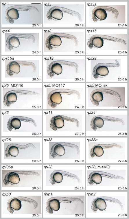
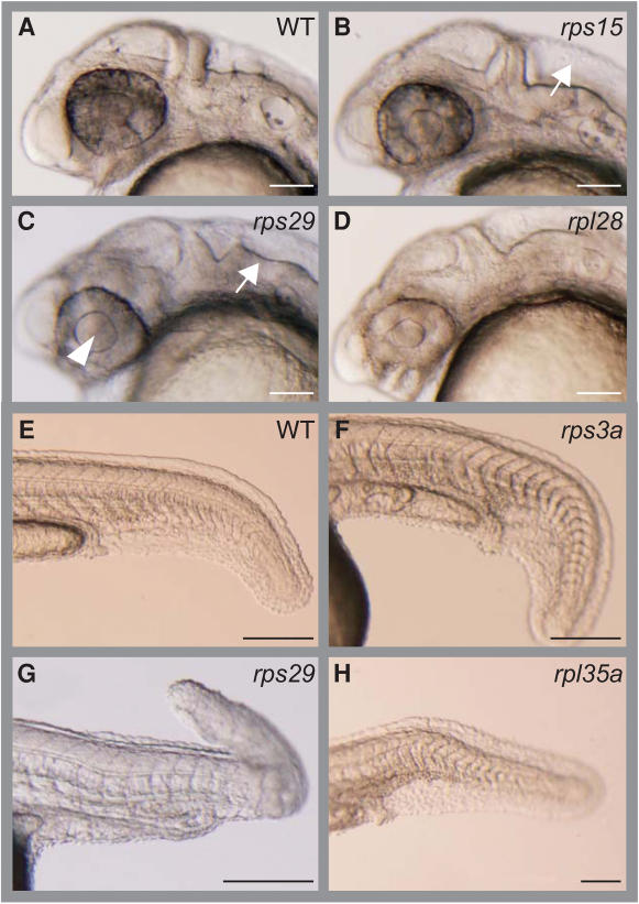
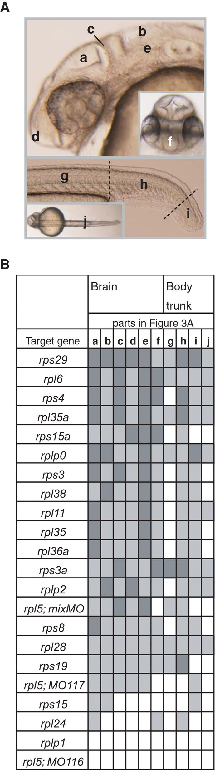
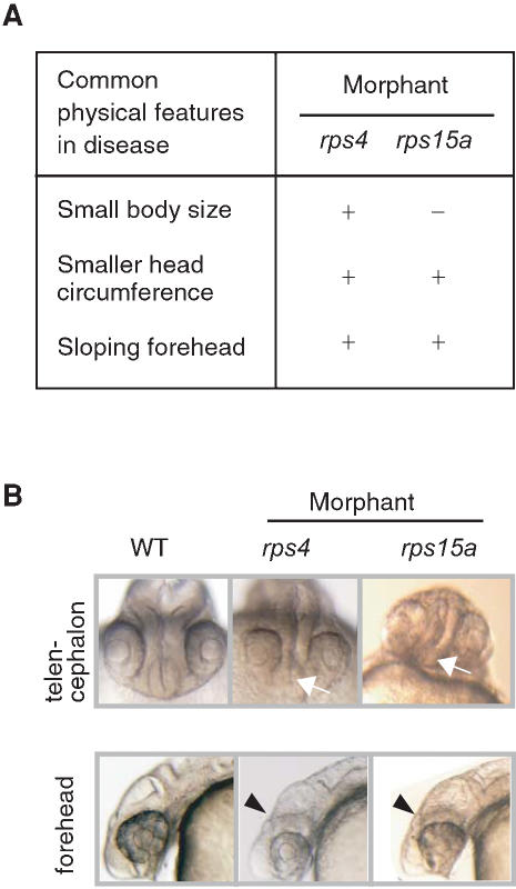
The ribosomal proteins (RPs) form the majority of cellular proteins and are mandatory for cellular growth. RP genes have been linked, either directly or indirectly, to various diseases in humans. Mutations in RP genes are also associated with tissue-specific phenotypes, suggesting a possible role in organ development during early embryogenesis. However, it is not yet known how mutations in a particular RP gene result in specific cellular changes, or how RP genes might contribute to human diseases. The development of animal models with defects in RP genes will be essential for studying these questions. In this study, we knocked down 21 RP genes in zebrafish by using morpholino antisense oligos to inhibit their translation. Of these 21, knockdown of 19 RPs resulted in the development of morphants with obvious deformities. Although mutations in RP genes, like other housekeeping genes, would be expected to result in nonspecific developmental defects with widespread phenotypes, we found that knockdown of some RP genes resulted in phenotypes specific to each gene, with varying degrees of abnormality in the brain, body trunk, eyes, and ears at about 25 hours post fertilization. We focused further on the organogenesis of the brain. Each knocked-down gene that affected the morphogenesis of the brain produced a different pattern of abnormality. Among the 7 RP genes whose knockdown produced severe brain phenotypes, 3 human orthologs are located within chromosomal regions that have been linked to brain-associated diseases, suggesting a possible involvement of RP genes in brain or neurological diseases. The RP gene knockdown system developed in this study could be a powerful tool for studying the roles of ribosomes in human diseases.


Conflict of interest statement

Competing Interests: The authors have declared that no competing interests exist.

Figures

Figure 1
Figure 1. Lateral Views of Wild-Type and MO-Injected Embryos
The genes targeted by MOs and the observation time are indicated for each image. ‘rpl5; MO116’ and ‘rpl5; MO117’ indicate two MOs designed and injected separately for two functional copies of rpl5 on the zebrafish genome. ‘rpl5; MOmix’ indicates the mixture of MO116 and MO117. Control MOs that included 5 mispaired bases were also used; one example of a control MO injection is shown (rpl38; misMO). Note that compared to the control, the rpl38 morphant is shorter and displays a light-colored eye and thin yolk sac extension. Scale bar, 500 µm.
Figure 2
Figure 2. Specific Morphological Changes in Brain and Body Trunk
Examples of morphants displaying characteristic deformations are shown. The targeted genes and the deformed areas of the morphants are indicated. (A, E) Wild type (WT). (B) rps15; enlarged 4th ventricle (white arrow). (C) rps29; enlarged lens (arrowhead) and undulated rhombencephalon (white arrow). (D) rpl28; protruded forehead. (F) rps3a; wider trunk and downward-curving tail. (G) rps29; wavy notochord and extremely bent tail. (H) rpl35a; sharply downward bent tail. Anterior is to the left. Bars: A∼D, 100 µm; E∼H, 200 µm.
Figure 3
Figure 3. Schematic Representation of the Extent of Abnormalities in Brain and Body Trunk
(A) 6 parts of the brain, 3 parts of the body trunk, and a dorsal view of the trunk are indicated in wild type embryo. a, optic tectum; b, 4th ventricle; c, midbrain-hindbrain boundary; d, anterior part of head; e, rhombencephalon; f, telencephalon; g, body along yolk sac extension; h, tail; i, tip of tail; j, dorsal view of the trunk. (B) The deformations were assessed using a 3-level severity scale; dark gray indicates a severe effect, light gray indicates a mild effect, and white represents no apparent effect. Detailed information and images of the abnormalities in the morphants are available at the zebrafish database (http://zebrafish.med.miyazaki-u.ac.jp).
Figure 4
Figure 4. Similarities between the Clinical Features of Human Brain Diseases and Defects in Zebrafish Morphants
(A) Common features shared between Abidi X-linked mental retardation and microhydranencephaly are listed on the left and the corresponding changes in rps4 and rps15a morphants are indicated on the right. (B) Telencephalon hypoplasia is seen in rps4 morphants, and reduction in size and deformity of the telencephalon is apparent in rps15a morphants (white arrows). Unclear subdivisions of the brain and flattened foreheads (arrowheads) can be seen in lateral views of these morphants.

References

    1. Wool IG. The structure and function of eukaryotic ribosomes. Annu Rev Biochem. 1979;48:719–754. - PubMed
    1. Nakao A, Yoshihama M, Kenmochi N. RPG: the Ribosomal Protein Gene database. Nucleic Acids Res. 2004;32:D168–170. - PMC - PubMed
    1. Noller HF. RNA structure: reading the ribosome. Science. 2005;309:1508–1514. - PubMed
    1. Klein DJ, Moore PB, Steitz TA. The roles of ribosomal proteins in the structure assembly, and evolution of the large ribosomal subunit. J Mol Biol. 2004;340:141–177. - PubMed
    1. Lambertsson A. The Minute genes in Drosophila and their molecular functions. Adv Genet. 1998;38:69–134. - PubMed

Publication types

MeSH terms

Substances