. 2006 Dec 20;1(1):e43.
doi: 10.1371/journal.pone.0000043.

Is adipose tissue a place for Mycobacterium tuberculosis persistence?

Affiliations

Is adipose tissue a place for Mycobacterium tuberculosis persistence?

Olivier Neyrolles et al. PLoS One. .

Abstract

Background: Mycobacterium tuberculosis, the etiological agent of tuberculosis (TB), has the ability to persist in its human host for exceptionally long periods of time. However, little is known about the location of the bacilli in latently infected individuals. Long-term mycobacterial persistence in the lungs has been reported, but this may not sufficiently account for strictly extra-pulmonary TB, which represents 10-15% of the reactivation cases.

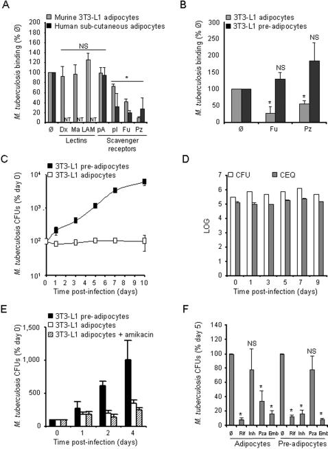
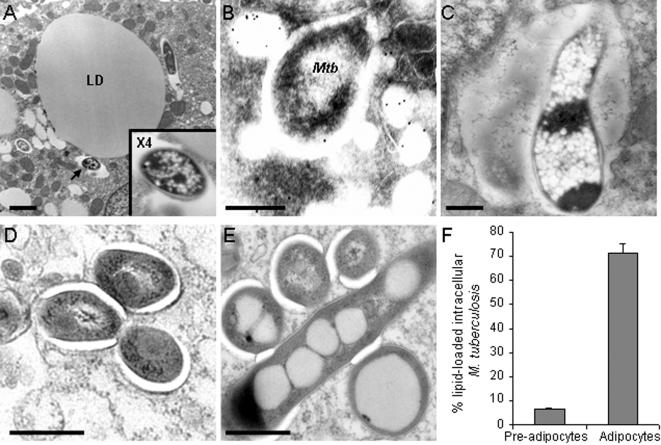
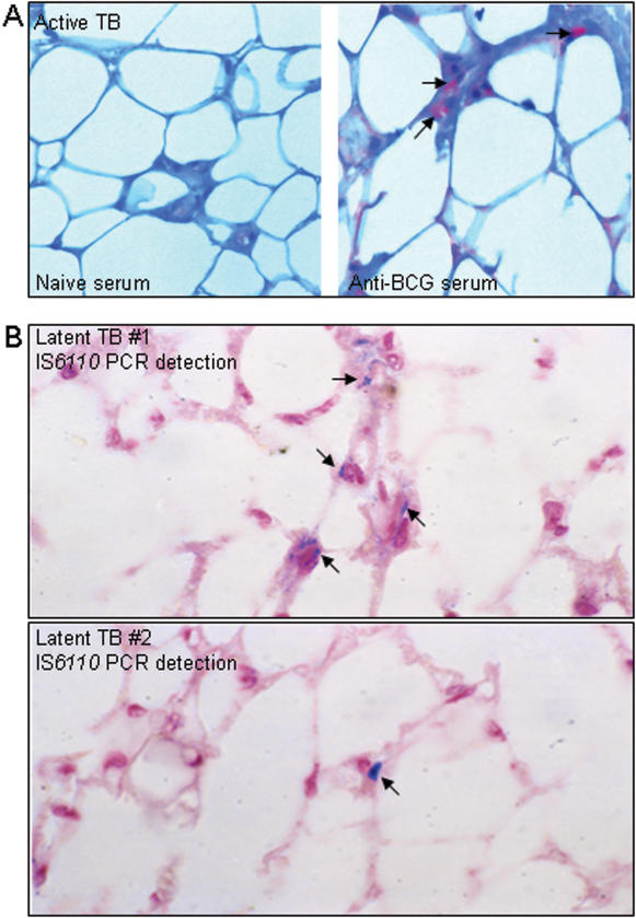
Methodology/principal findings: We applied in situ and conventional PCR to sections of adipose tissue samples of various anatomical origins from 19 individuals from Mexico and 20 from France who had died from causes other than TB. M. tuberculosis DNA could be detected by either or both techniques in fat tissue surrounding the kidneys, the stomach, the lymph nodes, the heart and the skin in 9/57 Mexican samples (6/19 individuals), and in 8/26 French samples (6/20 individuals). In addition, mycobacteria could be immuno-detected in perinodal adipose tissue of 1 out of 3 biopsy samples from individuals with active TB. In vitro, using a combination of adipose cell models, including the widely used murine adipose cell line 3T3-L1, as well as primary human adipocytes, we show that after binding to scavenger receptors, M. tuberculosis can enter within adipocytes, where it accumulates intracytoplasmic lipid inclusions and survives in a non-replicating state that is insensitive to the major anti-mycobacterial drug isoniazid.

Conclusions/significance: Given the abundance and the wide distribution of the adipose tissue throughout the body, our results suggest that this tissue, among others, might constitute a vast reservoir where the tubercle bacillus could persist for long periods of time, and avoid both killing by antimicrobials and recognition by the host immune system. In addition, M. tuberculosis-infected adipocytes might provide a new model to investigate dormancy and to evaluate new drugs for the treatment of persistent infection.


Conflict of interest statement

Competing Interests: The authors have declared that no competing interests exist.

Figures

Figure 1
Figure 1. M. tuberculosis persists in a non-replicating state inside adipocytes.
A. 3T3-L1 mouse (grey bars) and primary human (black bars) adipocytes were infected with M. tuberculosis, clinical isolate MT103, at a MOI of 1. Mycobacterial binding to the cells was measured after 4 h at 4°C in the presence of various inhibitors. Man, yeast mannan; LAM, lipoarabinomannan; pA, polyadenosinic acid; pI, polyinosinic acid; Fuc, fucoidan; PC, PAz-PC, a predominant form of oxidized low-density lipoprotein. Results are expressed as % of the binding without inhibitor (Ø). B. 3T3-L1 adipocytes (grey bars) and pre-adipocytes (black bars) were infected as described in A. in the presence of Fuc or PAz-PC. Mycobacterial binding was assessed and analysed as in A. C. 3T3-L1 pre-adipocytes (black squares) and adipocytes (open squares) were pulsed with M. tuberculosis MT103 at a MOI of 1 for 4 h at 37°C, then chased with fresh medium. Mycobacterial survival, expressed as % of the bacterial load at day 0, was monitored on a 10-day period. The results are standardized as % of the bacterial load at day 0 because of the differential M. tuberculosis binding to adipocytes and pre-adipocytes. D. 3T3-L1 adipocytes were infected as described in C. Viable counts (CFU) were quantified by plating serial dilutions of cell lysates (open bars), and total counts (CEQ) were quantified by qPCR (grey bars). E. 3T3-L1 adipocytes were infected as described in C. In order to kill extra-cellular bacteria cells were pre-incubated with 200 µg/ml amikacin 2 h prior lysis and plating. Results are expressed as % of bacterial load at day 0. F. 3T3-L1 adipocytes and pre-adipocytes were infected as described in C. Cells were treated with 1 µg/ml rifampicin (Rif), 0.1 µg/ml isoniazid (Inh), 25 µg/ml pyrazinamide (Pza) or 5 µg/ml ethambutol (Emb) at day 3, and bacterial loads were quantified at day 5. Results are expressed as % of bacterial load at day 5 in the absence of antibiotics (Ø). In all panels, results are the mean of three independent experiments and bars indicate ± sd. NT, not tested; NS, not significant, and *, p<0.05, as assessed by Mann-Whitney test of median comparison.
Figure 2
Figure 2. M. tuberculosis accumulates lipid droplets inside adipocytes.
3T3-L1 mouse adipocytes (A–C&E) and pre-adipocytes (D) were pulsed with M. tuberculosis MT103 at a MOI of 1 for 4 h, then chased with fresh medium for 24 h and observed by electron microscopy. In A., the arrow indicates a mycobacterial vacuole shown at higher magnification in the inset; LD, lipid droplet; bar = 2 µm. In B., perilipinA/B was immuno-stained with gold-conjugated antibodies. Mtb, M. tuberculosis; bar = 200 nm. In C, bar = 500 nm. In D&E, bar = 200 nm. F. Electron micrographs of 2 independent experiments were used to quantified lipid droplet accumulation inside mycobacteria when within adipocytes and pre-adipocytes. An average of 50 mycobacteria were observed in different fields in each sample.
Figure 3
Figure 3. Detection of M. tuberculosis in adipose tissue from individuals with latent or active TB.
A. Perinodal adipose tissue was taken from biopsy of the mediastinal lymph node from a patient with active TB. Bacilli were immuno-detected using an anti-BCG rabbit hyperimmune serum (right panel). As a control, a serial section was stained with serum from a naïve animal (left panel). B. Perinodal adipose tissue was taken at autopsy from two individuals (upper and lower panels) with no clinical sign of pulmonary TB. In situ PCR was used to detect IS6110. In A&B, arrows indicate positive signals.

References

    1. Kaufmann SH. How can immunology contribute to the control of tuberculosis? Nat Rev Immunol. 2001;1:20–30. - PubMed
    1. Frieden TR, Sterling TR, Munsiff SS, Watt CJ, Dye C. Tuberculosis. Lancet. 2003;362:887–899. - PubMed
    1. Fine PE, Small PM. Exogenous reinfection in tuberculosis. N Engl J Med. 1999;341:1226–1227. - PubMed
    1. van Rie A, Warren R, Richardson M, Victor TC, Gie RP, et al. Exogenous reinfection as a cause of recurrent tuberculosis after curative treatment. N Engl J Med. 1999;341:1174–1179. - PubMed
    1. Parrish NM, Dick JD, Bishai WR. Mechanisms of latency in Mycobacterium tuberculosis. Trends Microbiol. 1998;6:107–112. - PubMed

Publication types

MeSH terms