. 2007 Mar 1;579(Pt 2):515-26.
doi: 10.1113/jphysiol.2006.121889. Epub 2006 Dec 21.

Genioglossus premotoneurons and the negative pressure reflex in rats

Affiliations

Genioglossus premotoneurons and the negative pressure reflex in rats

Nancy L Chamberlin et al. J Physiol. .

Abstract

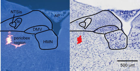
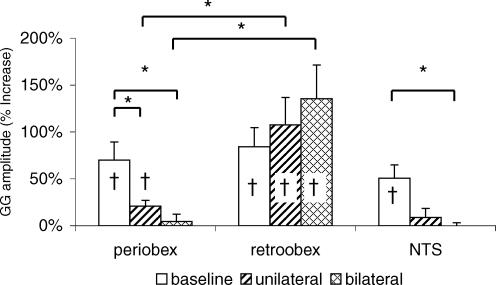
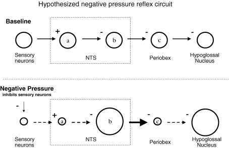
Reflex increases in genioglossus (GG) muscle activity in response to negative pharyngeal pressure are important for maintenance of upper airway patency in humans. However, little is known of the central circuitry that mediates this negative pressure reflex (NPR). We used two approaches to determine which GG premotoneurons relay negative pressure-related information to the hypoglossal motor nucleus. First, to identify GG premotoneurons, we injected pseudorabies virus (PRV152) into the GG muscle. We found that medullary GG premotoneurons were concentrated mainly in the reticular formation adjacent to the hypoglossal motor nucleus. Second, in order to determine whether these perihypoglossal neurons were involved in the NPR, we quantified GG EMG responses to negative pressure applied to the isolated upper airway in anaesthetized rats before and after microinjection of muscimol (9 nl; 0.25 mM), a GABA-A receptor agonist, into the perihypoglossal premotor field. Pressures as low as -4 cm H(2)O increased inspiratory phase-related GG activity. The NPR was abolished following bilateral injections of muscimol into the perihypoglossal premotor field at and up to 500 mum rostral to the obex. Muscimol in this location also increased the amplitude of basal, unstimulated phasic GG activity. By contrast, inhibition of neurons caudal to the obex decreased phasic GG activity but had no impact on the NPR. These results suggest that perihypoglossal GG premotoneurons near the obex mediate the NPR and those caudal to the obex are important mediators of respiratory-related GG activity but are not involved in the NPR.


Figures

Figure 1
Figure 1. Effect of local inhibition of neurons rostral to the obex on phasic GG and its response to negative pharyngeal pressure
A, example of increased phasic GG amplitude in response to negative pressure before application of muscimol (compare breaths 1–3 with 4–6). The dashed line shows the amplitude of baseline moving time averaged GG signal prior to application of negative pressure. B, example of increase in phasic GG amplitude and abolition of negative pressure reflex following bilateral injection of muscimol at the site shown in Fig. 2 (case 57). Scales for A and B are the same. The baseline airway pressure is zero.
Figure 2
Figure 2. Photomicrographs of a muscimol injection site in the periobex region
Left, fluorescence photomicrograph showing latex beads. Some beads entered a blood vessel branching medially. Right, brightfield photomicrograph following Nissl stain of the same section. Red area was digitally added to show injection site. AP, area postrema; DMV, dorsal motor nucleus of the vagus; HMN, hypoglossal motor nucleus; NTSis interstitial subnucleus of the nucleus of the solitary tract; ts, solitary tract.
Figure 3
Figure 3. Anatomical distribution of muscimol injection sites compared to GG premotoneurons
Orange, pink and red dots indicate positions of GFP-ir neurons in three representative rats 63–64 h following PRV152 injection into the right side of the GG. Small dots are hypoglossal motoneurons and larger dots are premotoneurons. Each drawing includes PRV-labelled neurons from three 40 μm sections in a 1: 4 series (i.e. every fourth section). Numbers indicate histologically verified locations where muscimol was injected in a separate series of rats. A, injection sites at levels 0–500 μm rostral to the obex (periobex). B, injection sites −100 to −600 μm caudal to the obex (retroobex). Numbers in blue type indicate NTS injections. AP, area postrema; DMV, dorsal motor nucleus of the vagus; HMN, hypoglossal motor nucleus. Scalebar is 500 μm.
Figure 5
Figure 5. Photomicrographs of GFP-ir neurons in aminergic, histaminergic and orexinergic cell groups
A, LC (locus coeruleus); B, A7 noradrenergic cell group; C, orexin field; arrowheads show 3 of the labelled neurons; D, tuberomammillary nucleus. E, digitally merged image of GFP (F) and TH (G) staining in LC. A–D. E–G, arrows show double-labelled neurons; case 44. Medial is to the left.
Figure 4
Figure 4. Photomicrographs of GFP-ir neurons following PRV152 injection
A, hypoglossal motoneurons and periobex premotoneurons. B, higher magnification of boxed area in A. C, ventrally located reticular neurons medial to the facial motor nucleus (7) and dorsal to the pyramids. D, higher magnification of boxed area in C. E, reticular neurons dorsal to the inferior olive (IO). F, pontine parvocellular reticular neurons (PCRt) dorsal to 7 and slightly rostral to the section in C. Scalebars: A, 500 μm; B, 200 μm.
Figure 6
Figure 6. Effect of local inhibition of neurons with muscimol on the NPR
Shown are the mean (±s.e.m.) changes in GG amplitude in 3 groups of rats during negative pressure under baseline conditions and following unilateral and bilateral muscimol injection. † Negative pressure application significantly increased GG amplitude (NPR). Note that muscimol injection into the periobex and NTS but not into the retroobex region significantly decreased the NPR (*P < 0.05).
Figure 7
Figure 7. Effects of local neuronal inhibition on phasic GG amplitude Changes in phasic GG amplitude
from baseline following unilateral and bilateral muscimol injection into the periobex and retroobex perihypoglossal reticular formation and the NTS. Muscimol injection in the periobex increased phasic GG activity whereas its injection in the retroobex decreased GG activity (**P < 0.01, *P < 0.1).
Figure 8
Figure 8. Proposed neural pathway for negative pressure GG activation
Depicted is the proposed sequence of events (left to right) following negative pharyngeal pressure. Under baseline conditions (zero transmural pressure; top), sensory neurons are tonically active and excite neurons in the NTS (a) that in turn inhibit NTS output neurons (b) that otherwise would inhibit periobex neurons. Periobex and hypoglossal motoneurons are active in phase with inspiration. Negative pressure (bottom) inhibits sensory neurons, which reduces activity of secondary sensory NTS neurons (a) which releases NTS output neurons (b) from inhibition. Periobex neuronal activity is reduced, thus disinhibiting hypoglossal motoneurons. Plus and minus signs indicate synaptic excitation and inhibition, respectively. Small and large circles indicate reductions and increases, respectively, in neuron firing rate in response to negative pressure.

References

    1. Aicher SA, Milner TA, Pickel VM, Reis DJ. Anatomical substrates for baroreflex sympathoinhibition in the rat. Brain Res Bull. 2000;51:107–110. - PubMed
    1. Aldes LD. Subcompartmental organization of the ventral (protrusor) compartment in the hypoglossal nucleus of the rat. J Comp Neurol. 1995;353:89–108. - PubMed
    1. Bailey EF, Huang YH, Fregosi RF. Anatomic consequences of intrinsic tongue muscle activation. J Appl Physiol. 2006;101:1377–1385. - PubMed
    1. Borke RC, Nau ME, Ringler RL., Jr Brain stem afferents of hypoglossal neurons in the rat. Brain Res. 1983;269:47–55. - PubMed
    1. Brouillette RT, Thach BT. A neuromuscular mechanism maintaining extrathoracic airway patency. J Appl Physiol. 1979;46:772–779. - PubMed

Publication types

LinkOut - more resources