

Retinoic acid-induced glandular differentiation of the oesophagus
- PMID: 17185354
- PMCID: PMC1994378
- DOI: 10.1136/gut.2006.097915
Retinoic acid-induced glandular differentiation of the oesophagus
Abstract
Background: Retinoic acid (RA) is a powerful differentiation agent. Barrett's oesophagus occurs when duodeno-gastro-oesophageal reflux causes squamous epithelium (SE) tissue to become columnar epithelium tissue by an unknown mechanism. The bile acid lithocholic acid (LCA) competes for the retinoid X receptor retinoid binding site. Hence, RA pathways may be implicated in Barrett's oesophagus.
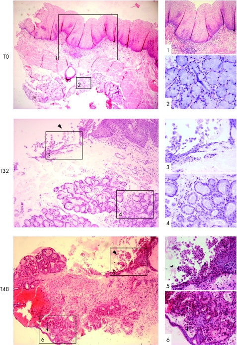
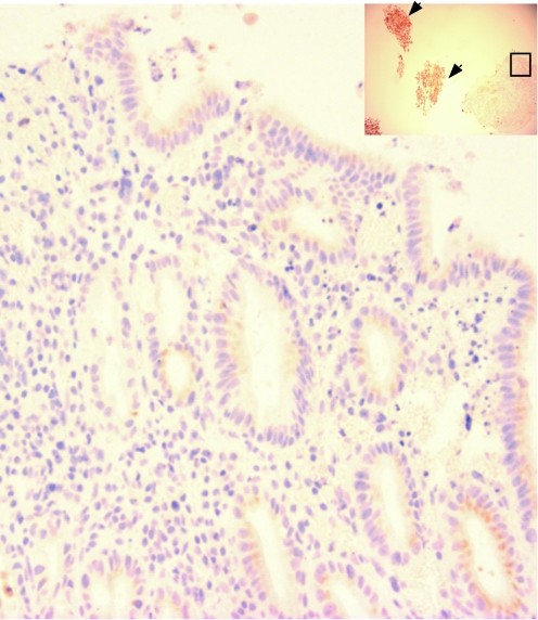
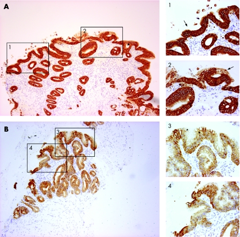
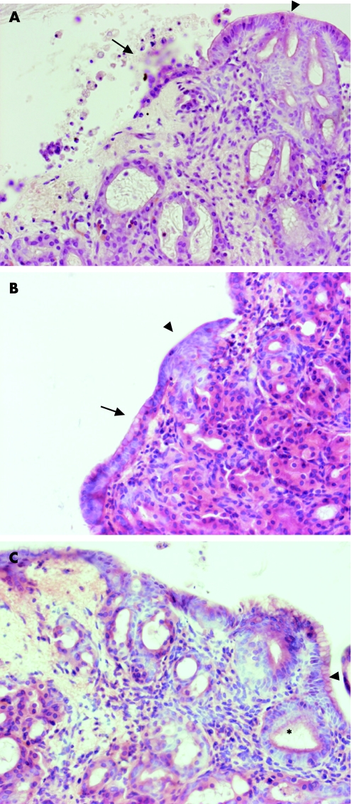
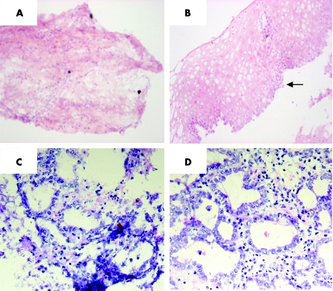
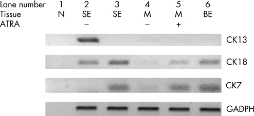
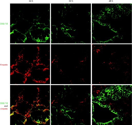
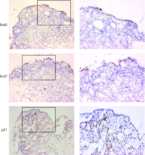
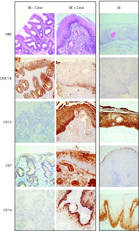
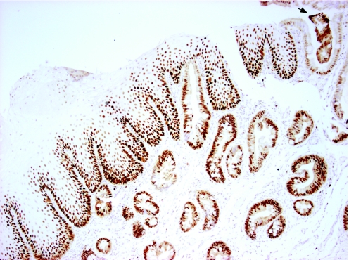
Methods: RA activity in tissues and cell lines treated with all-trans retinoic acid (ATRA) with or without LCA was assessed using a reporter. Expression of p21 was determined by real-time PCR in Barrett's oesophagus cell lines with or without LCA. SE and Barrett's oesophagus biopsy specimens were exposed to 100 muM of ATRA or 20 mM of a RA inhibitor, citral, in organ culture for >72 h. Characteristics of treated specimens, compared with untreated controls, were analysed by immunohistochemical analysis (cytokeratins (CKs), vimentin) and RT-PCR (CKs). Confocal microscopy assessed temporal changes in co-localisation of CK8/18 and vimentin. Cell proliferation was assessed by bromo-deoxyuridine incorporation and immunohistochemical analysis for Ki67 and p21.
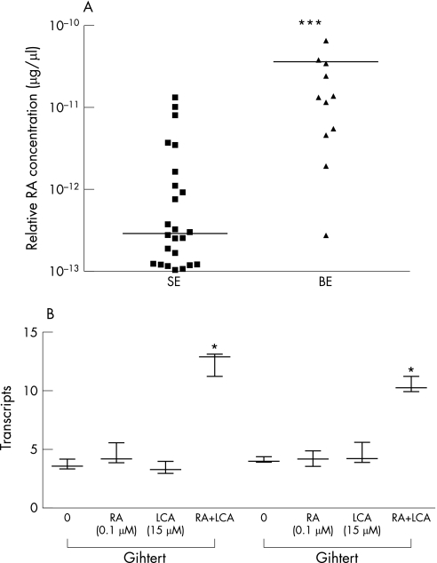
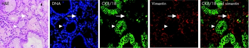
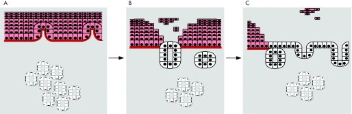
Results: RA biosynthesis was increased in Barrett's oesophagus compared with SE (p<0.001). LCA and ATRA caused a synergistic increase in RA signalling as shown by increased p21 (p<0.01). Morphological and molecular analysis of SE exposed to ATRA showed columnar differentiation independent of proliferation. Metaplasia could be induced from the stromal compartment alone and vimentin expression co-localised with CK8/18 at 24 h, which separated into CK8/18-positive glands and vimentin-positive stroma by 48 h. Citral-treated Barrett's oesophagus led to phenotypic and immunohistochemical characteristics of SE, which was independent of proliferation.
Conclusion: RA activity is increased in Barrett's oesophagus and is induced by LCA. Under conditions of altered RA activity and an intact stroma, the oesophageal phenotype can be altered independent of proliferation.
Conflict of interest statement
Competing interests: None.
Comment in
-
Open questions in oesophageal adenocarcinogenesis.Gut. 2007 Jul;56(7):897-8. doi: 10.1136/gut.2006.117135. Gut. 2007. PMID: 17566022 Free PMC article. Review.
-
Retinoids, bile acids and PPARs in Barrett's oesophagus.Gut. 2008 Jan;57(1):137. doi: 10.1136/gut.2007.134627. Gut. 2008. PMID: 18094207 No abstract available.
References
-
- Shaheen N, Ransohoff D F. Gastroesophageal reflux, Barrett's esophagus, and esophageal cancer: scientific review. JAMA 20022871972–1981. - PubMed
-
- Brown L M, Devesa S S. Epidemiologic trends in esophageal and gastric cancer in the United States. Surg Oncol Clin N Am 200211235–256. - PubMed
-
- Montgomery R K, Mulberg A E, Grand R J. Development of the human gastrointestinal tract: twenty years of progress. Gastroenterology 199916702–731. - PubMed
-
- Tosh D, Slack J M. How cells change their phenotype. Nat Rev Mol Cell Biol 20023187–194. - PubMed
Publication types
MeSH terms
Substances
Grants and funding
LinkOut - more resources
Full Text Sources
Research Materials