. 2007 Jan 2;104(1):270-5.
doi: 10.1073/pnas.0609412103. Epub 2006 Dec 21.

Identification of NVP-TAE684, a potent, selective, and efficacious inhibitor of NPM-ALK

Affiliations

Identification of NVP-TAE684, a potent, selective, and efficacious inhibitor of NPM-ALK

Anna V Galkin et al. Proc Natl Acad Sci U S A. .

Erratum in

  • Proc Natl Acad Sci U S A. 2007 Feb 6;104(6):2025

Abstract

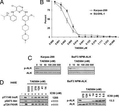
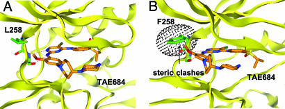
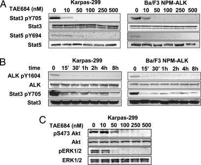
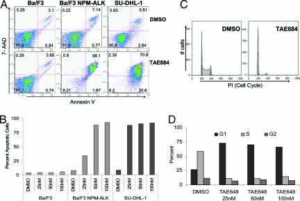
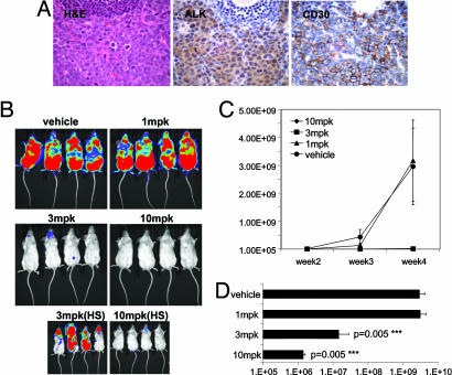
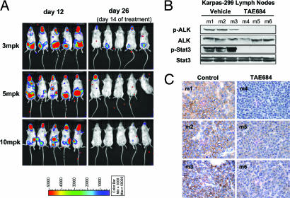
Constitutive overexpression and activation of NPM-ALK fusion protein [t(2:5)(p23;q35)] is a key oncogenic event that drives the survival and proliferation of anaplastic large-cell lymphomas (ALCLs). We have identified a highly potent and selective small-molecule ALK inhibitor, NVP-TAE684, which blocked the growth of ALCL-derived and ALK-dependent cell lines with IC(50) values between 2 and 10 nM. NVP-TAE684 treatment resulted in a rapid and sustained inhibition of phosphorylation of NPM-ALK and its downstream effectors and subsequent induction of apoptosis and cell cycle arrest. In vivo, NVP-TAE684 suppressed lymphomagenesis in two independent models of ALK-positive ALCL and induced regression of established Karpas-299 lymphomas. NVP-TAE684 also induced down-regulation of CD30 expression, suggesting that CD30 may be used as a biomarker of therapeutic NPM-ALK kinase activity inhibition.


Conflict of interest statement

The authors declare no conflict of interest.

Figures

Fig. 1.
Fig. 1.
Effects of TAE684 on NPM-ALK-dependent cell proliferation in vitro. (A) Structure of TAE684. (B) TAE684 inhibits cell proliferation of Karpas-299 and SU-DHL-1 cell lines. Cell proliferation was assayed by using the Bright-Glo Luciferase Assay System after 72 h of treatment with serial dilutions of TAE684. Obtained relative luminescence values were normalized to values from corresponding DMSO-treated wells and displayed as percent survival ± SE. (C) TAE684 blocks NPM-ALK autophosphorylation in Karpas-299 and Ba/F3 NPM-ALK cells after 4 h of treatment. (D) Effect of TAE684 on InsR signaling in H-4-II-E rat hepatoma cells. H-4-II-E cells were preincubated for 30 min with TAE684 at concentrations indicated before stimulation with recombinant insulin. Treatment of Ba/F3 NPM-ALK cells with TAE684 was performed in parallel. IC50 concentrations for InsR and ALK signaling inhibition were determined through the quantification of bands by using Bio-Rad's QuantityOne software package.
Fig. 2.
Fig. 2.
Structural basis for ALK kinase inhibition selectivity by TAE684. (A) Model of ALK in complex with TAE684, developed based on the published crystal structure of InsR in an “active” conformation by using homology modeling (MOE). TAE684 is expected to bind to the ATP-binding site by using a bidentate hydrogen bonding pair to the kinase “hinge” region of ALK. The orthomethoxy group attached to the 2-aniline substitutent is anticipated to project into a small groove located between the side chains of residues L258 and M259. (B) Position L258 is one of the major determinants for ALK selectivity of TAE684. Substitution of leucine with a bulkier amino acid such as phenylalanine (F258) induces a conformational change that leads to a steric clash of the hinge region with TAE684.
Fig. 3.
Fig. 3.
Effects of TAE684 on NPM-ALK downstream signaling events. (A and B) TAE684 inhibits NPM-ALK and STAT3 activation in Ba/F3 NPM-ALK and Karpas-299 cells in a concentration- and time-dependent manner (A and B, respectively). (C) Effects of TAE684 on ERK and Akt phosphorylation in Karpas-299 cells after 4 h of treatment.
Fig. 4.
Fig. 4.
TAE684 induces apoptosis and G1 phase arrest in NPM-ALK-expressing Ba/F3 cells and ALCL patient cell lines. (A) Ba/F3, Ba/F3 NPM-ALK, and SU-DHL-1 cells were treated with either DMSO or TAE684 50 nM for 48 h. Induction of apoptosis was assayed with Annexin V and 7-AAD staining. (B) Total percent of Annexin V-positive cells was determined after 48-h treatment with DMSO or increasing concentrations of TAE684. (C) Karpas-299 cells were pretreated with DMSO or 50 nM TAE684 for 48 h, fixed, and stained with PI for cell cycle analysis. (D) Cell cycle distribution of Karpas-299 cells after 48-h treatment with TAE684. Representative graph from one of three separate experiments is shown.
Fig. 5.
Fig. 5.
Effects of orally administered TAE684 on disease progression in an in vivo Karpas-299 lymphoma model. (A) Histopathology of an excised enlarged lymph node from a Fox Chase SCIDBeige mouse 4 weeks after an i.v. injection of one million Karpas-299 cells. Representative sections stained with H&E and against CD246 (ALK) or CD30 antigens are shown (magnification ×60). Images demonstrate strong infiltration of anaplastic, CD30- and ALK-positive Karpas-299 into the lymph node architecture. (B) Dose–response of the Karpas-299 lymphomas to 1, 3, and 10 mg/kg TAE684 or vehicle solution administered once daily. Dosing was initiated 3 days after mice received an i.v. injection of luciferase-expressing Karpas-299 cells. Representative low- and high-sensitivity bioluminescence images after 4 weeks of dosing are shown (n = 8 mice per group). (C) Effects of TAE684 or vehicle treatment on disease progression, estimated by weekly increases in bioluminescence signal with the Xenogen Imaging System (±SD). (D) Bioluminescence signal readout ± SD for mice shown in B after 4 weeks of treatment with either TAE684 or vehicle solution.
Fig. 6.
Fig. 6.
TAE684 treatment induced disease regression in established Karpas-299 lymphomas. (A) Treatment with 3, 5, and 10 mg/kg (mpk) TAE684 was initiated 12 days after Karpas-299 inoculation and disease establishment as evidenced by bioluminescent imaging, obtained before (day 12) and after 2 weeks (day 26) of dosing. (B and C) Mice (m1–m6) with established palpable Karpas-299 lymphomas were treated for 3 days with either 10 mg/kg TAE684 (m4–m6) or vehicle solution (m1–m3). Four hours after the third dose, mice were killed, and excised lymph nodes were analyzed for in vivo effects of TAE684 treatment on NPM-ALK and Stat3 phosphorylation by immunoblotting (B) or for CD30 expression by immunohistochemistry (C).

References

    1. Duyster J, Bai RY, Morris SW. Oncogene. 2001;20:5623–5637. - PubMed
    1. Pulford K, Morris SW, Turturro F. J Cell Physiol. 2004;199:330–358. - PubMed
    1. Kadin ME, Carpenter C. Semin Hematol. 2003;40:244–256. - PubMed
    1. Coluccia AM, Gunby RH, Tartari CJ, Scapozza L, Gambacorti-Passerini C, Passoni L. Expert Opin Ther Targets. 2005;9:515–532. - PubMed
    1. Morris SW, Kirstein MN, Valentine MB, Dittmer KG, Shapiro DN, Saltman DL, Look AT. Science. 1994;263:1281–1284. - PubMed