. 2007 Jan 2;104(1):353-8.
doi: 10.1073/pnas.0608807104. Epub 2006 Dec 21.

Amyloid precursor protein overexpression depresses excitatory transmission through both presynaptic and postsynaptic mechanisms

Affiliations

Amyloid precursor protein overexpression depresses excitatory transmission through both presynaptic and postsynaptic mechanisms

Jonathan T Ting et al. Proc Natl Acad Sci U S A. .

Abstract

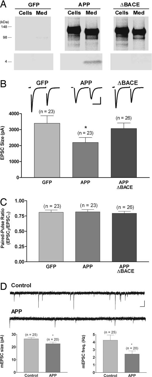
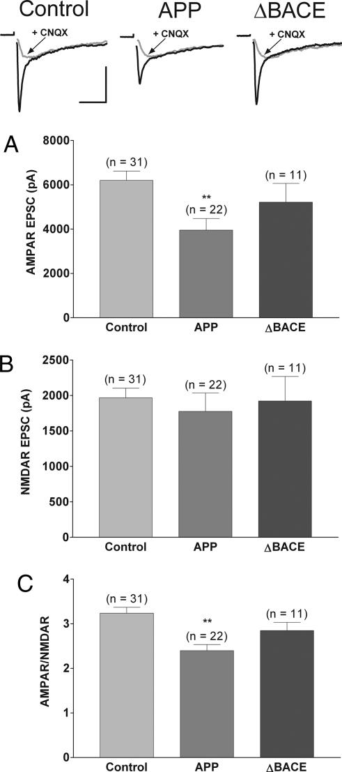
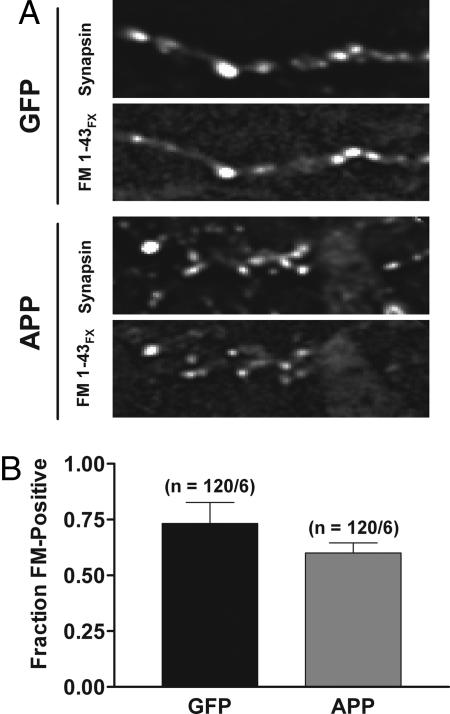
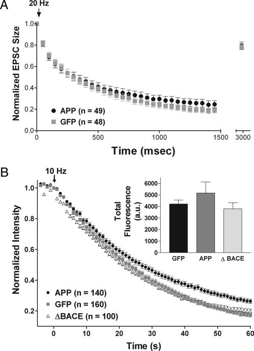
Overexpression of the amyloid precursor protein (APP) in hippocampal neurons leads to elevated beta-amyloid peptide (Abeta) production and consequent depression of excitatory transmission. The precise mechanisms underlying APP-induced synaptic depression are poorly understood. Uncovering these mechanisms could provide insight into how neuronal function is compromised before cell death during the early stages of Alzheimer's disease. Here we verify that APP up-regulation leads to depression of transmission in cultured hippocampal autapses; and we perform whole-cell recording, FM imaging, and immunocytochemistry to identify the specific mechanisms accounting for this depression. We find that APP overexpression leads to postsynaptic silencing through a selective reduction of alpha-amino-3-hydroxy-5-methyl-4-isoxazolepropionic acid (AMPA) receptor-mediated currents. This effect is likely mediated by Abeta because expression of mutant APP incapable of producing Abeta did not depress transmission. In addition, although we eliminate presynaptic silencing as a mechanism underlying APP-mediated inhibition of transmission, we did observe an Abeta-induced presynaptic deficit in vesicle recycling with sustained stimulation. These findings demonstrate that APP elevation disrupts both presynaptic and postsynaptic compartments.


Conflict of interest statement

The authors declare no conflict of interest.

Figures

Fig. 1.
Fig. 1.
Overexpression of wild-type APP but not a mutant APP that is unable to produce Aβ reduces excitatory postsynaptic current (EPSC) size without altering the paired-pulse ratio (PPR); overexpression of APP also reduces mEPSC amplitude and frequency. (A) Immunoprecipitation by using 6E10 of [35S]methionine-labeled APP (Upper) and Aβ (Lower) from cells and medium showed that infection with viral constructs encoding either APP or APPΔBACE directed expression of full-length, cell-associated APP and secreted APPsα in medium. Viral constructs encoding GFP served as a control for these experiments. Only neurons overexpressing wild-type APP generated significant levels of Aβ. (B) Average EPSC size was reduced in neurons overexpressing wild-type APP (0.62 of GFP-alone control; ∗, P < 0.05) but not in neurons overexpressing APPΔBACE. Traces show typical paired-pulse responses from each group with action currents blanked for clarity. (Scale bar, 1.5 nA, 10 ms.) (C) There was no difference in the PPR, measured as the relative sizes of the first and second responses to a pair of stimuli delivered 50 ms apart, in neurons overexpressing wild-type APP or APPΔBACE compared with GFP-alone-expressing neurons. (D) There was a small but significant decrease in mEPSC size (0.85 of control; ∗, P < 0.02) and a reduction in mEPSC frequency (0.56 of control; ∗, P < 0.04) in neurons overexpressing wild-type APP. For these mEPSC experiments, results from uninfected and GFP-expressing neurons, which were indistinguishable, were combined for the control group. Traces show typical spontaneous responses from control and APP-overexpressing neurons. (Scale bar, 20 pA, 100 ms.)
Fig. 2.
Fig. 2.
Overexpression of APP but not APPΔBACE reduces the AMPAR/NMDAR amplitude ratio by selectively decreasing AMPAR-mediated currents. (Top) Representative EPSCs recorded in Mg2+-free external solution ± 10 μM 6-cyano-7-nitroquinoxaline-2,3-dione (CNQX) with action currents blanked for clarity. (Scale bars, 3 nA, 20 ms.) (A) Summary of average AMPAR-mediated EPSC peak amplitude showing a significant reduction in neurons overexpressing APP (0.64 of control; ∗∗, P < 0.01). (B) Summary of average NMDAR-mediated EPSC peak amplitude showing no change in neurons overexpressing APP. (C) Summary of average AMPAR/NMDAR amplitude ratios for control neurons and neurons overexpressing APP (0.74 of control; ∗∗, P < 0.01). For these experiments, uninfected and GFP-expressing neurons, which were indistinguishable, were combined for the control group.
Fig. 3.
Fig. 3.
APP overexpression does not alter the fraction of presynaptically active synapses. (A) Raw fluorescent images show puncta labeled with antisynapsin antibody and fixable FM1-43FX in synapses expressing GFP alone (Upper) and in synapses overexpressing APP (Lower). (B) The fraction of synapsin-positive puncta that colabeled with FM1-43FX was not significantly different in neurons overexpressing APP.
Fig. 4.
Fig. 4.
Overexpression of APP has no effect on depression during a brief train, refilling of the RRP, or size of the cycling vesicle pool, but it reduces the rate of FM destaining, likely through an Aβ-mediated mechanism. (A) EPSC amplitudes in response to a 20-Hz train of stimuli lasting 1.5 s depressed at comparable rates in APP- and GFP-alone-expressing neurons. The amplitude of a single EPSC evoked 1.5 s after the end of the train was used to measure recovery from depletion of the pool of readily releasable vesicles. Each data point was normalized to the initial EPSC amplitude. (B) Time course of FM 4-64 fluorescence changes of puncta in neurons expressing APP, APPΔBACE, or GFP alone during electrical field stimulation at 10 Hz (arrow marks start of stimulation at time 0), plotted as mean fluorescence of each individual puncta normalized to the value immediately preceding stimulation. (Inset) Average total releasable fluorescence of individual sites; there was no difference among groups.

References

    1. LaFerla FM. Nat Rev Neurosci. 2002;3:862–872. - PubMed
    1. Mattson MP. Nature. 2004;430:631–639. - PMC - PubMed
    1. Selkoe DJ. Science. 2002;298:789–791. - PubMed
    1. Small DH, Mok SS, Bornstein JC. Nat Rev Neurosci. 2001;2:595–598. - PubMed
    1. Price DL, Sisodia SS. Annu Rev Neurosci. 1998;21:479–505. - PubMed

Publication types

LinkOut - more resources