. 2007 Jan 2;104(1):151-6.
doi: 10.1073/pnas.0607063104. Epub 2006 Dec 21.

The zinc-finger antiviral protein recruits the RNA processing exosome to degrade the target mRNA

Affiliations

The zinc-finger antiviral protein recruits the RNA processing exosome to degrade the target mRNA

Xuemin Guo et al. Proc Natl Acad Sci U S A. .

Erratum in

Abstract

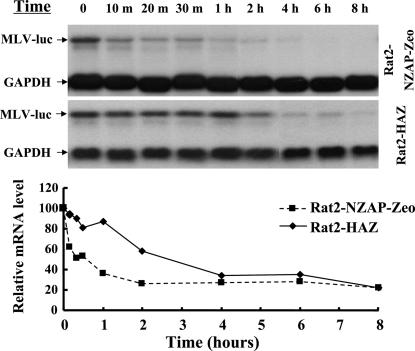
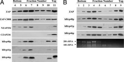
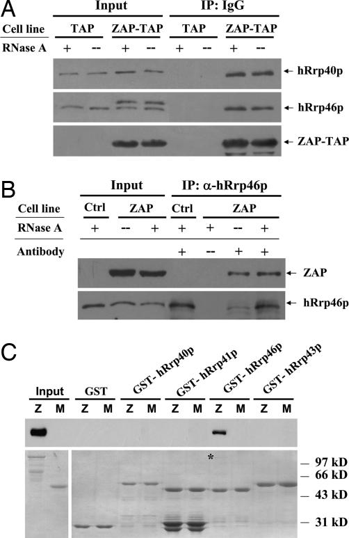
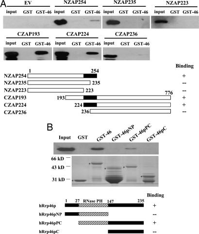
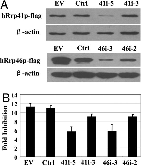
Zinc-finger antiviral protein (ZAP) is a host antiviral factor that specifically inhibits the replication of Moloney murine leukemia virus (MLV) and Sindbis virus (SIN) by preventing accumulation of the viral mRNA in the cytoplasm. In previous studies, we demonstrated that ZAP directly binds to its specific target mRNAs. In this article, we provide evidence indicating that ZAP recruits the RNA processing exosome to degrade the target RNA. ZAP comigrated with the exosome in sucrose or glycerol velocity gradient centrifugation. Immunoprecipitation of ZAP coprecipitated the exosome components. In vitro pull-down assays indicated that ZAP directly interacted with the exosome component hRrp46p and that the binding region of ZAP was mapped to amino acids 224-254. Depletion of the exosome component hRrp41p or hRrp46p with small interfering RNA significantly reduced ZAP's destabilizing activity. These findings suggest that ZAP is a trans-acting factor that modulates mRNA stability.


Conflict of interest statement

The authors declare no conflict of interest.

Figures

Fig. 1.
Fig. 1.
ZAP promotes target viral mRNA degradation in the cytoplasm. Rat2-HAZ and Rat2-NZAP-Zeo cells were infected with MLV-luc virus. Forty-eight hours after infection, actinomycin D was added to the media at a final concentration of 5 μg/ml to stop transcription. (Upper) At the indicated time points, the cells were lysed and the cytoplasmic RNA was extracted and subjected to Northern blotting. (Lower) The relative levels of the viral mRNA were measured by using a phosphorimager and were normalized by the GAPDH mRNA level.
Fig. 2.
Fig. 2.
ZAP cosediments with the exosome components. 293TRex cells expressing various versions of ZAP were lysed in the hypotonic buffer. The lysates were fractionated by sucrose (A) or glycerol (B) velocity gradient centrifugation. Equal aliquots of each fraction were subjected to SDS/PAGE and were Western blotted with the 9E10 anti-myc antibody or antibodies against the exosome components hRrp40p, hRrp41p, or hRrp46p. Total RNA was extracted from equal aliquots of each fraction, and the 28S and 18S rRNAs were detected by ethidium bromide staining
Fig. 3.
Fig. 3.
Interaction between ZAP and the exosome components. (A) The 293 cells expressing ZAP (1–332)-TAP or TAP alone were lysed in the lysis buffer in the presence (+) or absence (−) of 100 μg/ml RNase A. The proteins were precipitated with IgG Sepharose and were Western blotted with anti-protein A antibody to detect the TAP-tag or with the anti-hRrp40p or anti-hRrp46p antibody to detect the exosome components. Input, total cell lysates; TAP, TAP-expressing control cells; ZAP-TAP, ZAP (1–332)-TAP-expressing cells. (B) 293TRex-ZAP cells were treated with tetracycline to induce ZAP expression and lysed in the presence (+) or absence (−) of 100 μg/ml RNase A. The proteins were immunoprecipitated with preimmune (−) or anti-hRrp46p antisera and were Western blotted with the 9E10 anti-myc antibody. Input, total cell lysates; Ctrl, 293TRex control cells; ZAP, 293TRex-ZAP cells. (C) GST, GST-hRrp40p, GST-hRrp41p, GST-hRrp43p, and GST-hRrp46p expressed in E. coli were treated with RNase A at a final concentration of 100 μg/ml, immobilized onto glutathione-Sepharose 4B resin, and incubated with MBP-ZAP-myc in the presence of RNase A for 2 h at 4°C. The resins were washed, and the proteins were resolved by SDS/PAGE and detected by Western blotting by using the 9E10 anti-myc antibody (Upper) or Coomassie blue staining (Lower). Z, MBP-ZAP; M, MBP.
Fig. 4.
Fig. 4.
Mapping the interaction region for ZAP and hRrp46p. (A) Recombinant GST or GST-hRrp46p was immobilized onto glutathione-Sepharose 4B resin and incubated with the lysates of the indicated ZAP truncation mutants in the presence of RNase A (final concentration, 100 μg/ml) for 2 h at 4°C. The resins were washed, and the proteins were resolved by SDS/PAGE and detected by Western blotting by using the 9E10 anti-myc antibody. Input, total cells lysates. (B) The indicated recombinant GST proteins were immobilized onto glutathione-Sepharose 4B resin. 293TRex-ZAP cells were treated with tetracycline to induce ZAP expression, and the lysate was treated with RNase A (final concentration, 100 μg/ml) at 37°C for 30 min and then incubated with the resins for 2 h at 4°C. The resins were washed, and the proteins were resolved by SDS/PAGE and detected by Western blotting by using the 9E10 anti-myc antibody (Upper) or Coomassie blue staining (Lower). The positions of the GST proteins are indicated by asterisks.
Fig. 5.
Fig. 5.
Down-regulation of hRrp41p and hRrp46p with the interfering RNA reduces ZAP's activity. (A) The plasmids expressing the indicated FLAG-tagged exosome components were cotransfected into HEK293 cells with empty vector, control RNAi, or plasmids expressing the RNAi directed against the indicated exosome components. The relative expression levels of the FLAG-tagged exosome components were analyzed by Western blot analysis. (B) pMLV-luc, pRL-TK, and the indicated RNAi expressing plasmid were cotransfected into 293TRex-ZAP cells. Immediately after transfection, tetracycline was added to induce ZAP expression. Forty-eight hours after transfection, the luciferase activities were measured. The expression level of pRL-TK, a plasmid expressing Renilla luciferase and not responsive to ZAP, was used to normalize transfection efficiency. Fold inhibition is calculated as the normalized luciferase activity in the untreated cells divided by the normalized luciferase activity in the tetracycline-treated cells. The data are means + SD of three independent experiments.

References

    1. Bevilacqua A, Ceriani MC, Capaccioli S, Nicolin A. J Cell Physiol. 2003;195:356–372. - PubMed
    1. Guhaniyogi J, Brewer G. Gene. 2001;265:11–23. - PubMed
    1. Khodursky AB, Bernstein JA. Trends Genet. 2003;19:113–115. - PubMed
    1. Vasudevan S, Peltz SW. Curr Opin Cell Biol. 2003;15:332–337. - PubMed
    1. Vasudevan S, Peltz SW, Wilusz CJ. BioEssays. 2002;24:785–788. - PubMed

Publication types