. 2007 Feb;8(2):194-9.
doi: 10.1038/sj.embor.7400881. Epub 2006 Dec 22.

Acidic residues of yeast frataxin have an essential role in Fe-S cluster assembly

Affiliations

Acidic residues of yeast frataxin have an essential role in Fe-S cluster assembly

Françoise Foury et al. EMBO Rep. 2007 Feb.

Abstract

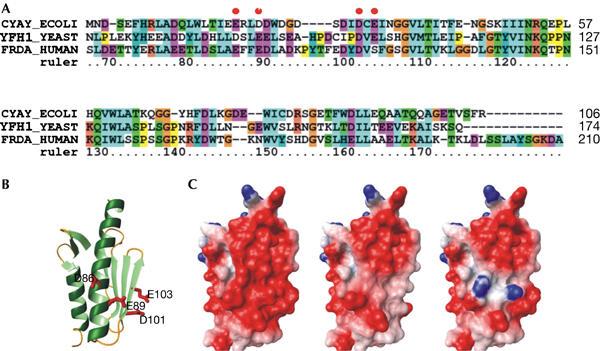
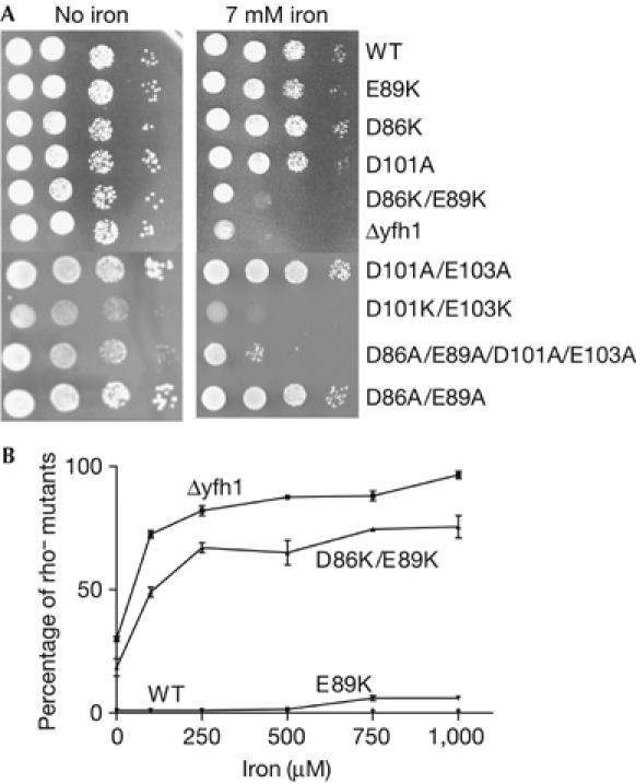
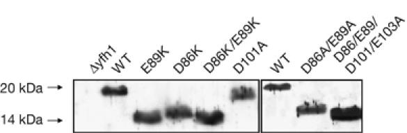
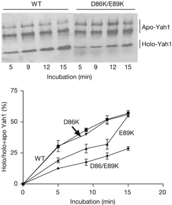
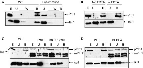
Friedreich ataxia is caused by decreased levels of frataxin, a mitochondrial acidic protein that is assumed to act as chaperone in the assembly of Fe-S clusters on the scaffold Isu protein. Frataxin has the in vitro capacity to form iron-loaded multimers, which also suggests an iron storage function. It has been reported that alanine substitution of residues in an acidic ridge of yeast frataxin (Yfh1) elicits loss of iron binding in vitro but has no effect on Fe-S cluster synthesis in vivo. Here, we show that a marked change in the electrostatic properties of a specific region of Yfh1 surface - by substituting two or four acidic residues by lysine or alanine, respectively - impairs Fe-S cluster assembly, weakens the interaction between Yfh1 and Isu1, and increases oxidative damage. Therefore, the acidic ridge is essential for the Yfh1 function and is likely to be involved in iron-mediated protein-protein interactions.


Figures

Figure 1
Figure 1
Sequence and structural comparisons. (A) Sequence alignment of the conserved domain of Escherichia coli (CyaY), yeast (Yfh1) and human frataxins. The numbering refers to the Yfh1 sequence. (B) Ribbon representation of the conserved domain of Yfh1 (2ga5). The side chains of the residues mutated in this article are shown in red. (C) Comparison of the electrostatic surfaces of Yfh1 wild type (left), D86A/E89A/D101A/E103A (middle) and D86K/E89K (right).
Figure 2
Figure 2
Iron sensitivity of wild-type and mutant cells. (A) Serial dilutions of cell suspensions were spotted onto plates containing glucose minimum medium and different concentrations of FeSO4, and cells were grown at 28°C for 3 days. (B) Cells were grown in liquid glucose-rich medium for 16 h in the presence of increasing concentrations of FeSO4 and spread for single colonies on minimum medium. The small colonies were identified as respiratory-deficient rho mutants. WT, wild type; Δyfh1, YFH1 gene-deleted strain.
Figure 3
Figure 3
Electrophoretic mobility of wild-type and mutant Yfh1 in SDS–polyacrylamide gels. A 40 μg portion of mitochondrial proteins was loaded onto 14% polyacrylamide gels. Yfh1 was detected by western blot analysis using a polyclonal Yfh1 antibody. D86/E89/D101/E103A is D86A/E89A/D101A/E103A. WT, wild type.
Figure 4
Figure 4
Conversion of 35S-radiolabelled apo- to holo-Yah1 in isolated energized mitochondria from wild-type and mutant strains. In native gel electrophoresis, the acidic mature form of Yah1 gives a fast migrating holo-form and two slowly migrating reduced and oxidized apo-forms (Leibrecht & Kessler, 1997). Exposure time of the autoradiography was 10 days. The experiment was carried out twice for each strain. WT, wild type.
Figure 5
Figure 5
Physical association of Yfh1 and Isu1 in wild-type and mutant cell extracts, using immunoaffinity chromatography with a polyclonal antibody raised against Yfh1. Cell extracts (E), unbound (U) and bound (B) fractions were analysed by SDS–polyacrylamide gel electrophoresis and western blotting using antibodies against Yfh1 and Isu1. (A) Interaction between Yfh1 and Isu1 (wild type) and replacement of Yfh1 antibody by the pre-immune serum (pre-immune). (B) EDTA (5 mM) was added during the washing steps. (C, D) The interaction between Yfh1 and Isu1 is weak in yfh1 mutants. pYfh1 (precursor) and mYfh1 (mature) are shown for the mutants and only mYfh1 is shown for the wild type. In the mutants, the lower band corresponds to a degradation product of Yfh1. DEDEA, D86A/E89A/D101A/E103A; W, washing step fractions; WT, wild type.

References

    1. Adamec J, Rusnak F, Owen WG, Naylor S, Benson LM, Gacy AM, Isaya G (2000) Iron-dependent self assembly of recombinant yeast frataxin: implications for Friedreich ataxia. Am J Hum Genet 67: 549–562 - PMC - PubMed
    1. Adinolfi S, Trifuoggi M, Politou AS, Martin S, Pastore A (2002) A structural approach to understanding the iron-binding properties of phylogenetically different frataxins. Hum Mol Genet 11: 1865–1877 - PubMed
    1. Aloria K, Schilke B, Andrew A, Craig EA (2004) Iron-induced oligomerization of yeast frataxin homologue Yfh1 is dispensable in vivo. EMBO Rep 5: 1096–1101 - PMC - PubMed
    1. Babcock M, de Silva D, Oaks R, Davis-Kaplan S, Jiralerspong S, Montermini L, Pandolfo M, Kaplan J (1997) Regulation of mitochondrial iron accumulation by Yfh1p, a putative homolog of frataxin. Science 276: 1709–1712 - PubMed
    1. Branda SS, Cavadini P, Adamec J, Kalousek F, Taroni F, Isaya G (1999) Yeast and human frataxin are processed to mature form in two sequential steps by the mitochondrial processing peptidase. J Biol Chem 274: 22763–22769 - PubMed

Publication types

MeSH terms