Comparative Study
. 2007 Feb;8(2):188-93.
doi: 10.1038/sj.embor.7400878. Epub 2006 Dec 22.

Herpes simplex virus eliminates host mitochondrial DNA

Affiliations
Comparative Study

Herpes simplex virus eliminates host mitochondrial DNA

Holly A Saffran et al. EMBO Rep. 2007 Feb.

Abstract

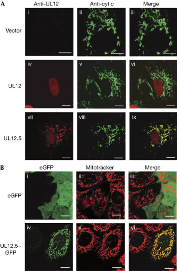
Mitochondria have crucial roles in the life and death of mammalian cells, and help to orchestrate host antiviral defences. Here, we show that the ubiquitous human pathogen herpes simplex virus (HSV) induces rapid and complete degradation of host mitochondrial DNA during productive infection of cultured mammalian cells. The depletion of mitochondrial DNA requires the viral UL12 gene, which encodes a conserved nuclease with orthologues in all herpesviruses. We show that an amino-terminally truncated UL12 isoform-UL12.5-localizes to mitochondria and triggers mitochondrial DNA depletion in the absence of other HSV gene products. By contrast, full-length UL12, a nuclear protein, has little or no effect on mitochondrial DNA levels. Our data document that HSV inflicts massive genetic damage to a crucial host organelle and show a novel mechanism of virus-induced shutoff of host functions, which is likely to contribute to the cell death and tissue damage caused by this widespread human pathogen.


Figures

Figure 1
Figure 1
HSV-1 depletes mt DNA and mt mRNA in infected Vero cells. Mt mRNA (A) and mt DNA (B) were monitored by northern and Southern blot hybridization, respectively, by using probes for COX2 and ND6. Infections with the indicated HSV-1 strains (10 PFU/cell) were allowed to proceed for 4, 8 and 12 h. Mt mRNA (C) and mt DNA (D) loss in Vero cells and 6-5 cells that express UL12 after HSV infection. COX2, cytochrome c oxidase subunit II; HSV-1, herpes simplex virus type 1; mt, mitochondrial; ND6, nicotinamide adenine dinucleotide dehyrogenase subunit 6; PFU, plaque forming unit; WT, wild type.
Figure 2
Figure 2
UL12.5 localizes to mitochondria. (A) HeLa cells were transfected with empty vector (i–iii), plasmids expressing UL12 (iv–vi) or UL12.5 (vii–ix) and then analysed by immunofluorescence for UL12/UL12.5 (i,iv,vii; red) and cytochrome c (ii,v,viii; green). The merged images (iii,vi,ix) document the colocalization of UL12.5 with cytochrome c. (B) UL12.5–eGFP fusion protein localizes to mitochondria. Live HeLa cells were transfected with plasmids encoding eGFP (i–iii) or UL12.5–eGFP (iv–vi) and stained with MitoTracker Red. The merged images (iii,vi) document the localization of UL12.5–eGFP to mitochondria. Scale bars, 10 μm. eGFP, enhanced green fluorescent protein.
Figure 3
Figure 3
UL12.5 is sufficient for mt DNA depletion. (A) Cytoplasmic PicoGreen foci localize to mitochondria. 143B cells co-transfected with empty vector and pcDNA-mOrange plasmid were stained with PicoGreen (green) and MitoTracker Far Red (red). The mOrange signal is not shown. Panel ii shows a higher magnification of the boxed region in panel i, documenting the association of PicoGreen-stained mt DNA nucleoids with the MitoTracker signal. (B) 143B cells were co-transfected with pcDNA-mOrange and empty vector (i,iv), plasmids expressing UL12 (ii,v) or UL12.5 (iii,vi). Colonies arising from individual cells expressing mOrange were examined by staining with PicoGreen and Mitotracker Far Red two days after plating. The mOrange signal is not shown. Panels iv, v and vi show a higher magnification of the boxed regions in panels i, ii and iii, respectively. Scale bars, 10 μm. mt, mitochondrial.
Figure 4
Figure 4
Isolation of rho0 143B cell lines. (A) Clonal cell lines isolated after transient expression of UL12.5 were scored for mt DNA content by staining with PicoGreen and MitoTracker Red. (i) Control 143B cells, (ii) clone E4, which retains mt DNA and (iii,iv) clones A8 and E2, which lack mt DNA. Scale bars, 10 μm. (B) Rho0 143B cells show the nutritional requirements of cells lacking mt DNA. The indicated cell lines were cultured in the presence (+) and absence (−) of supplemented pyruvate and uridine. mt, mitochondrial; rho0, cells depleted of mt DNA.

References

    1. Ashley N, Harris D, Poulton J (2005) Detection of mitochondrial DNA depletion in living human cells using PicoGreen staining. Exp Cell Res 303: 432–446 - PubMed
    1. Beal MF (2005) Mitochondria take center stage in aging and neurodegeneration. Ann Neurol 58: 495–505 - PubMed
    1. Chandrasekaran K, Mehrabian Z, Li XL, Hassel B (2004) RNase-L regulates the stability of mitochondrial DNA-encoded mRNAs in mouse embryo fibroblasts. Biochem Biophys Res Commun 325: 18–23 - PubMed
    1. Dimauro S (2004) Mitochondrial medicine. Biochim Biophys Acta 1659: 107–114 - PubMed
    1. Fernandez-Silva P, Enriquez JA, Montoya J (2003) Replication and transcription of mammalian mitochondrial DNA. Exp Physiol 88: 41–56 - PubMed

Publication types