. 2006 Dec;79(6):1030-9.
doi: 10.1086/509900. Epub 2006 Oct 24.

Mutations of presenilin genes in dilated cardiomyopathy and heart failure

Affiliations

Mutations of presenilin genes in dilated cardiomyopathy and heart failure

Duanxiang Li et al. Am J Hum Genet. 2006 Dec.

Abstract

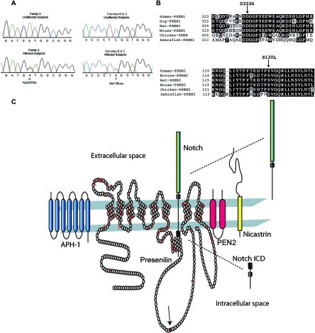
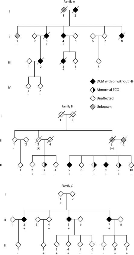
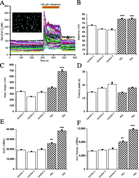
Two common disorders of the elderly are heart failure and Alzheimer disease (AD). Heart failure usually results from dilated cardiomyopathy (DCM). DCM of unknown cause in families has recently been shown to result from genetic disease, highlighting newly discovered disease mechanisms. AD is the most frequent neurodegenerative disease of older Americans. Familial AD is caused most commonly by presenilin 1 (PSEN1) or presenilin 2 (PSEN2) mutations, a discovery that has greatly advanced the field. The presenilins are also expressed in the heart and are critical to cardiac development. We hypothesized that mutations in presenilins may also be associated with DCM and that their discovery could provide new insight into the pathogenesis of DCM and heart failure. A total of 315 index patients with DCM were evaluated for sequence variation in PSEN1 and PSEN2. Families positive for mutations underwent additional clinical, genetic, and functional studies. A novel PSEN1 missense mutation (Asp333Gly) was identified in one family, and a single PSEN2 missense mutation (Ser130Leu) was found in two other families. Both mutations segregated with DCM and heart failure. The PSEN1 mutation was associated with complete penetrance and progressive disease that resulted in the necessity of cardiac transplantation or in death. The PSEN2 mutation showed partial penetrance, milder disease, and a more favorable prognosis. Calcium signaling was altered in cultured skin fibroblasts from PSEN1 and PSEN2 mutation carriers. These data indicate that PSEN1 and PSEN2 mutations are associated with DCM and heart failure and implicate novel mechanisms of myocardial disease.


Figures

Figure  1.
Figure 1.
Presenilin sequencing electropherograms, sequence alignments, and protein structure, with known mutations and components of the γ-secretase complex. A, DNA-sequencing electropherograms for PSEN1 and PSEN2. The sequencing electropherogram for family A demonstrated heterozygosity in PSEN1 at nucleotide 1539, which changed the wild-type aspartic acid to glycine. For families B and C, heterozygosity was demonstrated in PSEN2 at nucleotide 756, which changed the wild-type serine to a leucine. B, PSEN1 and PSEN2 sequence alignments. The amino acid sequences of PSEN1 and PSEN2 from various species are shown and indicate that the mutations occurred in highly conserved residues. C, Presenilin 1 protein structure and mutation locations and the γ-secretase complex. The putative transmembrane structure of the 467-aa presenilin 1 protein is shown, including known AD mutations,, denoted by the crossed symbols. Most AD mutations reside in the transmembrane domains, whereas the DCM mutation (solid symbol with arrow) identified in family A resides in the large, intracytoplasmic loop. The proposed topology of the 448-aa presenilin 2 protein (not shown) closely resembles that of presenilin 1. The PSEN2 mutation identified in families B and C resides at position 130 in its first extracellular domain, near a previously reported AD mutation at aa 122, which is also in the first extracellular domain. PSEN1 is shown accompanied by APH-1, nicastrin, and PEN2, proteins that constitute the γ-secretase complex and are required for γ-secretase activity., Notch is shown as a representative member of the type I membrane protein family. Notch, following extracellular binding of its ligands Delta or Jagged, undergoes presenilin-mediated γ-secretase cleavage, which generates its active, intracellular signaling domain that translocates to the nucleus and acts as a transcriptional regulator in diverse systems, including cardiovascular development and disease. APP and several other transmembrane protein substrates, including Erb-B4 and E- and N-cadherins, are similarly processed.
Figure  2.
Figure 2.
Pedigrees of families A, B, and C with presenilin mutations. Diamonds represent either males or females. Symbols with a diagonal line represent deceased family members. Black symbols indicate family members affected with DCM with or without heart failure, half–filled symbols represent those with abnormal ECGs, gray symbols represent family members who were of unknown status, and white symbols represent family members who were unaffected. The presence (+) or absence (−) of PSEN1 (family A) or PSEN2 (families B and C) mutations are indicated for those from whom DNA was available. For family B, a plus sign within parentheses (+) indicates obligate carriers of the PSEN2 mutation.
Figure  3.
Figure 3.
Histological examination of LV myocardium from an affected member of family A and controls. Histological examination was performed on LV myocardial tissue from the explanted heart taken at the time of cardiac transplantation of an affected member (II.3) of family A, who carried the PSEN1 mutation. A, Regions of myocardial dropout and irregular fibrosis, consistent with remote ischemic damage (Gomori’s modified trichrome; 2×). B–D, Pleomorphic hyperchromatic myocardial nuclei. Panel B demonstrates the diffuse nature of this change (20×). Panels C and D demonstrate rectangular boxcar nuclei (hematoxylin and eosin staining; 60×). E, Control ventricular myocardium, from a patient with amyloid cardiomyopathy, that demonstrates birefringent amyloid deposits (Congo Red under polarized light; 40×). F, Myocardium from subject II.3, in which no amyloid was observed (Congo Red under polarized light; 40×). G, Immunoperoxidase staining of control tissue from a patient with AD and congophilic amyloid angiopathy, which demonstrates strong reactivity with the antioligomeric antibody (AHB0052) (40×). H, Ventricular myocardium from subject II.3, which demonstrated no evidence of reactivity with AHB0052 (40×).
Figure  4.
Figure 4.
Histamine-induced alteration of intracellular calcium concentration ([Ca2+]i) in cultured skin fibroblasts derived from control subjects and those with a PSEN1 or PSEN2 mutation. A, Representative experiment from cells loaded with fura-2/AM. A photomicrograph from one dish of control cells is shown (inset). The baseline was recorded for 300 s before exposure to 100 μM histamine for 150 s. Each tracing represents the measured [Ca2+]i from a single fibroblast. B, Baseline [Ca2+]i measured before the application of histamine. Baseline [Ca2+]i was higher in fibroblasts cultured from patients with the PSEN1 or PSEN2 mutations than in fibroblasts cultured from control subjects. The data shown include the mean (± SEM) responses from control patient 1 (six dishes; 453 cells), control patient 2 (four dishes; 241 cells), control patient 3 (three dishes; 98 cells), a PSEN1 mutation carrier (eight dishes; 440 cells), and a PSEN2 mutation carrier (eight dishes; 561 cells). There was an average of 62 cells per dish, with a range of 21–92 cells. C, Maximal [Ca2+]i response to histamine. The histamine-induced [Ca2+]i response was increased in fibroblasts with the PSEN2 mutation compared with those of three controls. The response from fibroblasts with the PSEN1 mutation was different from one of three controls (P<.0001). The results shown are the peak responses in [Ca2+]i to 100 μM histamine. D, Elapsed time to the maximal [Ca2+]i responses following histamine application. No difference was observed between the fibroblasts derived from control patients and those with a PSEN1 or PSEN2 mutation. The elapsed time was measured in seconds from the histamine application to the maximal response of [Ca2+]i. The perfusion flow rate was high compared with the dish volume; therefore, cells were exposed to 100 μM histamine in <3 s. E, Area under the curve (AUC) of the histamine-induced increase in [Ca2+]i. The total AUC of the histamine-induced increase in [Ca2+]i was higher in fibroblasts from patients with the PSEN1 and PSEN2 mutation than in fibroblasts from control patients. F, AUC during the plateau phase of the histamine-induced increase in [Ca2+]i. The mean plateau phase [Ca2+]i response in fibroblasts cultured from patients with the PSEN1 or PSEN2 mutations was greater than the responses from fibroblasts cultured from control patients. The AUC during the plateau phase of the histamine application was measured from 390 to 450 s. A triple asterisk (***) indicates P<.0001 in three of three controls (Bonferroni/Dunn t test); a double asterisk (**) indicates P<.0001 in two of three controls.

References

Web Resources

    1. OHSU Familial Dilated Cardiomyopathy Research Project, http://www.fdc.to/
    1. Online Mendelian Inheritance in Man (OMIM), http://www.ncbi.nlm.nih.gov/Omim/ (for AD, PSEN1, PSEN2, DCM, LMNA, MYH7, TNNT2, SCN5A, CSRP3, and PLN) - PubMed

References

    1. Hunt SA (2005) ACC/AHA 2005 guideline update for the diagnosis and management of chronic heart failure in the adult: a report of the American College of Cardiology/American Heart Association Task Force on Practice Guidelines (Writing Committee to Update the 2001 Guidelines for the Evaluation and Management of Heart Failure). J Am Coll Cardiol 46:e1–e8210.1016/j.jacc.2005.08.022 - DOI - PubMed
    1. Nussbaum RL, Ellis CE (2003) Alzheimer’s disease and Parkinson’s disease. N Engl J Med 348:1356–136410.1056/NEJM2003ra020003 - DOI - PubMed
    1. Sherrington R, Rogaev EI, Liang Y, Rogaeva EA, Levesque G, Ikeda M, Chi H, et al (1995) Cloning of a gene bearing missense mutations in early-onset familial Alzheimer’s disease. Nature 375:754–76010.1038/375754a0 - DOI - PubMed
    1. Hardy J, Crook R (2001) Presenilin mutations line up along transmembrane α-helices. Neurosci Lett 306:203–20510.1016/S0304-3940(01)01910-3 - DOI - PubMed
    1. Rogaev EI, Sherrington R, Rogaeva EA, Levesque G, Ikeda M, Liang Y, Chi H, Lin C, Holman K, Tsuda T, Mar L, Sorbi S, Nacmias B, Piacentini S, Amaducci L, Chumakov I, Cohen D, Lannfelt L, Fraser PE, Rommens JM, St George-Hyslop PH (1995) Familial Alzheimer’s disease in kindreds with missense mutations in a gene on chromosome 1 related to the Alzheimer’s disease type 3 gene. Nature 376:775–77810.1038/376775a0 - DOI - PubMed

Publication types

MeSH terms