. 2006 Dec;79(6):1081-8.
doi: 10.1086/509044. Epub 2006 Oct 3.

Mutant desmocollin-2 causes arrhythmogenic right ventricular cardiomyopathy

Affiliations

Mutant desmocollin-2 causes arrhythmogenic right ventricular cardiomyopathy

Arnd Heuser et al. Am J Hum Genet. 2006 Dec.

Abstract

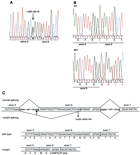
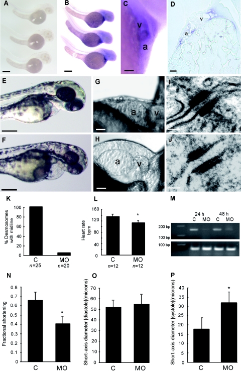
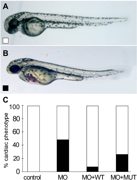
Arrhythmogenic right ventricular cardiomyopathy (ARVC) is a genetically heterogeneous heart-muscle disorder characterized by progressive fibrofatty replacement of right ventricular myocardium and an increased risk of sudden cardiac death. Mutations in desmosomal proteins that cause ARVC have been previously described; therefore, we investigated 88 unrelated patients with the disorder for mutations in human desmosomal cadherin desmocollin-2 (DSC2). We identified a heterozygous splice-acceptor-site mutation in intron 5 (c.631-2A-->G) of the DSC2 gene, which led to the use of a cryptic splice-acceptor site and the creation of a downstream premature termination codon. Quantitative analysis of cardiac DSC2 expression in patient specimens revealed a marked reduction in the abundance of the mutant transcript. Morpholino knockdown in zebrafish embryos revealed a requirement for dsc2 in the establishment of the normal myocardial structure and function, with reduced desmosomal plaque area, loss of the desmosome extracellular electron-dense midlines, and associated myocardial contractility defects. These data identify DSC2 mutations as a cause of ARVC in humans and demonstrate that physiologic levels of DSC2 are crucial for normal cardiac desmosome formation, early cardiac morphogenesis, and cardiac function.


Figures

Figure  1.
Figure  1.
DSC2 mutation c.631-2A→G causes ARVC. A, Partial nucleotide sequence of DSC2 intron 5–exon 6 junction. The genomic sequence shows a heterozygous A→G transition in the splice-acceptor site position of intron 5. B, Partial nucleotide sequence of cloned wild-type (WT) and mutant (MUT) transcripts derived from DSC2 cDNA of the mutation carrier (pCR2.1 plasmid) (TOPO-TA cloning [Invitrogen]). The mutant transcript shows a deletion of the first 25 bp of exon 6, which causes a frameshift and a premature stop codon after 10 novel aa residues (MGILQNFHCP*). C, Schematic view of the splicing mechanism in normal and mutant DSC2. The mutation c.631-2A→G (arrowhead) affects the splice-acceptor site of intron 5 and activates an alternative cryptic splice site (arrow) in exon 6. The consequence is a 25-bp deletion that shifts the reading frame and generates a premature stop codon.
Figure  2.
Figure  2.
Clinical features of the mutation carrier. A, Twelve-lead resting ECG (50 mm/s). Note the T-wave inversions in the leads V1–V6 and the epsilon wave (arrow) in V1. B, ECG (25 mm/s) of a ventricular tachycardia with left bundle branch block morphology. C, Image of an angiogram showing an enlarged right ventricle in 30° right-anterior oblique, with evidence of diastolic bulging (arrowheads) at the right ventricle inflow tract and diffuse inferior hypokinesia (arrows).
Figure  3.
Figure  3.
Expression analysis of the mutant DSC2. A, Allele-specific quantitative real-time PCR of DSC2 wild-type (WT) and mutant (MUT) mRNA of cardiac tissue derived from patient (P) and control (C) samples. Note the reduced expression (3%) of the mutant allele in cardiac tissue in the patient. B, Western-blot analysis of a right ventricular biopsy sample obtained from the patient with the DSC2 mutation c.631-2A→G. Note that the amount of DSC2 in the patient is reduced when compared with control myocardium. Blots were probed with antibodies for DSC2 (Progen, 1:10,000) and α-tubulin (Sigma-Aldrich, 1:5,000) as loading control.
Figure  4.
Figure  4.
Zebrafish dsc2 expression and knockdown. AD, Zebrafish dsc2 expression. Whole-mount in situ hybridizations with use of full-length sense (A) and antisense (B) zebrafish dsc2 riboprobe show weak staining in the heart and brain at 48 hpf with the antisense probe, whereas the sense control riboprobe does not reveal any staining. Scale bars indicate 100 μM. Higher magnification image of the heart (C) and longitudinal section through the ventricle (D) of whole-mount in situ stained embryos confirm cardiac expression. Scale bars indicate 25 μM. a = Atrium; v = ventricle. EP, Knockdown of dsc2 in zebrafish. E, Phenotype at 48 hpf of an embryo injected with mismatch control oligo compared with a morphant (injected with 25 ng of morpholino dsc2-1) embryo at the same stage (F). Scale bars indicate 100 μM. High-resolution pictures of the heart of morphant embryo (H) compared with control-injected embryo (G). Scale bars indicate 25 μM. Transmission EM of cardiac desmosomes from a control embryo (I) and a morphant embryo (J) at 48 hpf. Scale bars indicate 50 nm. K, Percentage of desmosomes with midline in control (C) and morphant (MO) embryos. L, Heart rate measured at room temperature in morphant embryos compared with control injected embryos (P<.05). M, RT-PCR of zebrafish dsc2, to determine morpholino efficiency. N, Fractional shortening of the ventricular chamber of control and morphant fish at 48 hpf. Diastolic (O) and systolic (P) short-axis diameter measured in controls and morphant embryos (P<.05).
Figure  5.
Figure  5.
Rescue of dsc2 morphants (MO). A, Wild-type phenotype. B, Morphant phenotype. C, Rescue of dsc2 morphant phenotype (blackened bars) is seen with wild-type (WT) (unblackened bars) but not with mutant (MUT) DSC2 mRNA. Results are based on data from three different experiments with 25 ng of morpholino and 1 pg of the respective capped mRNAs.

References

Web Resources

    1. GenBank, http://www.ncbi.nlm.nih.gov/Genbank/ (for DSC2 transcript variant [accession number NM_024422.2] and zebrafish DNA from clone [accession number BX649302])
    1. Online Mendelian Inheritance in Man (OMIM), http://www.ncbi.nlm.nih.gov/Omim/ (for ARVC/D 1–10, DSC2, DSG2, PPKS1, and LAH)

References

    1. Marcus FI, Fontaine GH, Guiraudon G, Frank R, Laurenceau JL, Malergue C, Grosgogeat Y (1982) Right ventricular dysplasia: a report of 24 adult cases. Circulation 65:384–398 - PubMed
    1. Fontaine G, Fontaliran F, Hebert JL, Chemla D, Zenati O, Lecarpentier Y, Frank R (1999) Arrhythmogenic right ventricular dysplasia. Annu Rev Med 50:17–3510.1146/annurev.med.50.1.17 - DOI - PubMed
    1. Thiene G, Nava A, Corrado D, Rossi L, Pennelli N (1988) Right ventricular cardiomyopathy and sudden death in young people. N Engl J Med 318:129–133 - PubMed
    1. Nava A, Thiene G, Canciani B, Scognamiglio R, Daliento L, Buja G, Martini B, Stritoni P, Fasoli G (1988) Familial occurrence of right ventricular dysplasia: a study involving nine families. J Am Coll Cardiol 12:1222–1228 - PubMed
    1. Wlodarska EK, Konka M, Kepski R, Zaleska T, Ploski R, Ruzyllo W, Janion M, Jaworska K, Rydlewska-Sadowska W, Hoffman P (2004) Familial form of arrhythmogenic right ventricular cardiomyopathy. Kardiol Pol 60:1–14 - PubMed

Publication types