. 2007 Mar;204(1):273-82.
doi: 10.1016/j.expneurol.2006.11.003. Epub 2006 Dec 21.

At-level neuropathic pain is induced by lumbosacral ventral root avulsion injury and ameliorated by root reimplantation into the spinal cord

Affiliations

At-level neuropathic pain is induced by lumbosacral ventral root avulsion injury and ameliorated by root reimplantation into the spinal cord

A J Bigbee et al. Exp Neurol. 2007 Mar.

Abstract

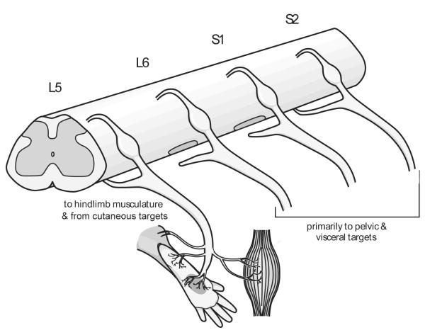
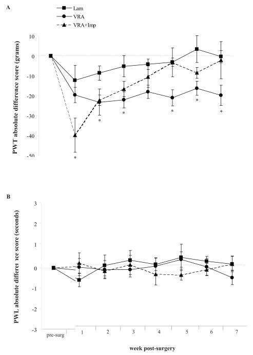
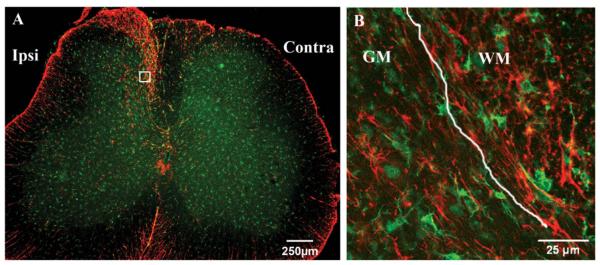
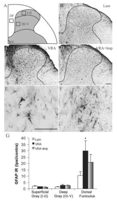
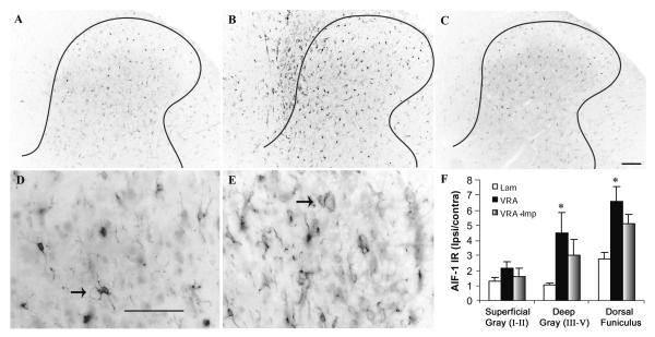
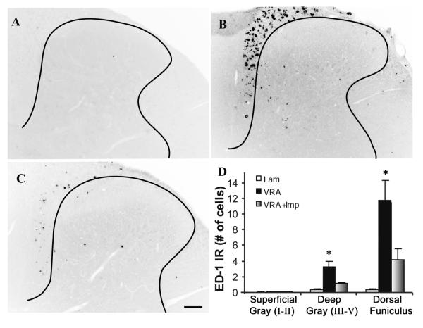
Neuropathic pain is common after traumatic injuries to the cauda equina/conus medullaris and brachial plexus. Clinically, this pain is difficult to treat and its mechanisms are not well understood. Lesions to the ventral roots are common in these injuries, but are rarely considered as potential contributors to pain. We examined whether a unilateral L6-S1 ventral root avulsion (VRA) injury in adult female rats might elicit pain within the dermatome projecting to the adjacent, uninjured L5 spinal segment. Additionally, a subset of subjects had the avulsed L6-S1 ventral roots reimplanted (VRA+Imp) into the lateral funiculus post-avulsion to determine whether this neural repair strategy elicits or ameliorates pain. Behavioral tests for mechanical allodynia and hyperalgesia were performed weekly over 7 weeks post-injury at the hindpaw plantar surface. Allodynia developed early and persisted post-VRA, whereas VRA+Imp rats exhibited allodynia only at 1 week post-operatively. Hyperalgesia was not observed at any time in any experimental group. Quantitative immunohistochemistry showed increased levels of inflammatory markers in laminae III-V and in the dorsal funiculus of the L5 spinal cord of VRA, but not VRA+Imp rats, specific to areas that receive projections from mechanoreceptive, but not nociceptive, primary afferents. These data suggest that sustained at-level neuropathic pain can develop following a pure motor lesion, whereas the pain may be ameliorated by acute root reimplantation. We believe that our findings are of translational research interest, as root implantation surgery is emerging as a potentially useful strategy for the repair of cauda equina/conus medullaris injuries.


Figures

Figure 1
Figure 1. The unilateral lumbosacral ventral root avulsion (VRA) model
The L6 and S1 ventral roots were avulsed unilaterally from the CNS/PNS interface of the spinal cord. For VRA+Imp rats, both ventral roots were reimplanted into the lateral funicuclus (not shown). Note that the injured L6 and S1 efferents project primarily to pelvic floor and visceral targets, while the intact L5 efferents project more so to hindlimb musculature and receive afferents from cutaneous targets. Behavioral tests for allodynia and hyperalgesia were performed at the L5 dermatome on the central footpads of the hindlimbs (gray shaded region).
Figure 2
Figure 2. Neuropathic pain following unilateral L6-S1 VRA and VRA + Imp
The paw withdrawal thresholds (PWT; in grams of force) and paw withdrawal latencies (PWL; in seconds) were determined as indicators of mechanical allodynia (A) and hyperalgesia (B), respectively, prior to surgery and weekly thereafter for 7 wks. Data are presented as an absolute difference score (i.e., the ipsilateral minus the contralateral values normalized to presurgical baseline levels). A) For mechanical allodynia, VRA is significantly reduced relative to Lam at 2,3,5,6 and 7 weeks, while VRA+Imp differs from Lam only at 1 wk. B) For hyperalgesia, no differences between groups were observed. Data shown are the mean values ± SEM. *, significantly different from Lam within each time point at p<0.05.
Figure 3
Figure 3. Unilateral L6-S1 VRA elicits an inflammatory response in the L5 ipsilateral dorsal horn
A) Astrocytes (GFAP; red) and microglia (AIF-1; green) in the L5 spinal cord at 8 wks post L6-S1 VRA. Note that the inflammatory response is restricted to the ipsilateral dorsal horn quadrant. A magnified region from the inset is shown in B), where astrocytes (red) and microglia (green) are found in both the gray matter (GM) and white matter (WM), and exhibit characteristic morphological properties. Calibration bars are 250- and 25 μm, respectively. The white line in (B) indicates the WM/GM border.
Figure 4
Figure 4. Increased activation of astrocytes in the ipsilateral L5 dorsal horn at 8 wks following unilateral L6-S1 VRA
A) Three regions of interest for quantitative immunohistochemistry were examined in the superficial gray matter (laminae I-II), deep gray matter (laminae III-V), and the white matter dorsal funiculus (DF). The central canal (CC) is also shown for orientation purposes. The boxes represent an area of 10,000 μm2. (B-D) GFAP immunoreactivity (IR) is shown in the ipsilateral L5 dorsal horn quadrants for Lam (B), VRA (C), and VRA+Imp subjects (D). Note the prominent upregulation of GFAP in the VRA DF (B) relative to Lam (A), while VRA+Imp is qualitatively similar to Lam. The black border identifies the gray matter/white matter border. Representative high magnification (40x) images for GFAP from VRA gray matter (E) and white matter (F) are also shown. Calibration bars are 100 μm. (G) VRA GFAP IR is significantly increased in the DF relative to Lam (p<0.05), while VRA+Imp is not different from Lam. GFAP IR in laminae I-II and laminae III-V did not significantly differ between groups. Values represent the area of GFAP-positive IR / 10,000 μm2 for the ipsilateral region relative to the same region on the contralateral side. Data shown are the mean values ± SEM. *, significantly different from Lam within each time point at p<0.05.
Figure 5
Figure 5. Increased microglial activation in the ipsilateral L5 dorsal horn at 8 wks following unilateral L6-S1 VRA
AIF-1 IR identifies microglia in the ipsilateral dorsal horn in Lam, VRA, and VRA+Imp rats. Note the low-intensity levels of staining in Lam (A) and VRA+Imp rats (C), while the intensity is substantially increased in VRA rats (B) in both laminae III-V and the DF. (D) AIF-1 positive microglia in the VRA laminae III-V show characteristic morphology with extended processes (arrow), while in the DF white matter (E), many AIF-1 positive cells show a more macrophage-type morphology (arrow) indicative of an active inflammatory response. Calibration bars are 100 μm. (F) In the VRA group, AIF-I IR was significantly increased in laminae III-V and in the DF relative to Lam, while VRA+Imp was not. There were no differences between groups in laminae I-II. Data shown are the mean values ± SEM. *, significantly different from Lam within each time point at p<0.05.
Figure 6
Figure 6. Macrophage response in the L5 ipsilateral dorsal horn of VRA rat at 8 wks post-avulsion of the L6-S1 ventral roots
(A) Few, if any, ED-1 positive macrophages were observed in Lam. (B) Macrophage staining was prominent in the L5 DF of VRA rats, and also, but to a lesser degree, in laminae III-V. (C) The macrophage response was ameliorated in the VRA+Imp rats. The calibration bar is 100 μm. (D) The number of ED-1 positive macrophages in VRA rats was significantly increased in both laminae III-V and in the DF relative to Lam (p<0.05), while VRA+Imp was not different from Lam in those regions. There were no group differences in laminae I-II. Data shown are the mean number of ED-1 positive cells / 10,000 μm2 region of interest ± SEM. *, significantly different from Lam within each time point at p<0.05.

Similar articles

Cited by

References

    1. Aldskogius H. Microglia in neuroregeneration. Microsc Res Tech. 2001;54:40–46. - PubMed
    1. Anneser JM, Berthele A, Borasio GD, Castro-Lopes JM, Zieglgansberger W, Tolle TR. Axotomy of the sciatic nerve differentially affects expression of metabotropic glutamate receptor mRNA in adult rat motoneurons. Brain Res. 2000;868:215–221. - PubMed
    1. Back SK, Kim JS, Hong SK, Na HS. Ascending pathways for mechanical allodynia in a rat model of neuropathic pain. Neuroreport. 2003;14:1623–1626. - PubMed
    1. Berman JS, Birch R, Anand P. Pain following human brachial plexus injury with spinal cord root avulsion and the effect of surgery. Pain. 1998;75:199–207. - PubMed
    1. Cairns BE, Gambarota G, Svensson P, Arendt-Nielsen L, Berde CB. Glutamate-induced sensitization of rat masseter muscle fibers. Neuroscience. 2002;109:389–399. - PubMed

Publication types

MeSH terms