. 2007 Feb 16;366(2):574-85.
doi: 10.1016/j.jmb.2006.11.068. Epub 2006 Dec 1.

Point mutations in the HIV-1 matrix protein turn off the myristyl switch

Affiliations

Point mutations in the HIV-1 matrix protein turn off the myristyl switch

Jamil S Saad et al. J Mol Biol. .

Abstract

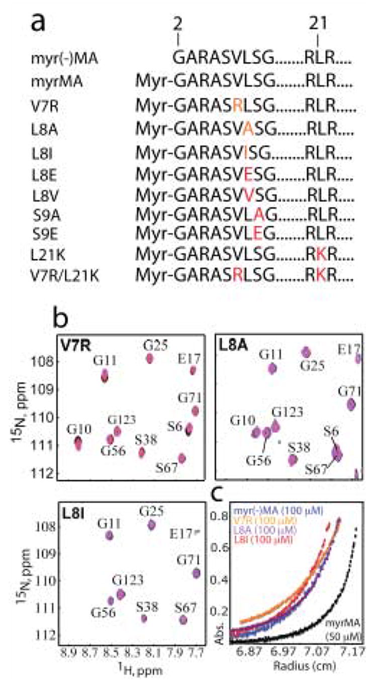
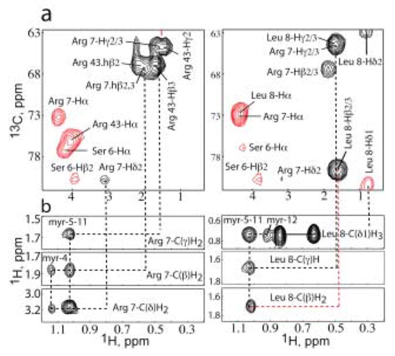
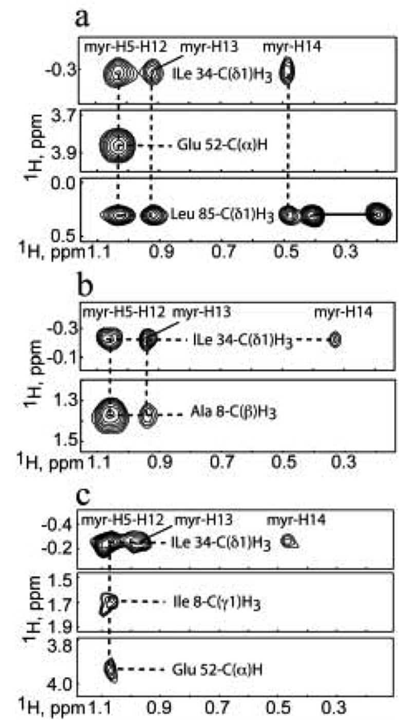
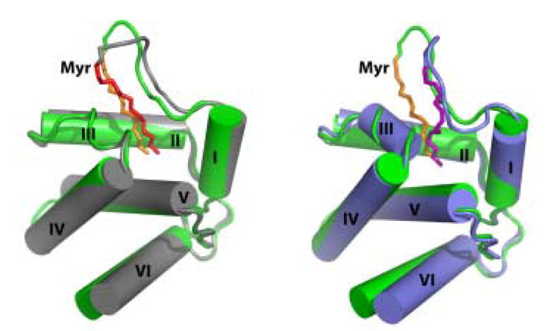
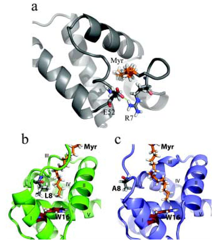
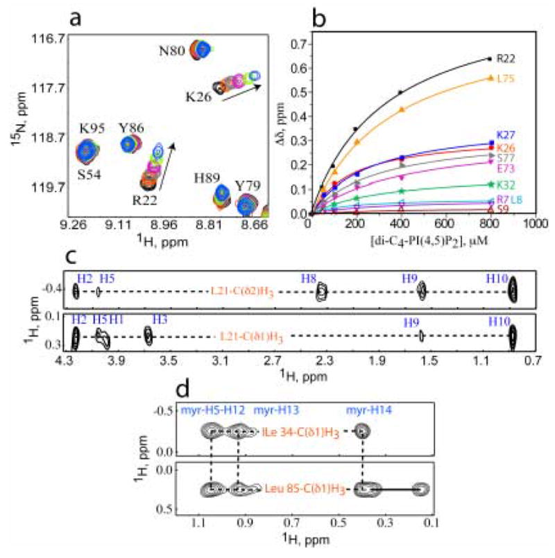
During the late phase of human immunodeficiency virus type-1 (HIV-1) replication, newly synthesized retroviral Gag proteins are targeted to lipid raft regions of specific cellular membranes, where they assemble and bud to form new virus particles. Gag binds preferentially to the plasma membrane (PM) of most hematopoietic cell types, a process mediated by interactions between the cellular PM marker phosphatidylinositol-(4,5)-bisphosphate (PI(4,5)P(2)) and Gag's N-terminally myristoylated matrix (MA) domain. We recently demonstrated that PI(4,5)P(2) binds to a conserved cleft on MA and promotes myristate exposure, suggesting a role as both a direct membrane anchor and myristyl switch trigger. Here we show that PI(4,5)P(2) is also capable of binding to MA proteins containing point mutations that inhibit membrane binding in vitro, and in vivo, including V7R, L8A and L8I. However, these mutants do not exhibit PI(4,5)P(2) or concentration-dependent myristate exposure. NMR studies of V7R and L8A MA reveal minor structural changes that appear to be responsible for stabilizing the myristate-sequestered (myr(s)) species and inhibiting exposure. Unexpectedly, the myristyl group of a revertant mutant with normal PM targeting properties (V7R,L21K) is also tightly sequestered and insensitive to PI(4,5)P(2) binding. This mutant binds PI(4,5)P(2) with twofold higher affinity compared with the native protein, suggesting a potential compensatory mechanism for membrane binding.


Figures

Figure 1
Figure 1
(a) A representation of the MA sequence showing positions of the mutations in red. (b) Overlay of 2D 1H-15N HSQC spectra collected at different concentrations [600–900 μM (black), 300 μM red, 150 μM (blue), 50 μM (magenta)] for V7R, L8A, and L8I samples. (c) Representative sedimentation profiles obtained for myr(-)MA, WT and mutant myrMA proteins [(26,000 rpm, 20°C, 100 μM)]. For WT myrMA, best fits were obtained for a monomer-trimer equilibrium affording association constant (Kassoc) of 2.5 ± 0.6 × 10⁸ M−2.27 Sedimentation equilibrium curves for myr(-)MA, V7R, L8A, and L8I fit best to monomeric species.
Figure 2
Figure 2
Representative NMR data obtained for V7R. (a) 4D 15N,13C-edited NOE data showing intramolecular and intermolecular NOEs for residues Arg-7 (left) and Leu-8 (right). NOE cross-peaks to the myristate group are not observed because the myristate group is not 13C-labeled. (b) 13C-edited/12C-double-half-filtered NOE data showing unambiguously assigned intermolecular NOEs between unlabeled myristyl group and 13C-labeled protein. Solid lines denote 1H-12C breakthrough doublets NOE peaks.
Figure 3
Figure 3
(a) Representative 13C-edited/12C-double-half-filtered NOE data showing unambiguously assigned intermolecular NOEs between unlabeled myristyl group and 13C-labeled V7R (a), L8A (b), and L8I (c). Solid and dashed lines denote 1H-12C breakthrough doublets and intermolecular NOE peaks, respectively.
Figure 4
Figure 4
Stereoviews showing the best-fit backbone superpositions of the 20 refined structures calculated for V7R (top) and L8A (bottom) proteins. Myristate group is shown in red.
Figure 5
Figure 5
Representative structures of V7R (grey) and L8A (slate) superimposed with WT myrMA protein (green). Myristate groups of WT myrMA, V7R- and L8A proteins are shown in orange, red and purple, respectively. NMR data revealed that Helix VI is flexible for native and mutant myrMA proteins.
Figure 6
Figure 6
Cartoon and stick representation showing minor conformational adjustments in the N terminus of V7R and L8A proteins compared with WT myrMA. (a) For V7R, a salt bridge between Arg-7 and Glu-52 stabilizes the myr(s) form. (b) When compared with WT myrMA (green), substitution of Leu-8 with Ala (slate, c) creates a better cavity for the myristate group (orange) and allows for the terminal methyl of myristate group to pack closer to Trp-16 (firebrick).
Figure 7
Figure 7
(a) Overlay of 2D 1H-15N HSQC spectra upon titration of V7R with di-C4-PI(4,5)P2 [50 μM, 35 °C; di-C4-PI(4,5)P2:MA = 0:1 (black), 1:1 (red) , 2:1 (grey), 4:1 (magenta), 8:1 (green), 16:1 (blue)]. (b) 15N NMR chemical shift titration data, which fit to 1:1 binding isotherms (Kd = 261 ± 18 μM). Representative 13C-edited/12C-double-half-filtered NOE data showing unambiguously assigned intermolecular NOEs between (c) Leu-21 and di-C4-PI(4,5)P2, and (d) Ile-34 and Leu-85 to the myristate group. Solid and dashed lines denote 1H-12C breakthrough doublets and intermolecular NOE peaks, respectively.
Figure 8
Figure 8
Subcellular localization of MA mutants. HeLa cells were transfected with WT pNL4-3 or derivatives encoding the indicated MA mutants. Cells were fixed, permeabilized, and stained with anti-p17 monoclonal antibody and Texas red-conjugated anti-mouse IgG. Cells were mounted and examined using a Delta Vision RT microscope and the images were deconvolved. Two representative fields for WT, V7R, and V7R/L21K are shown.

References

    1. Coffin JM, Hughes SH, Varmus HE. Retroviruses. Cold Spring Harbor Laboratory Press; Plainview, N.Y.: 1997. - PubMed
    1. Morita E, Sundquist WI. Retrovirus Budding. Annual Review of Cell and Develpomental Biology. 2004;20:395–425. - PubMed
    1. von Schwedler UK, Stuchell M, Muller B, Ward DM, Chung HY, Morita E, Wang HE, Davis T, He GP, Cimbora DM, Scott A, Krausslich HG, Kaplan J, Morham SG, Sundquist WI. The protein network of HIV budding. Cell. 2003;114:701–713. - PubMed
    1. Dong X, Li H, Derdowski A, Ding L, Burnett A, Chen X, Peters TR, Dermody TS, Woodruff E, Wang JJ, Spearman P. AP-3 directs the intracellular trafficking of HIV-1 Gag and plays a key role in particle assembly. Cell. 2005;120:663–674. - PubMed
    1. Hermida-Matsumoto L, Resh MD. Localization of Human Immunodeficiency virus Type 1 Gag and env at the Plasma Membrane by Confocal Imagine. Journal of Virology. 2000;74:8670–8679. - PMC - PubMed

Publication types

Substances