

Absence of surface expression of feline infectious peritonitis virus (FIPV) antigens on infected cells isolated from cats with FIP
- PMID: 17188823
- PMCID: PMC7127496
- DOI: 10.1016/j.vetmic.2006.11.026
Absence of surface expression of feline infectious peritonitis virus (FIPV) antigens on infected cells isolated from cats with FIP
Abstract
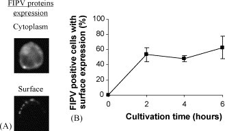
Feline infectious peritonitis virus (FIPV) positive cells are present in pyogranulomas and exudates from cats with FIP. These cells belong mainly to the monocyte/macrophage lineage. How these cells survive in immune cats is not known. In this study, FIPV positive cells were isolated from pyogranulomas and exudates of 12 naturally FIPV-infected cats and the presence of two immunologic targets, viral antigens and MHC I, on their surface was determined. The majority of the infected cells were confirmed to be cells from the monocyte/macrophage lineage. No surface expression of viral antigens was detected on FIPV positive cells. MHC I molecules were present on all the FIPV positive cells. After cultivation of the isolated infected cells, 52+/-10% of the infected cells re-expressed viral antigens on the plasma membrane. In conclusion, it can be stated that in FIP cats, FIPV replicates in cells of the monocyte/macrophage lineage without carrying viral antigens in their plasma membrane, which could allow them to escape from antibody-dependent cell lysis.
Figures

Similar articles
-
Diagnosis of Feline Infectious Peritonitis: A Review of the Current Literature.Viruses. 2019 Nov 15;11(11):1068. doi: 10.3390/v11111068. Viruses. 2019. PMID: 31731711 Free PMC article. Review.
-
Characterization of antiviral T cell responses during primary and secondary challenge of laboratory cats with feline infectious peritonitis virus (FIPV).BMC Vet Res. 2019 May 22;15(1):165. doi: 10.1186/s12917-019-1909-6. BMC Vet Res. 2019. PMID: 31118053 Free PMC article.
-
Pathogenesis of oral type I feline infectious peritonitis virus (FIPV) infection: Antibody-dependent enhancement infection of cats with type I FIPV via the oral route.J Vet Med Sci. 2019 Jun 21;81(6):911-915. doi: 10.1292/jvms.18-0702. Epub 2019 Apr 23. J Vet Med Sci. 2019. PMID: 31019150 Free PMC article.
-
Antibody-dependent enhancement of feline infectious peritonitis virus infection in feline alveolar macrophages and human monocyte cell line U937 by serum of cats experimentally or naturally infected with feline coronavirus.J Vet Med Sci. 1998 Jan;60(1):49-55. doi: 10.1292/jvms.60.49. J Vet Med Sci. 1998. PMID: 9492360
-
Strategies for combating FIPV infection: antiviral agents and vaccines.Res Vet Sci. 2025 Aug;192:105709. doi: 10.1016/j.rvsc.2025.105709. Epub 2025 May 27. Res Vet Sci. 2025. PMID: 40446698 Review.
Cited by
-
Diagnosis of Feline Infectious Peritonitis: A Review of the Current Literature.Viruses. 2019 Nov 15;11(11):1068. doi: 10.3390/v11111068. Viruses. 2019. PMID: 31731711 Free PMC article. Review.
-
A review of feline infectious peritonitis virus infection: 1963-2008.J Feline Med Surg. 2009 Apr;11(4):225-58. doi: 10.1016/j.jfms.2008.09.008. Epub 2009 Feb 28. J Feline Med Surg. 2009. PMID: 19254859 Free PMC article. Review. No abstract available.
-
The paradox of feline coronavirus pathogenesis: a review.Adv Virol. 2011;2011:109849. doi: 10.1155/2011/109849. Epub 2011 Aug 21. Adv Virol. 2011. PMID: 22312333 Free PMC article.
-
Myosins 1 and 6, myosin light chain kinase, actin and microtubules cooperate during antibody-mediated internalisation and trafficking of membrane-expressed viral antigens in feline infectious peritonitis virus infected monocytes.Vet Res. 2014 Feb 12;45(1):17. doi: 10.1186/1297-9716-45-17. Vet Res. 2014. PMID: 24517254 Free PMC article.
-
Feline infectious peritonitis. ABCD guidelines on prevention and management.J Feline Med Surg. 2009 Jul;11(7):594-604. doi: 10.1016/j.jfms.2009.05.008. J Feline Med Surg. 2009. PMID: 19481039 Free PMC article. Review.
References
-
- Cammarata Parodi M., Cammarata G., Paltrinieri S., Lavazza A., Ape F. Using direct immunofluorescence to detect coronaviruses in peritoneal and pleural effusions. J. Small Anim. Pract. 1993;34:609–613.
-
- Dewerchin H.L., Cornelissen E., Nauwynck H.J. Feline infectious peritonitis virus infected monocytes internalize viral membrane-bound proteins upon antibody addition. J. Gen. Virol. 2006;87:1685–1690. - PubMed
-
- Favoreel, H.W., 1999. Antibody-induced clearance of viral and cellular plasma membrane proteins from pseudorabies virus-infected cells, and its possible role in immune evasion. PhD Thesis, University of Ghent.
Publication types
MeSH terms
Substances
LinkOut - more resources
Full Text Sources
Research Materials
Miscellaneous
