. 2006 Dec 28;24(6):891-901.
doi: 10.1016/j.molcel.2006.11.027.

The checkpoint clamp activates Mec1 kinase during initiation of the DNA damage checkpoint

Affiliations

The checkpoint clamp activates Mec1 kinase during initiation of the DNA damage checkpoint

Jerzy Majka et al. Mol Cell. .

Abstract

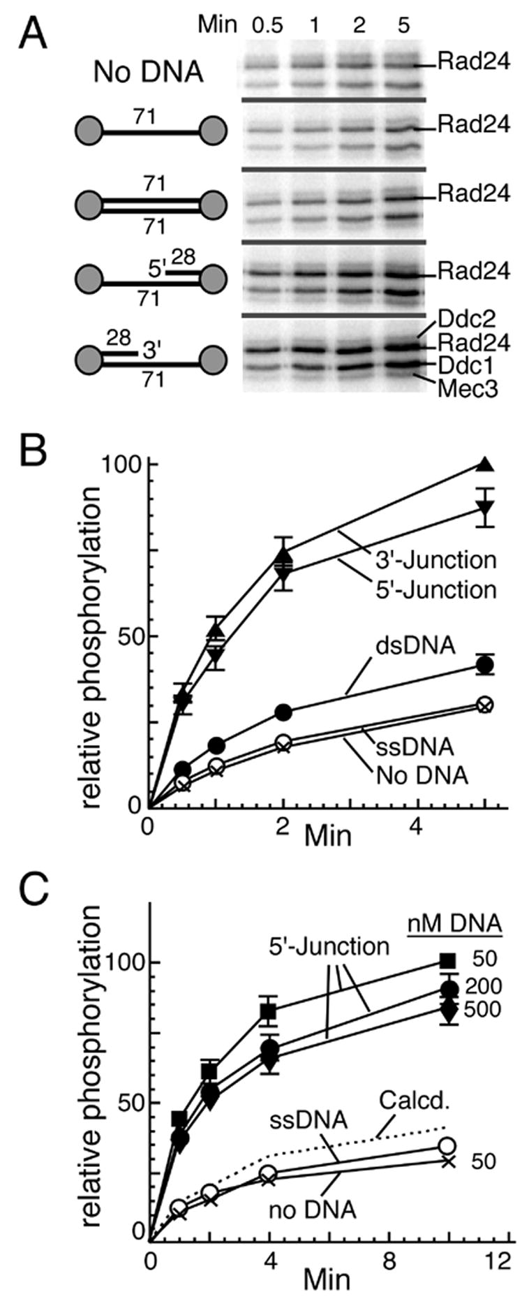
Yeast Mec1/Ddc2 protein kinase, the ortholog of human ATR/ATRIP, plays a central role in the DNA damage checkpoint. The PCNA-like clamp Rad17/Mec3/Ddc1 (the 9-1-1 complex in human) and its loader Rad24-RFC are also essential components of this signal transduction pathway. Here we have studied the role of the clamp in regulating Mec1, and we delineate how the signal generated by DNA lesions is transduced to the Rad53 effector kinase. The checkpoint clamp greatly activates the kinase activity of Mec1, but only if the clamp is appropriately loaded upon partial duplex DNA. Activated Mec1 phosphorylates the Ddc1 and Mec3 subunits of the clamp, the Rad24 subunit of the loader, and the Rpa1 and Rpa2 subunits of RPA. Phosphorylation of Rad53, and of human PHAS-1, a nonspecific target, also requires a properly loaded clamp. Phosphorylation and binding studies with individual clamp subunits indicate that the Ddc1 subunit mediates the functional interactions with Mec1.


Figures

Figure 1
Figure 1. Mec1 forms a heterodimer with Ddc2
(A) Purification of Mec1/Ddc2. Colloidal Coomassie-stained 7% SDS-polyacrylamide gel of the preparation after glutathione-affinity chromatography (lane 1), treatment with rhinoviral protease (lane 2), and heparin agarose and gel filtration chromatography (lane 3). (B) Sedimentation velocity analysis of the Mec1/Ddc2 complex. Mec1/Ddc2 was sedimented in buffer D400 at protein concentrations of 0.2 and 0.4 mg/ml at 5°C and 35,000 rpm. Apparent sedimentation coefficient distributions g(s*) are given as well as the non-linear least square fits (smooth lines) to the sum of two Gaussian functions, centered at sapp = 3.8S and sapp = 6.2S, with residuals depicted below the graph. Sedimentation and diffusion coefficients were used to calculate the molecular mass of Mec1/Ddc2 complex (See supplemental for full details). Open arrow points to a minor species at sapp = 3.8S, presumably excess Ddc2 in the preparation.
Figure 2
Figure 2. Phosphorylation by Mec1/Ddc2 is stimulated by a loaded checkpoint clamp
(A) Scheme of the assay. The 3 kb Bluescript ssDNA contained ten roughly equally spaced 28-mer primers. (B) Phosphorylation reactions were carried out at 30 °C for 30 min in buffer containing 125 mM NaCl (See Experimental procedures for details). All reactions contained 5 nM Mec1/Ddc2. Other factors and DNA when present were: 2.5 nM deca-primed ssDNA (25 nM primers), 500 nM RPA, 30 nM Rad24-RFC, and 100 nM Rad17/3/1. Rpa2 band intensities indicate the average number of phosphate groups per Rpa2 molecule (Pi/Rpa2). (C) Clamp and clamp loader specificity for Mec1 stimulation. Assays were as in (B) with 100 nM of the indicated clamp and 30 nM of the indicated loader. Abbreviations are: 17/3/1, Rad17/3/1; Rad24, Rad24-RFC, Ctf18, Ctf18-RFC; Elg1, Elg1-RFC; Rfc1, RFC; Rad24-KE in lane 17 indicates Rad24K115E-RFC defective for loading of Rad17/3/1 (Majka et al., 2004). Total phosphorylation intensities in lanes 1–15 and in 16,17 are relative to the positive controls (lanes 4 and 16, respectively).
Figure 3
Figure 3. The clamp mediates Mec1/Ddc2 activity on DNA substrates lacking RPA
(A) Phosphorylation reactions (100 μl) containing the indicated effector oligonucleotide, Rad17/3/1, Rad24-RFC, and Mec1/Ddc2, but no RPA, were carried out in buffer containing 100 mM NaCl at 30 °C. At the indicated time points, 20 μl aliquots were analyzed by SDS-10%-PAGE followed by phosphorimaging. (B) Quantitation of the data in (A). Combined radioactivity of bands corresponding to Rad24, Ddc1 and Mec3 proteins was determined and set to 100% for the highest phosphorylation. 5′-and 3′-Junction designates 5′- and 3′- primer-template junction DNA, respectively, as shown in (A). (C) Reactions as in (A) contained 30 nM Mec1/Ddc2, 30 nM Rad24-RFC, 50 nM Rad17/3/1, and increasing concentrations of DNA as indicated. The dashed line labeled “calcd.” represents the theoretical response for 500 nM DNA if Rad17/3/1 and Mec1/Ddc2 distributed independently over the available DNA templates. Error bars are defined in Experimental procedures
Figure 4
Figure 4. Efficient phosphorylation of RPA and Rad53-kd by Mec1 requires proper loading of Rad17/3/1
(A) Time course of phosphorylation reactions containing the indicated effector oligonucleotide, RPA, Rad17/3/1, Rad24-RFC, and Mec1/Ddc2, were carried out in buffer containing 100 mM NaCl at 30 °C. Analysis was by SDS-10%-PAGE followed by phosphorimaging. See Experimental procedures for details. (B) Quantitation of Rpa2 phosphorylation of the data in (A) and of additional control experiments: no DNA (open circles), ssDNA (filled circles), dsDNA (filled squares). (C) Assays as in (A) were carried for 5 min at 30 C, but contained in addition 125 nM Rad53-kd. DNA was omitted (lane 1) or was 3′-junction DNA (3′-J, lanes 2,3) or 5′-junction DNA (5′-J, lanes 4,5). Rad24-RFC was omitted in lanes 2 and 4. (D) Standard time course phosphorylation reactions containing Rad17/3/1, Rad24-RFC, Rad53-kd and Mec1/Ddc2, and the indicated effector oligonucleotide or no DNA, with or without RPA as indicated, were carried out in buffer containing 100 mM NaCl at 30 °C. See Experimental procedures for details and error analysis.
Figure 5
Figure 5. Phosphorylation of PHAS-1 by Mec1 requires proper loading of Rad17/3/1
(A) Time course of phosphorylation reactions containing the indicated effector oligonucleotide coated with RPA where indicated, Rad17/3/1, Rad24-RFC, Mec1/Ddc2, and 4 μM PHAS-1 were carried out in buffer containing 100 mM NaCl at 30 °C. Analysis was by SDS-10%-PAGE followed by phosphorimaging. Error bars are defined in Experimental procedures. (B) ATP-dependent lag phase reflects slow clamp loading. Complete reactions as in (A) with the exception of one component were preincubated at 30 C for 1 min and started by addition of the omitted component as indicated in the figure. (C) The minimal activation system does not show a ATP-dependent lag phase. Complete system is taken from (B). The minimal reaction containing no DNA, 100 nM Ddc1, 30 nM Mec1, and 4 μM PHAS-1 and 30 mM NaCl was preincubated at 30 C for 1 min and started by addition of ATP.
Figure 6
Figure 6. Low salt bypasses the requirement for a loaded Rad17/3/1 clamp in vitro
(A) A standard phosphorylation assay containing 5′-junction DNA, RPA, Rad17/3/1, Rad24-RFC, Mec1/Ddc2, and Rad53-kd was carried out at the indicated mM NaCl level for 5 min at 30 C. The indicated mM NaCl includes contributions from protein storage buffers. Individual components were omitted from the assay as indicated. Reactions were analyzed by SDS-10%-PAGE followed by phosphorimaging for quantitation of Rad53-kd phosphorylation. (B) Assays contained 100 nM of Rad17/3/1 or of the indicated clamp subunits, 30 nM Mec1/Ddc2 and 125 nM Rad53-kd for 5 min at 30 C. Note the difference in X-scale between (A) and (B). Error bars are defined in Experimental procedures (C) Pull-down assays were carried out as described in Experimental procedures with 160 nM proteins in buffer containing either 40 or 75 mM NaCl. Precipitated proteins were resolved on SDS-8%-PAGE followed by Coomassie staining. Arrowheads indicate non-specific bands present in the Mec1/Ddc2-only or clamp (subunit)-only pulldowns.
Figure 7
Figure 7. Mechanism of Mec1 kinase activation
Loading of the Rad17/3/1 clamp activates Mec1. The various types of phosphorylations by Mec1 are indicated, but not all targets are shown. Whether the clamp loader is released after loading, as indicated in the central complex, has not been established. The Ddc1-only bypass pathway of Mec1 activation, surrounded by a dotted line, is only operative at low salt in vitro and appears not to operate in the cell.

Similar articles

Cited by

References

    1. Bartrand AJ, Iyasu D, Brush GS. DNA stimulates Mec1-mediated phosphorylation of replication protein A. J Biol Chem. 2004;279:26762–26767. - PubMed
    1. Bellaoui M, Chang M, Ou J, Xu H, Boone C, Brown GW. Elg1 forms an alternative RFC complex important for DNA replication and genome integrity. EMBO J. 2003;22:4304–4313. - PMC - PubMed
    1. Binz SK, Sheehan AM, Wold MS. Replication protein A phosphorylation and the cellular response to DNA damage. DNA Repair (Amst) 2004;3:1015–1024. - PubMed
    1. Brush GS, Kelly TJ. Phosphorylation of the replication protein A large subunit in the Saccharomyces cerevisiae checkpoint response. Nucleic Acids Res. 2000;28:3725–3732. - PMC - PubMed
    1. Brush GS, Morrow DM, Hieter P, Kelly TJ. The ATM homologue MEC1 is required for phosphorylation of replication protein A in yeast. Proc Natl Acad Sci U S A. 1996;93:15075–15080. - PMC - PubMed

Publication types

MeSH terms