. 2007 Mar 15;92(6):1938-51.
doi: 10.1529/biophysj.106.093500. Epub 2006 Dec 22.

Subunit dependence of Na channel slow inactivation and open channel block in cerebellar neurons

Affiliations

Subunit dependence of Na channel slow inactivation and open channel block in cerebellar neurons

Teresa K Aman et al. Biophys J. .

Abstract

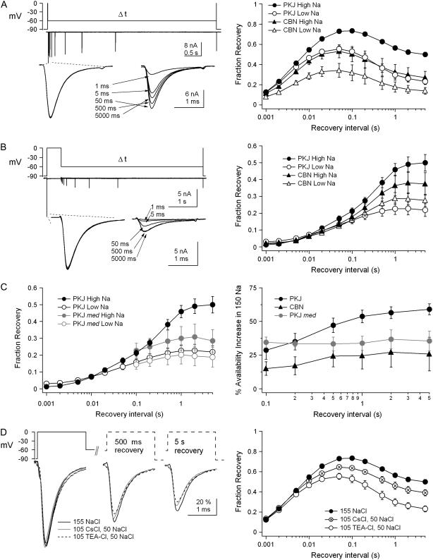
Purkinje and cerebellar nuclear neurons both have Na currents with resurgent kinetics. Previous observations, however, suggest that their Na channels differ in their susceptibility to entering long-lived inactivated states. To compare fast inactivation, slow inactivation, and open-channel block, we recorded voltage-clamped, tetrodotoxin-sensitive Na currents in Purkinje and nuclear neurons acutely isolated from mouse cerebellum. In nuclear neurons, recovery from all inactivated states was slower, and open-channel unblock was less voltage-dependent than in Purkinje cells. To test whether specific subunits contributed to this differential stability of inactivation, experiments were repeated in Na(V)1.6-null (med) mice. In med Purkinje cells, recovery times were prolonged and the voltage dependence of open-channel block was reduced relative to control cells, suggesting that availability of Na(V)1.6 is quickly restored at negative potentials. In med nuclear cells, however, currents were unchanged, suggesting that Na(V)1.6 contributes little to wild-type nuclear cells. Extracellular Na(+) prevented slow inactivation more effectively in Purkinje than in nuclear neurons, consistent with a resilience of Na(V)1.6 to slow inactivation. The tendency of nuclear Na channels to inactivate produced a low availability during trains of spike-like depolarization. Hyperpolarizations that approximated synaptic inhibition effectively recovered channels, suggesting that increases in Na channel availability promote rebound firing after inhibition.


Figures

FIGURE 1
FIGURE 1
Properties of inactivation in Purkinje and cerebellar nuclear cells. (A) Voltage protocols and representative traces for recovery from 2-ms (upper) and 500-ms (middle) conditioning steps. Recovery interval, 1–5000 ms. Overlay of representative traces from 500-ms conditioning and test steps at a higher gain (lower). (B) Fraction of available channels versus recovery interval. After 2-ms conditioning steps, recovery was biexponential. Purkinje cells (PKJ), N = 11; cerebellar nuclear cells (CBN), N = 10. After 500-ms conditioning steps, channels recovered in three exponential phases: fast, intermediate, and slow. PKJ, N = 8; CBN, N = 9. Parameters of the fits are given in Table 1. (C, upper) Voltage protocols and representative traces for entry into inactivated states. Conditioning pulse, 0.5–2500 ms. Current amplitudes were tested after a 100-ms interval to allow recovery from fast inactivation. (C, lower). Overlay of representative traces from conditioning and test steps at a higher gain. The current evoked by the briefest (1-ms) conditioning step deactivates upon repolarization, so the current terminates earlier than in the other traces. (D) Fraction of available channels versus conditioning pulse interval. Data are fitted with double exponentials (PKJ, N = 7; CBN, N = 7). Parameters of the fits are given in Table 1.
FIGURE 2
FIGURE 2
Resurgent Na current in wild-type and med neurons. (A) Voltage protocol to elicit resurgent current and representative traces from Purkinje and cerebellar nuclear cells. Currents are normalized to the peak of the transient Na current elicited by a step to 0 mV. (B, left and center bar groups) Average transient (0 mV) and resurgent current (−30 mV) amplitudes from PKJ wild-type (WT) (N = 78), PKJ med (N = 19), CBN WT (N = 48), and CBN med (N = 8) neurons (left axis). Note that transient current is divided by 10, for an axis range of 0–15 nA, whereas the resurgent-current amplitude range is 0–1.5 nA. (B, right bar group) Resurgent current normalized to the transient current at 0 mV (right axis).
FIGURE 3
FIGURE 3
Recovery from and entry into inactivated states in wild-type and med neurons. (A and C) Wild-type and med mean fraction of available channels versus recovery interval for (A) Purkinje neurons (2 ms: WT, N = 21; med, N = 7; 500 ms: WT, N = 13; med, N = 7) and (C) cerebellar nuclear neurons (2 ms: WT, N = 20; med, N = 8; 500 ms: WT, N = 17, med, N = 5). Channels recovered from a 2-ms step according to a double exponential and from a 500-ms step according to a triple exponential. Parameters of the fits are given in Table 1. Gray circles in C are the data from med Purkinje neurons, for comparison. For some data points, error bars are smaller than the symbols. (B and D) Wild-type and med average fraction of available channels versus conditioning pulse interval for (B) Purkinje neurons (WT, N = 12; med, N = 8) and (D) cerebellar nuclear neurons (WT, N = 15; med, N = 5). Channels entered slow-inactivated states in two phases (Table 1). Gray circles in D are the data from med Purkinje neurons, for comparison. For some data points, error bars are smaller than the symbols.
FIGURE 4
FIGURE 4
Interaction between open-channel block and slow inactivation in Purkinje cells. (A, left) Voltage protocols and representative traces for detecting the extent of block during long (50- to 500-ms) conditioning steps to different potentials, as labeled. (A, right) Normalized peak resurgent current versus conditioning pulse duration. Data are fitted with a single exponential, with parameters given in Results. For the +60-mV step, N = 6; for the +30-mV step, N = 7, and for the 0-mV step, N = 8. (B, upper) Voltage protocol and representative traces to test the effect of conditioning voltage on slow inactivation. (B, lower) Reference and test steps at higher gain, illustrating the onset of the difference in availability at longer conditioning durations. Test currents evoked after all four conditioning voltages are shown; arrows indicate largest and smallest amounts of recovery and are labeled with the corresponding conditioning voltages. (C) Plot of the percent availability increase after steps to −30, +30, and +60 mV relative to that after steps to 0 mV, as a function of conditioning duration. All recovery intervals were 100 ms. For intervals <100 ms, N = 8; for intervals >100 ms, N = 10. Dotted line indicates no change relative to 0 mV.
FIGURE 5
FIGURE 5
Voltage dependence of open-channel block. (A) Normalized peak resurgent current versus conditioning pulse duration for cerebellar nuclear neurons. Data from Purkinje cells are reproduced for comparison. Data are fitted with single exponentials, with parameters given in Results. For the +30-mV step, N = 5; for the 0-mV step, N = 6. (B) Voltage protocol and representative traces to measure the voltage dependence of block. (C) Resurgent current normalized to the resurgent current evoked after a step to +50 mV and fitted with a straight line for WT Purkinje cells (N = 8), WT nuclear cells (N = 6), and med Purkinje cells (N = 9).
FIGURE 6
FIGURE 6
Effect of permeant and impermeant ions on inactivation. (A and B, upper left) Voltage protocols and representative traces for recovery at −60 mV from 2-ms (A) and 500-ms (B) steps to 0 mV. (A and B, lower left) Responses to conditioning and test steps at higher gain. (A and B, right) Fraction of available channels versus recovery interval for Purkinje and cerebellar nuclear neurons in high and low Na for 2-ms conditioning steps for PKJ high Na (N = 11), PKJ low Na (N = 6), CBN high Na (N = 10), and CBN low Na (N = 8), and for 500-ms conditioning steps for PKJ high Na (N = 8), PKJ low Na (N = 7), CBN high Na (N = 6), and CBN low Na (N = 5). (C, left) Data as in B, right, but for med PKJ cells. High Na (N = 6), low Na (N = 6). Data from wild-type PKJ cells are superimposed for comparison. (C, right) Increase in availability in high Na relative to low Na for wild-type PKJ, med PKJ, and wild-type CBN cells. (D, left) Voltage protocol and representative traces for currents in 100 mM NaCl, CsCl, or TEA-Cl from Purkinje cells. (D, right) Fraction of available channels versus recovery interval for 155 NaCl, (N = 11), 105 CsCl and 50 NaCl (N = 5), and 105 TEA-Cl and 50 NaCl (N = 6).
FIGURE 7
FIGURE 7
Na-channel inactivation in cerebellar nuclear cells induced by quasiphysiological voltage steps. (A) Voltage protocol and representative traces for a cerebellar nuclear cell, mimicking depolarizations occurring during spontaneous firing at 20 Hz, a 250-ms period of inhibition, and a rebound burst of three action potentials at 100 Hz. (B) Peak transient Na currents normalized to the amplitude of the first current, plotted time locked to the traces in A (N = 5). For the rebound burst, symbols designate the voltage of the preceding hyperpolarization. (C, left) Voltage protocol and representative traces for rebound burst (B, boxed area) at high gain. (C, right) Rebound currents from B plotted versus recovery voltage (N = 5). The data set for each rebound current was fit with a straight line (rebound current 1, 2%/mV; rebound current 2, 0.5%/mV; rebound current 3, 0.3%/mV).

References

    1. Thach, W. 1968. Discharge of Purkinje and cerebellar nuclear neurons during rapidly alternating arm movements in the monkey. J. Neurophysiol. 31:785–797. - PubMed
    1. McDevitt, C. J., T. J. Ebner, and J. R. Bloedel. 1987. Relationships between simultaneously recorded Purkinje cells and nuclear neurons. Brain Res. 425:1–13. - PubMed
    1. Llinas, R., and M. Sugimori. 1980. Electrophysiological properties of in vitro Purkinje cell somata in mammalian cerebellar slices. J. Physiol. 305:171–195. - PMC - PubMed
    1. Jahnsen, H. 1986. Electrophysiological characteristics of neurones in the guinea-pig deep cerebellar nuclei in vitro. J. Physiol. 372:129–147. - PMC - PubMed
    1. Raman, I. M., and B. P. Bean. 1997. Resurgent sodium current and action potential formation in dissociated cerebellar Purkinje neurons. J. Neurosci. 17:4517–4526. - PMC - PubMed

Publication types

LinkOut - more resources